From flies' eyes to our ears: mutations in a human class III myosin cause progressive nonsyndromic hearing loss DFNB30
- PMID: 12032315
- PMCID: PMC124268
- DOI: 10.1073/pnas.102091699
From flies' eyes to our ears: mutations in a human class III myosin cause progressive nonsyndromic hearing loss DFNB30
Abstract
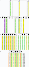
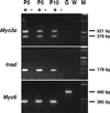
Normal vision in Drosophila requires NINAC, a class III myosin. Class III myosins are hybrid motor-signaling molecules, with an N-terminal kinase domain, highly conserved head and neck domains, and a class III-specific tail domain. In Drosophila rhabdomeres, NINAC interacts with actin filaments and with a PDZ scaffolding protein to organize the phototransduction machinery into a signaling complex. Recessive null mutations in Drosophila NINAC delay termination of the photoreceptor response and lead to progressive retinal degeneration. Here, we show that normal hearing in humans requires myosin IIIA, the human homolog of NINAC. In an extended Israeli family, nonsyndromic progressive hearing loss is caused by three different recessive, loss-of-function mutations in myosin IIIA. Of 18 affected relatives in Family N, 7 are homozygous and 11 are compound heterozygous for pairs of mutant alleles. Expression of mammalian myosin IIIA is highly restricted, with the strongest expression in retina and cochlea. The involvement of homologous class III myosins in both Drosophila vision and human hearing is an evolutionary link between these sensory systems.
Figures


Similar articles
-
Differential localizations of and requirements for the two Drosophila ninaC kinase/myosins in photoreceptor cells.J Cell Biol. 1992 Feb;116(3):683-93. doi: 10.1083/jcb.116.3.683. J Cell Biol. 1992. PMID: 1730774 Free PMC article.
-
Identification of a novel homozygous mutation in MYO3A in a Chinese family with DFNB30 non-syndromic hearing impairment.Int J Pediatr Otorhinolaryngol. 2016 May;84:43-7. doi: 10.1016/j.ijporl.2016.02.036. Epub 2016 Mar 5. Int J Pediatr Otorhinolaryngol. 2016. PMID: 27063751
-
Mutation spectrum of MYO7A and evaluation of a novel nonsyndromic deafness DFNB2 allele with residual function.Hum Mutat. 2008 Apr;29(4):502-11. doi: 10.1002/humu.20677. Hum Mutat. 2008. PMID: 18181211
-
Are class III and class IX myosins motorized signalling molecules?Biochim Biophys Acta. 2000 Mar 17;1496(1):52-9. doi: 10.1016/s0167-4889(00)00008-2. Biochim Biophys Acta. 2000. PMID: 10722876 Review.
-
Hearing. A gene for deaf ears?Curr Biol. 1995 Jul 1;5(7):716-8. doi: 10.1016/s0960-9822(95)00142-4. Curr Biol. 1995. PMID: 7583112 Review.
Cited by
-
Clinical findings associated with a de novo partial trisomy 10p11.22p15.3 and monosomy 7p22.3 detected by chromosomal microarray analysis.Case Rep Genet. 2011;2011:131768. doi: 10.1155/2011/131768. Epub 2011 Dec 8. Case Rep Genet. 2011. PMID: 23074670 Free PMC article.
-
Germline variants in patients diagnosed with pediatric soft tissue sarcoma.Acta Oncol. 2024 Jul 22;63:586-591. doi: 10.2340/1651-226X.2024.40730. Acta Oncol. 2024. PMID: 39037077 Free PMC article.
-
Genomic analysis of a heterogeneous Mendelian phenotype: multiple novel alleles for inherited hearing loss in the Palestinian population.Hum Genomics. 2006 Jan;2(4):203-11. doi: 10.1186/1479-7364-2-4-203. Hum Genomics. 2006. PMID: 16460646 Free PMC article.
-
Myosin IIIa boosts elongation of stereocilia by transporting espin 1 to the plus ends of actin filaments.Nat Cell Biol. 2009 Apr;11(4):443-50. doi: 10.1038/ncb1851. Epub 2009 Mar 15. Nat Cell Biol. 2009. PMID: 19287378 Free PMC article.
-
Small fish, big prospects: using zebrafish to unravel the mechanisms of hereditary hearing loss.Hear Res. 2020 Nov;397:107906. doi: 10.1016/j.heares.2020.107906. Epub 2020 Feb 6. Hear Res. 2020. PMID: 32063424 Free PMC article.
References
-
- Petit C, Levilliers J, Hardelin J P. Annu Rev Genet. 2001;35:589–646. - PubMed
Publication types
MeSH terms
Substances
Associated data
- Actions
- Actions
- Actions
- Actions
- Actions
- Actions
Grants and funding
LinkOut - more resources
Full Text Sources
Other Literature Sources
Medical
Molecular Biology Databases

