. 2002 Jun 3;21(11):2798-806.
doi: 10.1093/emboj/21.11.2798.

A novel regulatory element determines the timing of Mos mRNA translation during Xenopus oocyte maturation

Affiliations

A novel regulatory element determines the timing of Mos mRNA translation during Xenopus oocyte maturation

Amanda Charlesworth et al. EMBO J. .

Abstract

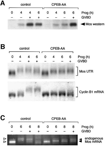
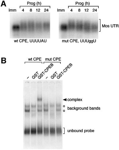
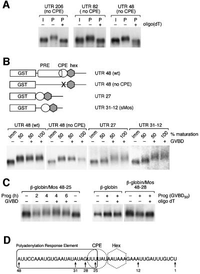
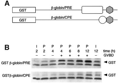
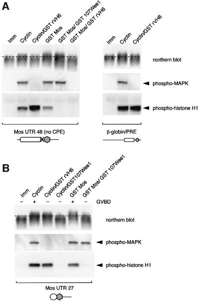
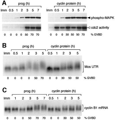
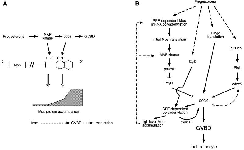
Progression through vertebrate oocyte maturation requires that pre-existing, maternally derived mRNAs be translated in a strict temporal order. The mechanism that controls the timing of oocyte mRNA translation is unknown. In this study we show that the early translational induction of the mRNA encoding the Mos proto-oncogene is mediated through a novel regulatory element within the 3' untranslated region of the Mos mRNA. This novel element is responsive to the MAP kinase signaling pathway and is distinct from the late acting, cdc2-responsive, cytoplasmic polyadenylation element. Our findings suggest that the timing of maternal mRNA translation is controlled through signal transduction pathways targeting distinct 3' UTR mRNA elements.


Figures

None
Fig. 1. CPEB-independent control of Mos mRNA translational activation. (A) Western blot of endogenous Mos protein. Immature oocytes were injected with 3–6 ng RNA encoding Xenopus CPEB-AA or water as a negative control, and left for 16 h to express the protein. Oocytes were stimulated by addition of progesterone. Samples were taken for both control and CPEB-AA-treated oocytes when the control oocytes had reached GVBD50 (4 h) and GVBD100 (6 h). Control oocytes were segregated at GVBD50 based on whether they had (+) or had not (–) completed GVBD. Expression of CPEB-AA significantly delayed GVBD but did not prevent low-level Mos protein accumulation prior to GVBD. This effect on Mos accumulation was observed in four independent experiments. At later time points some CPEB-AA-expressing oocytes underwent GVBD. Segregation of these oocytes based on GVBD status revealed that oocytes escaping the CPEB-AA block to maturation then accumulated high levels of Mos protein equivalent to control, post-GVBD, oocytes. (B) Northern blot showing the differential effect of CPEB-AA on polyadenylation of Mos UTR and cyclin B1 mRNA. Total RNA was prepared from the same oocyte samples described in (A) and analyzed for the polyadenylation status of either the co-injected wild-type terminal 48-nt Mos 3′ UTR (Mos UTR) or the endogenous cyclin B1 mRNA. Polyadenylation (retarded mobility) of the Mos UTR was observed in CPEB-AA-expressing oocytes. This experiment was repeated three times with similar results. In contrast, cyclin B1 polyadenylation was inhibited in CPEB-AA-expressing oocytes. (C) Polyadenylation of endogenous Mos mRNA analyzed by RT–PCR. In this experiment GVBD50 was at 6 h and GVBD100 was at 8 h. Polyadenylation (retarded mobility) of the Mos mRNA was observed in CPEB-AA-expressing oocytes following progesterone stimulation. A representative experiment is shown.
None
Fig. 2. CPE-independent polyadenylation of the Mos 3′ UTR. (A) Wild-type (Howard et al., 1999) and CPE-disrupted Mos UTRs correlating to the terminal 321 nt of the Mos mRNA 3′ UTR were injected into immature oocytes, and the ability of progesterone to induce polyadenylation (retarded mobility) was analyzed by northern blot. At the times indicated, pools of 10 oocytes were taken and RNA was extracted. Half of the oocytes had undergone GVBD after 4 h of culture. (B) Wild-type or mutant Mos UTR probes were analyzed for interaction with Xenopus CPEB by RNA electrophoretic mobility shift assay (EMSA). A specific CPEB binding complex was only observed with the wild-type Mos UTR probe.
None
Fig. 3. Identification of a novel Mos 3′ UTR regulatory element. (A) Truncation analysis of the Mos 3′UTR. Immature oocytes were injected with different lengths of the CPE-disrupted Mos 3′ UTR, and the ability of progesterone to induce polyadenylation (retarded mobility) was analyzed by northern blot. Total RNA was prepared from pools of oocytes when 50% of progesterone-stimulated oocytes had undergone GVBD. CPE-independent polyadenylation was retained within the last 48 nt of the Mos UTR. (B) Upper panel, schematic representation of the UTR constructs employed. The GST open reading frame (open box) was fused to the Mos 3′ UTR. The polyadenylation hexanucleotide (gray hexagon), CPE (open circle) and PRE (open rectangle) are also indicated. Mutational disruption of the CPE is indicated by an ‘X’. UTR 27 lacks Mos UTR sequence 5′ of the CPE, and sMos encompasses nucleotides 31–12 of the wild-type Mos UTR (Stebbins-Boaz et al., 1996). Lower panel, northern blot of wild-type and mutant Mos UTR-injected oocytes. When 50% of the oocytes had undergone GVBD, oocytes were segregated into pools that had either had a white spot at the animal pole (GVBD+) or had not (GVBD–), and total RNA was extracted. Total RNA was also prepared when all the oocytes had matured (100% GVBD) and from time-matched immature (Imm) oocytes. Wild-type 48 UTR and 48 UTR (no CPE) polyadenylation (retarded mobility) occurred prior to GVBD. CPE-directed polyadenylation of the Mos UTR 27 RNA and sMos RNA was observed predominantly after GVBD. (C) Northern blot of β-globin/Mos UTR chimeras. Total RNA was prepared from wild-type and mutant β-globin UTR-injected oocytes at the indicated times. Fifty percent of the oocytes had undergone GVBD (GVBD50) at 4 h. Where indicated, oligo(dT) and RNase H were used to remove any poly(A) tails from the UTR reporter constructs (oligo dT +). (D) Schematic representation of the relative positions of the polyadenylation response element (rectangle), CPE (circle) and polyadenylation hexanucleotide (dotted hexagon) sequences within the last 48 nt of the Mos UTR. The numbers indicate the position relative to the site of poly(A) addition (designated 1).
None
Fig. 4. The PRE directs temporally early mRNA translation in response to progesterone. (A) Schematic representation of the GST reporter RNA constructs. The relative positions of the polyadenylation hexanucleotide (gray hexagon), CPE (open circle) and PRE (open rectangle) are indicated. (B) GST western blot of protein lysates prepared from oocytes injected with equivalent amounts of GST β-globin/PRE or GST β-globin/CPE reporter RNAs.
None
Fig. 5. The polyadenylation response element (PRE) is the target of MAP kinase signaling. Immature oocytes were injected with RNA encoding GST107Wee1 to inhibit cdc2 activity or GSTrVH6 to inhibit MAP kinase activity, and left overnight to express the protein (Howard et al., 1999). Oocytes were then injected with RNA specifying PRE- or CPE-containing Mos UTRs (A and B, respectively) or a PRE-containing β-globin UTR (A, right panel), and left untreated (Imm) or co- injected with RNA encoding GST Mos to stimulate MAP kinase signaling, or cyclin B1 protein to activate cdc2 signaling (Howard et al., 1999). Pools of oocytes injected with the PRE-containing construct were prepared when 50% of the stimulated oocytes had undergone GVBD. Because PRE-mediated polyadenylation is temporally early, only oocytes that had not undergone GVBD were selected. Pools of oocytes injected with the CPE-containing construct were prepared when 100% of the stimulated oocytes had undergone GVBD, since CPE-mediated polyadenylation occurs predominantly after GVBD. Total RNA was prepared and protein lysates were taken from these pools. The polyadenylation status of the injected Mos UTRs was assessed by northern blot analyses (retarded mobility is indicative of polyadenylation). MAP kinase activity was assessed by western blot using phosphoMAP kinase antibodies, and cdc2 activity was assessed by western blot with phosphohistone H1 antibodies following an in vitro kinase assay (see Materials and methods) from the same samples utilized for northern blot analyses. The presence of the CPE sequence is represented schematically by an open circle, the PRE by a rectangle and the polyadenylation hexanucleotide by a gray hexagon.
None
Fig. 6. Mos polyadenylation temporally correlates with MAP kinase activation. Oocytes were injected with the terminal 321 nt Mos UTR reporter RNA and then left either untreated (Imm) or stimulated to mature (prog or cyclin protein) as indicated. Maturation kinetics of each oocyte population is indicated (% GVBD). Protein lysates and RNA extraction were prepared from pools of oocytes at the indicated times. Immature (Imm) oocyte samples were taken after 4 h. (A) Time course of progesterone- (prog) or cyclin-stimulated MAP kinase activation (upper panel), and cdc2 activation (lower panel). The MAP kinase activity in the sample was assessed by western blot using phosphoMAP kinase antibodies and cdc2 activity was measured by radiolabel incorporation into histone H1 (see Materials and methods). Samples were also analyzed by northern blot for polyadenylation (retarded mobility) of the 321-nt wild-type Mos UTR (B) and for endogenous cyclin B1 mRNA polyadenylation (C). While cyclin B1 injection advanced cdc2 activation and cyclin B1 mRNA polyadenylation, the injected oocytes reached 50% GVBD at the same rate as progesterone-treated oocytes (3 h).
None
Fig. 7. (A) Sequential model of Mos mRNA translational control in response to progesterone stimulation. In this model, PRE-directed Mos mRNA translational activation precedes CPE-directed translation as a consequence of progesterone-stimulated MAP kinase activation preceding cdc2 activation. Solid arrows denote causal relationships between processes. (B) Signal transduction pathways that converge upon cdc2 activation and GVBD in progesterone-stimulated Xenopus oocytes. Positive feedback loops are indicated by dotted lines. See text for details.

Similar articles

Cited by

References

    1. Abrieu A., Brassac,T., Galas,S., Fisher,D., Labbe,J.C. and Doree,M. (1998) The Polo-like kinase Plx1 is a component of the MPF amplification loop at the G2/M-phase transition of the cell cycle in Xenopus eggs. J. Cell Sci., 111, 1751–1757. - PubMed
    1. Ballantyne S., Daniel,D.L.J. and Wickens,M. (1997) A dependent pathway of cytoplasmic polyadenylation reactions linked to cell cycle control by c-mos and CDK1 activation. Mol. Biol. Cell, 8, 1633–1648. - PMC - PubMed
    1. Barkoff A.F., Dickson,K.S., Gray,N.K. and Wickens,M. (2000) Translational control of cyclin B1 mRNA during meiotic maturation: coordinated repression and cytoplasmic polyadenylation. Dev. Biol., 220, 97–109. - PubMed
    1. Castro A., Peter,M., Magnaghi-Jaulin,L., Vigneron,S., Galas,S., Lorca,T. and Labbe,J.C. (2001) Cyclin B/cdc2 induces c-Mos stability by direct phosphorylation in Xenopus oocytes. Mol. Biol. Cell, 12, 2660–2671. - PMC - PubMed
    1. Charlesworth A., Welk,J. and MacNicol,A. (2000) The temporal control of Wee1 mRNA translation during Xenopus oocyte maturation is regulated by cytoplasmic polyadenylation elements within the 3′ untranslated region. Dev. Biol., 227, 706–719. - PubMed

Publication types

MeSH terms