. 2002 Jun;22(12):4268-79.
doi: 10.1128/MCB.22.12.4268-4279.2002.

Distinct domains of the GATA-1 cofactor FOG-1 differentially influence erythroid versus megakaryocytic maturation

Affiliations

Distinct domains of the GATA-1 cofactor FOG-1 differentially influence erythroid versus megakaryocytic maturation

Alan B Cantor et al. Mol Cell Biol. 2002 Jun.

Abstract

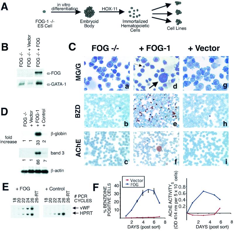
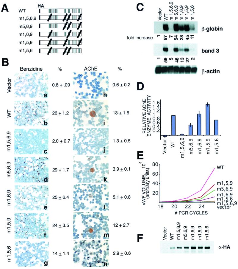
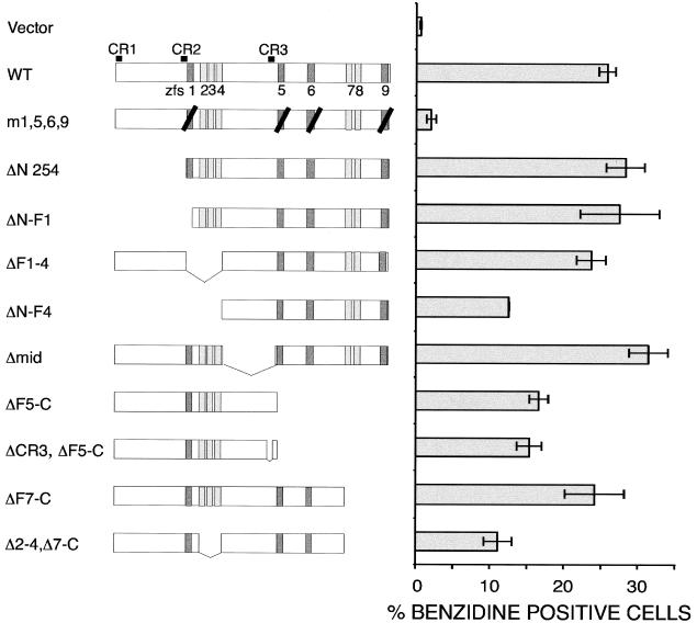
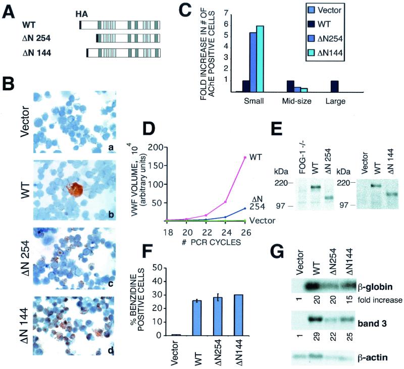
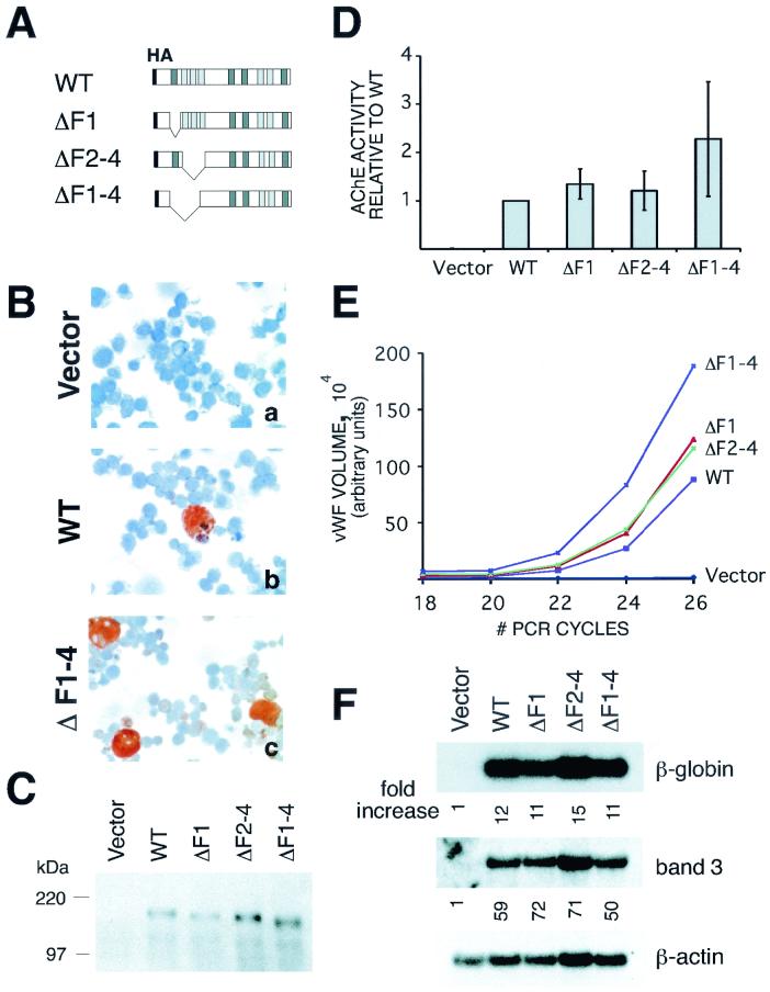
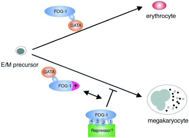
FOG family zinc finger proteins play essential roles in development through physical interaction with GATA factors. FOG-1, like its interacting partner GATA-1, is required for normal differentiation of erythroid and megakaryocytic cells. Here, we have developed a functional assay for FOG-1 based on its ability to rescue erythroid and megakaryocytic maturation of a genetically engineered FOG-1(-/-) cell line. We demonstrate that interaction through only one of FOG-1's four GATA-binding zinc fingers is sufficient for rescue, providing evidence against a model in which FOG-1 acts to bridge multiple GATA-binding DNA elements. Importantly, we find that distinct regions of FOG-1 differentially influence erythroid versus megakaryocyte maturation. As such, we propose that FOG-1 may modulate the fate of a bipotential erythroid/megakaryocytic precursor cell.


Figures

FIG. 1.
FIG. 1.
FOG-1-mediated rescue of erythroid and megakaryocytic terminal maturation of a FOG-1−/− hematopoietic cell line. (A) Schematic depiction of derivation of the FOG-1−/− cell line. (B) Western blot analysis of nuclear extracts from uninfected FOG-1−/− cells or FOG-1−/− cells infected with vector alone or FOG-1 cDNA packaged retroviruses. The blots were probed with a polyclonal antibody against FOG-1 and then stripped and reprobed with an antibody against GATA-1. (C) Comparison of the morphology of uninfected FOG-1−/− cells (a, b, and c) with those infected with retroviruses encoding FOG-1 cDNA (d, e, and f) or vector alone (g, h, and i) by May-Grunwald-Giemsa (MG/G) staining (a, d, and g), benzidine (BZD) (o-dianisodine) staining (b, e, and h), or AChE staining (c, f, and i). Hemoglobinized cells stain a dark brown color with BZD, and megakaryocytes develop an orange color with AChE staining. Small and large arrows indicate maturing erythrocytes and megakaryocytes, respectively, in panel d. Original magnification, ×1,000 (panels a, d, and g) or ×400 (remaining panels). (D) Northern blot analysis for erythroid markers β-globin and band 3 from retrovirally infected cells. The cDNA encoding a GFP/Cre fusion was used as a negative control. The fold increase in the signal compared to the uninfected FOG-1−/− cells and normalized to the β-actin loading controls is shown below each panel. (E) Semiquantitative RT-PCR analysis for the megakaryocyte marker vWF or ubiquitously expressed HPRT in cells rescued with FOG-1 cDNA or a control plasmid (GFP/Cre fusion). (F). Time course of erythroid and megakaryocytic maturation of rescued cells transduced with vector (shown in red) or FOG-1 (shown in blue). The percentages of BZD+ cells and total AChE enzymatic activity (normalized to uninfected cells) are indicated. Day zero of the time course represents the day the cells were sorted for GFP expression (2 days following retroviral infection). The results represent those of two independent experiments. OD 414 nm, optical density at 414 nm.
FIG. 2.
FIG. 2.
GATA-binding requirements for FOG-1-mediated rescue of erythropoiesis and megakaryopoiesis. (A) Schematic representation of mutant FOG-1 constructs. Darker shaded boxes represent GATA-binding zinc fingers. Black diagonal lines indicate substitution of alanine for the tyrosine residue that immediately precedes the final cysteine of each zinc finger structure. (B) Benzidine and AChE stains of FOG-1−/− cells rescued with each of the mutant constructs. Original magnification, ×400. (C) Northern blot analysis for β-globin and band 3 from the FOG-1−/− cells rescued with each of the mutant constructs. The fold increase in signal for each construct relative to the vector-alone control and normalized to the β-actin samples is shown below each panel. (D) Total AChE enzymatic activity from cells rescued with each of the mutant constructs. Total AChE activity was measured in triplicate from 5 × 10⁵ of the rescued cells. Basal activity measured from uninfected FOG-1−/− cells was subtracted, and the data are displayed relative to the wild-type construct. (E) Semiquantitative RT-PCR analysis of cells rescued with each of the mutant constructs. RT-PCR using vWF-specific and HPRT-specific primers was performed utilizing [32P]dCTP for radioisotope incorporation. The products were separated by polyacrylamide gel electrophoresis, and the labeled bands were quantified using a PhosphorImager. The data are displayed as vWF signal normalized to the HPRT control signal. (F) Western blot analysis of rescued cells for expression of mutant constructs. Nuclear extracts were prepared from a portion of the rescued cells 1 day after sorting for GFP expression. Equivalent amounts of total protein were separated by sodium dodecyl sulfate-polyacrylamide gel electrophoresis and immunoblotted with an anti-HA antibody.
FIG. 3.
FIG. 3.
Lack of an essential subdomain of FOG-1 required for terminal erythroid maturation of the FOG-1−/− cell line (excluding a GATA interacting domain). A schematic representation of each construct is shown next to the percentage of benzidine-positive cells obtained from at least two independent rescue experiments and displayed as the mean value ± the standard error of the mean. Three regions of high sequence conservation (outside of the zinc fingers) between FOG-1 and other members of the FOG family are indicated above the top of the wild-type construct as CR1, CR2, and CR3. Darker shaded boxes represent GATA-binding zinc fingers, and black diagonal lines represent substitution of alanine for the tyrosine residue immediately preceding the final cysteine residue of the zinc finger.
FIG. 4.
FIG. 4.
Amino terminus of FOG-1 is required for rescue of terminally differentiated megakaryocytes. (A) Schematic representation of constructs. (B) AChE stains of FOG-1−/− cells rescued with the mutant FOG-1 constructs. Original magnification, ×600. (C) Fold increase in the number of AChE+ cells compared to the wild type for each of the constructs. The data have been categorized for small, medium, and large cells. (D) Semiquantitative RT-PCR for vWF mRNA from FOG-1−/− cells rescued with each of the mutant constructs. The data have been normalized to the HPRT controls included in each sample. (E) Western blot analysis from nuclear extracts prepared from FOG-1−/− cells rescued with the different N-terminal deletion constructs using an antibody directed against the C terminus of FOG-1 (Santa Cruz Biotechnology; A-20) (right panel) or the HA epitope (left panel). Equivalent amounts of total protein were loaded into each lane for sodium dodecyl sulfate-polyacrylamide gel electrophoresis. The migration of molecular mass standards is indicated. (F) Percentage of benzidine-positive cells from FOG-1−/− cells rescued with each of the mutant constructs. Error bars represent ± standard error of the mean. (G) Northern blot analysis for β-globin and band 3 mRNAs from FOG-1−/− cells rescued with each construct. The fold increase in signal (after normalization to the β-actin control) relative to vector alone is shown beneath each panel.
FIG. 5.
FIG. 5.
Deletion of zinc fingers 1 to 4 enhances FOG-1-mediated rescue of megakaryopoiesis but not erythropoiesis. (A) Schematic representation of FOG-1 deletion constructs. (B) AChE staining of FOG-1−/− cells rescued with each of the constructs. Magnification, ×600. (C) Western blot analysis of a portion of FOG-1−/− cells rescued with each of the constructs using an anti-HA antibody. Nuclear extracts were prepared from GFP+ cells one day following FACS, and equivalent amounts of total protein for each construct were loaded for sodium dodecyl sulfate-polyacrylamide gel electrophoresis. (D) AChE enzymatic activity from infected cells. AChE enzymatic activity was measured in triplicate for 5 × 10⁵ of the rescued cells obtained 6 days following FACS. Background activity from the uninfected FOG-1−/− cells was subtracted, and the activity is reported relative to the wild-type molecule. (E) Semiquantitative RT-PCR analysis for vWF mRNA from FOG-1−/− cells rescued with each of the constructs. The vWF signals were normalized to the HPRT control values for each sample. (F) Northern blot analysis for β-globin and band 3 mRNAs from FOG-1−/− cells rescued with each of the constructs. Fold increase in signal (after normalization to the β-actin control for each sample) relative to the vector alone is shown below each panel.
FIG. 6.
FIG. 6.
Models of FOG-1's lineage-selective roles in erythropoiesis and megakaryopoiesis. Model for the lineage-selective effects of distinct regions of FOG-1. For erythroid differentiation, a simple interaction between GATA-1 and FOG-1 is both necessary and sufficient to drive erythroid maturation from a bipotential precursor cell. For megakaryocytic differentiation, activity provided by the amino terminus of FOG-1 (shown in magenta), in addition to an interaction with GATA-1, is required. Further complex control serving to restrict megakaryocyte differentiation may be provided by interaction between an unknown factor (shown in green) and zinc fingers 1 to 4 of FOG-1.

Similar articles

Cited by

References

    1. Andrews, N. C., and D. Faller. 1991. A rapid micropreparation technique for extraction of DNA-binding proteins from limiting numbers of mammalian cells. Nucleic Acids Res. 19:2499. - PMC - PubMed
    1. Bulger, M., and M. Groudine. 1999. Looping versus linking: toward a model for long-distance gene activation. Genes Dev. 13:2465-2477. - PubMed
    1. Crispino, J. D., M. Lodish, J. P. Mackay, and S. H. Orkin. 1999. Use of altered specificity mutants to probe a specific protein-protein interaction in differentiation: the GATA-1:FOG complex. Mol. Cell 3:219-228. - PubMed
    1. Crispino, J. D., M. Lodish, B. L. Thurberg, S. H. Litovsky, T. Collins, J. D. Molkentin, and S. H. Orkin. 2001. Proper coronary vascular development and heart morphogenesis depend on interaction of GATA-4 with FOG factors. Genes Dev. 15:839-844. - PMC - PubMed
    1. Cubadda, Y., P. Heitzler, R. P. Ray, M. Bourouis, P. Ramain, E. Gelbart, P. Simpson, and M. Haenlin. 1997. u-shaped encodes a zinc finger protein that regulates the proneural genes achaete and scute during the formation of bristles in Drosophila. Genes Dev. 11:3083-3095. - PMC - PubMed

Publication types

MeSH terms