. 2002 Jun;22(12):4020-32.
doi: 10.1128/MCB.22.12.4020-4032.2002.

Constitutive association of BRCA1 and c-Abl and its ATM-dependent disruption after irradiation

Affiliations

Constitutive association of BRCA1 and c-Abl and its ATM-dependent disruption after irradiation

Nicolas Foray et al. Mol Cell Biol. 2002 Jun.

Abstract

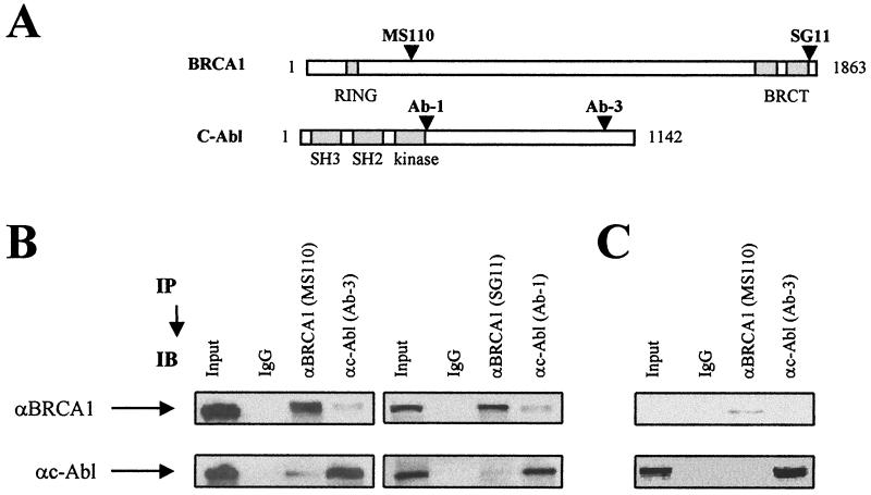
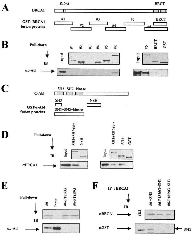
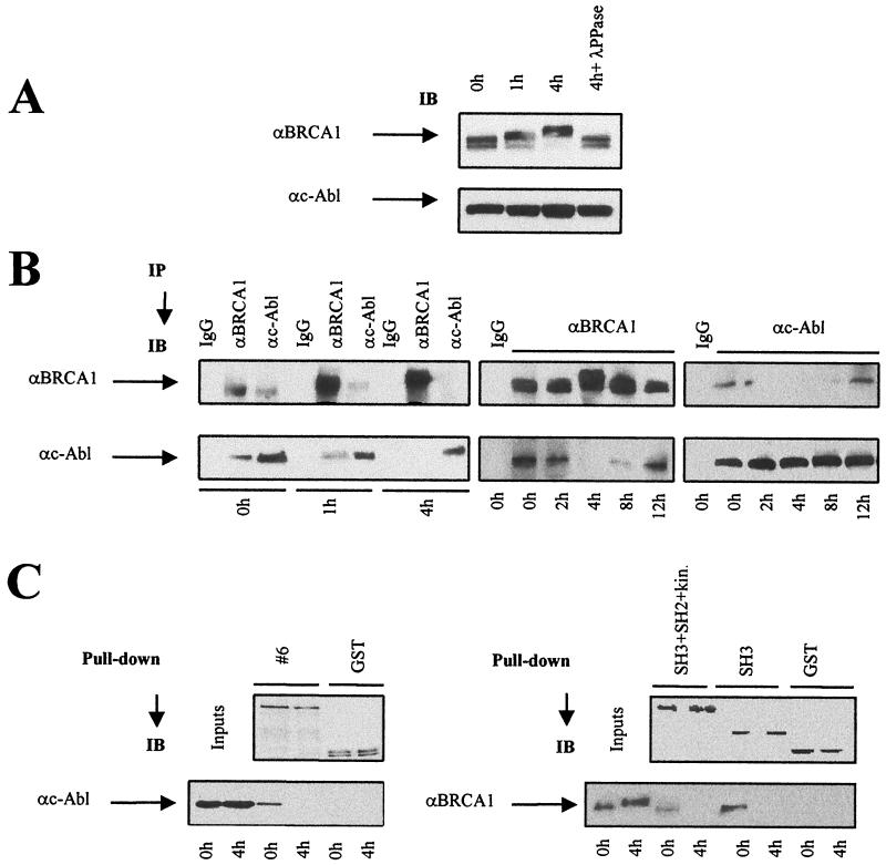
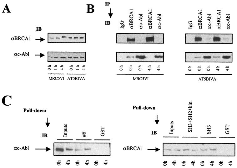
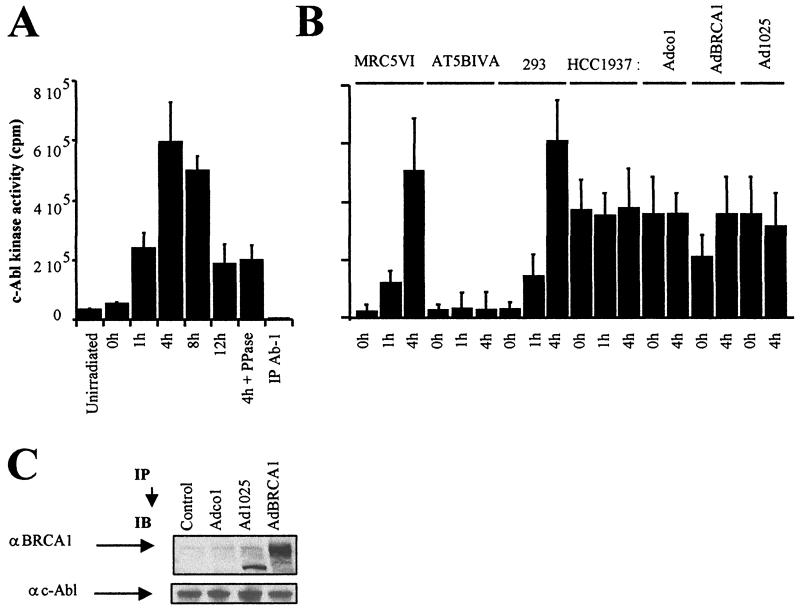
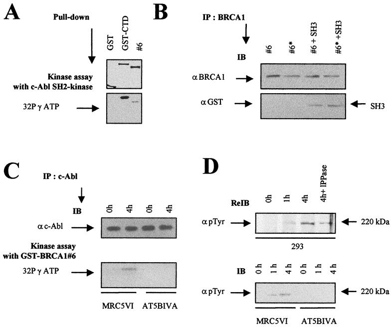
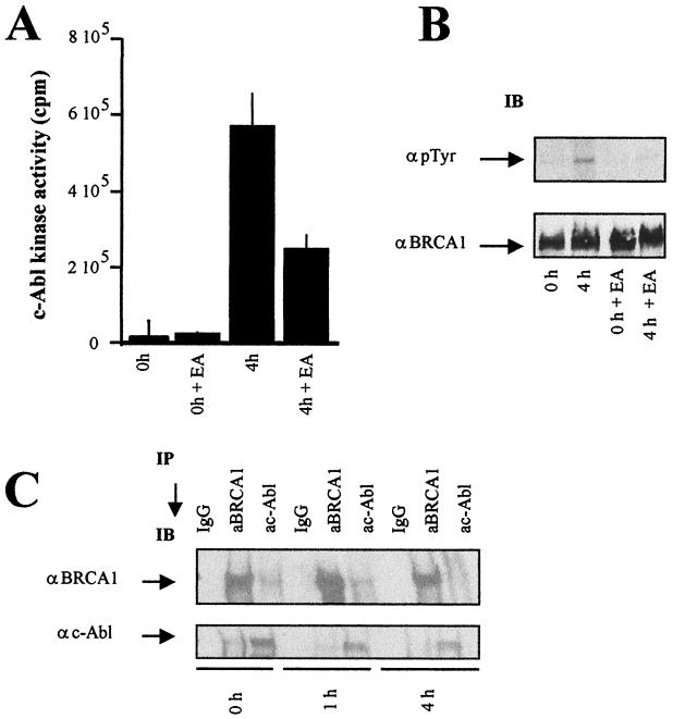
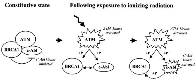
BRCA1 plays an important role in mechanisms of response to double-strand breaks, participating in genome surveillance, DNA repair, and cell cycle checkpoint arrests. Here, we identify a constitutive BRCA1-c-Abl complex and provide evidence for a direct interaction between the PXXP motif in the C terminus of BRCA1 and the SH3 domain of c-Abl. Following exposure to ionizing radiation (IR), the BRCA1-c-Abl complex is disrupted in an ATM-dependent manner, which correlates temporally with ATM-dependent phosphorylation of BRCA1 and ATM-dependent enhancement of the tyrosine kinase activity of c-Abl. The BRCA1-c-Abl interaction is affected by radiation-induced modification to both BRCA1 and c-Abl. We show that the C terminus of BRCA1 is phosphorylated by c-Abl in vitro. In vivo, BRCA1 is phosphorylated at tyrosine residues in an ATM-dependent, radiation-dependent manner. Tyrosine phosphorylation of BRCA1, however, is not required for the disruption of the BRCA1-c-Abl complex. BRCA1-mutated cells exhibit constitutively high c-Abl kinase activity that is not further increased on exposure to IR. We suggest a model in which BRCA1 acts in concert with ATM to regulate c-Abl tyrosine kinase activity.


Figures

FIG. 1.
FIG. 1.
Coimmunoprecipitation of BRCA1 and c-Abl. (A) Positions of the epitope domains of the anti-BRCA1 and the anti-c-Abl antibodies used in this study. The MS110 and SG11 antibodies recognize the N-terminal and the C-terminal domain of BRCA1, respectively; the Ab-1 and Ab-3 antibodies recognize the kinase-adjacent domain and the C-terminal domain of c-Abl, respectively. (B) Nuclear extracts from exponentially growing 293 cells were subjected to BRCA1 and c-Abl immunoprecipitation (IP). The immunoprecipitates were analyzed by immunoblotting (IB). The masses of endogenous BRCA1 and c-Abl are 220 and 140 kDa, respectively. αBRCA1, anti-BRCA1 antibody; αc-Abl, anti-c-Abl antibody. (C) Nuclear extracts from exponentially growing HCC1937 cells were subjected to BRCA1 and c-Abl immunoprecipitation.
FIG. 2.
FIG. 2.
Localization of the domains involved in the BRCA1-c-Abl complex. (A) Positions of the GST-BRCA1 fusion proteins. (B) Equal amounts (10 μg) of GST fusion proteins (except for GST controls [15 μg]) were analyzed by Coomassie blue-stained gels (top) and incubated with 293 cell nuclear extracts, and the resulting GST adsorbates were subjected to immunoblotting (IB) using anti-c-Abl (αc-Abl; Ab-3) antibody (bottom). The GST-BRCA1#3 protein was found to be unstable, as shown previously (34). (C) Positions of the GST-c-Abl fusion proteins. (D) Equal amounts (5 μg) of GST fusion proteins shown by Coomassie blue-stained gels (top) were incubated with 293 cell nuclear extracts, and the resulting GST adsorbates were subjected to immunoblotting using anti-BRCA1 (αBRCA1; MS110) antibody. (E) Equal amounts (5 μg) of GST fusion proteins shown by Coomassie blue-stained gels (top; the second lane is empty) were incubated with 293 cell nuclear extracts, and the resulting GST adsorbates were subjected to immunoblotting using anti-c-Abl (Ab-3) antibody (bottom). (F) Eluted wild-type and mutated GST-BRCA1#6 (P1856G and P1859G) proteins were mixed with eluted GST-SH3 proteins and subjected to BRCA1 immunoprecipitation using anti-BRCA1 (Ab-5 and Ab-3) antibodies. The presence of the GST-SH3 proteins in the adsorbates was examined by immunoblotting using anti-GST (αGST) antibody.
FIG. 3.
FIG. 3.
Disruption of the BRCA1-c-Abl complex after irradiation. Control and irradiated (20 Gy) 293 cells were collected at the indicated times. (A) Nuclear extracts were subjected to BRCA1 and c-Abl immunoblotting (IB) with MS110 and Ab-3 antibodies, respectively. λ-PPase, λ-phosphatase. (B) The same batch of extracts was subjected to BRCA1 and c-Abl immunoprecipitation (IP) with MS110 and Ab-3 antibodies, respectively. Preimmune mouse IgGs were used as controls. αc-Abl, anti-c-Abl; αBRCA1, anti-BRCA1. (C) As described in the legend to Fig. 2, purified GST-BRCA1#6, GST-SH3+SH2+kinase, GST-SH3, and GST fusion proteins were incubated with nuclear extracts from irradiated 293 cells; GST adsorbates were collected and subjected to BRCA1 or c-Abl immunoblotting using the same antibodies used for Fig. 2.
FIG. 4.
FIG. 4.
Disruption of the BRCA1-c-Abl complex is ATM dependent. Control and irradiated (20 Gy) MRC5VI and AT5BIVA fibroblast cells were collected at the indicated times. (A) Nuclear extracts were subjected to BRCA1 and c-Abl immunoblotting (IB) with MS110 and Ab-3 antibodies, respectively. αc-Abl, anti-c-Abl; αBRCA1, anti-BRCA1. (B) The same batch of extracts was subjected to BRCA1 and c-Abl immunoprecipitation (IP) with the same antibodies and preimmune IgGs used for Fig. 3. (C) Equal amounts (10 μg) of purified GST and GST-BRCA1#6 fusion proteins (see the Coomassie blue-stained gel in Fig. 2) were incubated with nuclear extracts from irradiated AT5BIVA cells subjected or not to 4 h of incubation. The resulting GST adsorbates were analyzed by immunoblotting using the anti-c-Abl (Ab-3) antibody. Equal amounts (5 μg) of purified GST, GST-SH3+SH2+kinase, and GST-SH3 fusion protein (see the Coomassie blue-stained gel in Fig. 2) were incubated with nuclear extracts from irradiated AT5BIVA cells subjected or not to 4 h of incubation. The resulting adsorbates were analyzed by immunoblotting with the anti-BRCA1 (MS110) antibody.
FIG. 5.
FIG. 5.
Tyrosine kinase activity of c-Abl after irradiation. (A) Tyrosine kinase activity of c-Abl measured from c-Abl immunoprecipitates (IP) of nuclear extracts from 293 cells exposed to 20 Gy and collected after irradiation at the indicated times. (B) Tyrosine kinase activity of c-Abl in nuclear extracts from 293, MRC5VI, AT5BIVA (ATM−/−), and HCC1937 (BRCA1-mutated) cells exposed to 20 Gy and incubated for the times indicated. HCC1937 cells were infected by empty cassette (Adco1), wild-type BRCA1 (AdBRCA1) adenoviruses, or adenoviruses expressing only the first 1,321 residues of BRCA1 (Ad1025). All the data presented are the mean plus standard error of triplicate experiments. The assay was controlled, with each sample using an anti-c-Abl (Ab-1) antibody that inhibits kinase activity, as recommended by the manufacturers (see Materials and Methods). We verified that the levels of c-Abl protein immunoprecipitated in all samples were similar (data not shown). (C) As a control for adenovirus expression, nuclear extracts from exponentially growing adenovirus-infected HCC1937 cells were subjected to immunoblotting (IB) using anti-BRCA1 (αBRCA1; MS110) and anti-c-Abl (αc-Abl; Ab-3) antibodies. It is noteworthy that the Ad1025 adenovirus produces a 156-kDa truncated BRCA1 protein.
FIG. 6.
FIG. 6.
C-terminal BRCA1 is a substrate for c-Abl tyrosine kinase. (A) c-Abl tyrosine kinase assay of GST-BRCA1#6 using c-Abl SH2 kinase. Purified c-Abl SH2 kinase (1 μg) was incubated with equal amounts (5 μg) of GST, GST-CTD, and GST-BRCA1#6 fusion proteins as indicated in Materials and Methods (top). The resulting GST adsorbates were subjected to SDS-PAGE and phosphorimager analysis (bottom). (B) SH3 binding of c-Abl-phosphorylated GST-BRCA1#6 substrates. Equal amounts of GST-BRCA1#6, whether tyrosine phosphorylated (#6*) or not (#6) as described below, were subjected to BRCA1 immunoprecipitation (IP) using anti-BRCA1 (αBRCA1; Ab-5 and Ab-3) antibodies. The substrates were mixed with equal amounts of eluted GST-SH3 proteins. The presence of the GST-SH3 proteins in the adsorbates was examined by immunoblotting (IB) using anti-GST (αGST) antibody. (C) c-Abl tyrosine kinase assay of GST-BRCA1#6 using endogenous c-Abl. c-Abl immunoprecipitates of nuclear extracts from MRC5VI and AT5BIVA (ATM−/−) cells exposed to 20 Gy were collected after irradiation at the indicated times (top) in a kinase assay with the GST-BRCA1#6 fusion protein (5 μg) as a substrate. The resulting GST adsorbates were subjected to SDS-PAGE and phosphorimager analysis (bottom). (D) The nitrocellulose membrane used for BRCA1 immunoblotting of 293 cells extracts shown in Fig. 3A was rehybridized by anti-p-Tyr (α p Tyr) tyrosine antibody (top). Nuclear extracts from unirradiated and irradiated MRC5VI and AT5BIVA cells were subjected to p-Tyr immunoblotting (bottom).
FIG. 7.
FIG. 7.
Effect of c-Abl kinase inhibition on the BRCA1-c-Abl complex. (A) c-Abl tyrosine kinase activity measured following c-Abl immunoprecipitation of nuclear extracts from untreated and EA-treated 293 cells exposed to 20 Gy and collected at the indicated times postirradiation. The error bars indicate standard deviations. (B) Nuclear extracts from irradiated control or EA-treated 293 cells were subjected to p-Tyr and BRCA1 immunoblotting (IB). α p Tyr, anti-p-Tyr; αBRCA1, anti-BRCA1. (C) Nuclear extracts from control and EA-treated cells were subjected to BRCA1 and c-Abl immunoprecipitation (IP) for BRCA1-c-Abl interaction using MS110 and Ab-3 antibodies, respectively. Preimmune mouse IgGs were used as controls. αc-Abl, anti-c-Abl. The experiments shown in panels B and C were carried out using 4% polyacrylamide gels in contrast to the experiment shown in Fig. 3A, where higher-percentage polyacrylamide gels were used. The difference in mobility shift postirradiation is therefore less pronounced in panels B and C.
FIG. 8.
FIG. 8.
Model for the association and disruption of the BRCA1-c-Abl complex and its role. BRCA1 and c-Abl interact constitutively, and c-Abl tyrosine kinase activity is inhibited. After irradiation, ATM kinase is activated, phosphorylating BRCA1 and activating c-Abl, causing disruption of the complex. Consequently, the c-Abl tyrosine kinase activity is stimulated, leading to phosphorylation of a minor subset of BRCA1.

Similar articles

Cited by

References

    1. Aprelikova, O. N., B. S. Fang, E. G. Meissner, S. Cotter, M. Campbell, A. Kuthiala, M. Bessho, and R. A. Jensen. 1999. BRCA1-associated growth arrest is RB-dependent. Proc. Natl. Acad. Sci. USA 96:11866-11871. - PMC - PubMed
    1. Barila, D., and G. Superti-Furga. 1998. An intermolecular SH3-domain interaction regulates c-Abl activity. Nat. Genet. 18:280-282. - PubMed
    1. Baskaran, R., M. E. Dahmus, and J. Y. Wang. 1993. Tyrosine phosphorylation of mammalian RNA polymerase II carboxyl-terminal domain. Proc. Natl. Acad. Sci. USA 90:11167-11171. - PMC - PubMed
    1. Baskaran, R., L. D. Wood, L. L. Whitaker, C. E. Canman, S. E. Morgan, Y. Xu, C. Barlow, D. Baltimore, A. Wynshaw-Boris, M. B. Kastan, and J. Y. J. Wang. 1997. Ataxia telangiectasia mutant protein activates c-Abl tyrosine kinase in response to ionizing radiation. Nature 387:516-519. - PubMed
    1. Bressan, D. A., B. K. Baxter, and J. H. Petrini. 1999. The Mre11-Rad50-Xrs2 protein complex facilitates homologous recombination-based double-strand break repair in Saccharomyces cerevisiae. Mol. Cell. Biol. 11:7681-7687. - PMC - PubMed

Publication types

MeSH terms

LinkOut - more resources