. 2002 Jun;22(11):3875-91.
doi: 10.1128/MCB.22.11.3875-3891.2002.

Role of SHP-2 in fibroblast growth factor receptor-mediated suppression of myogenesis in C2C12 myoblasts

Affiliations

Role of SHP-2 in fibroblast growth factor receptor-mediated suppression of myogenesis in C2C12 myoblasts

Maria I Kontaridis et al. Mol Cell Biol. 2002 Jun.

Abstract

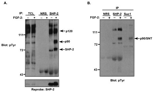
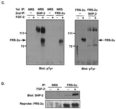
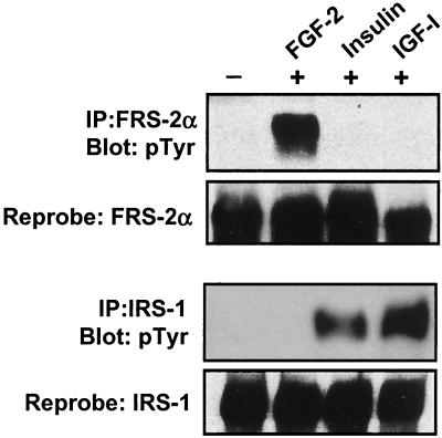
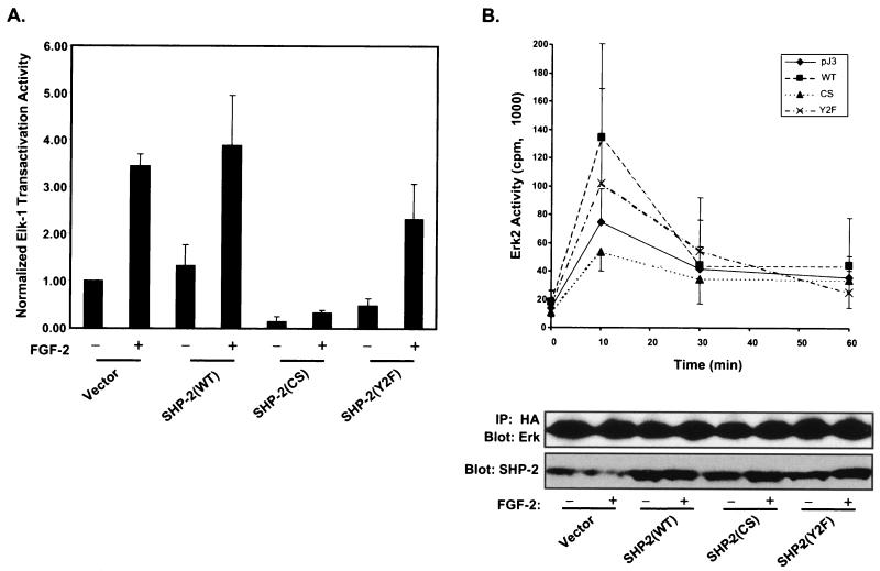
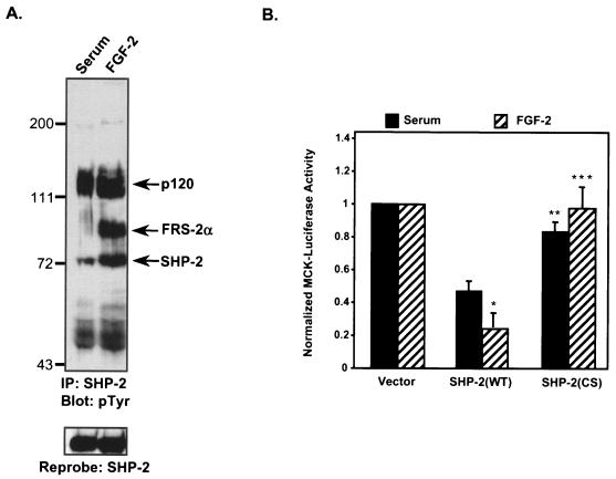
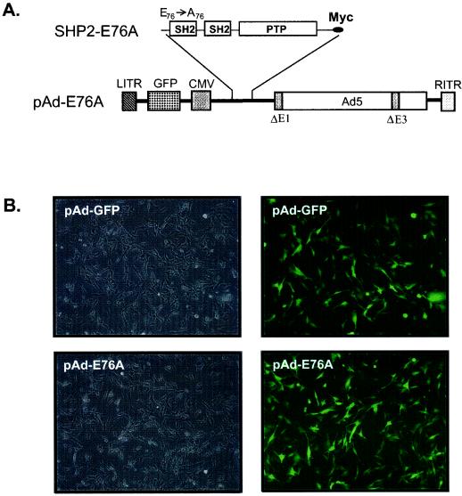
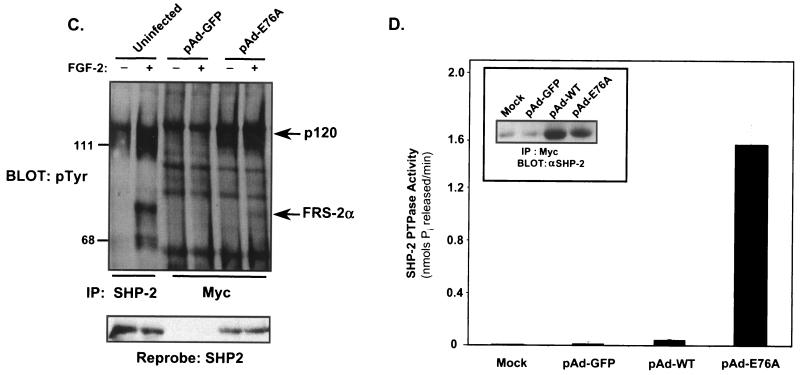
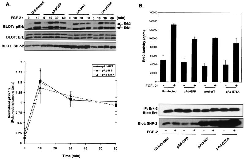
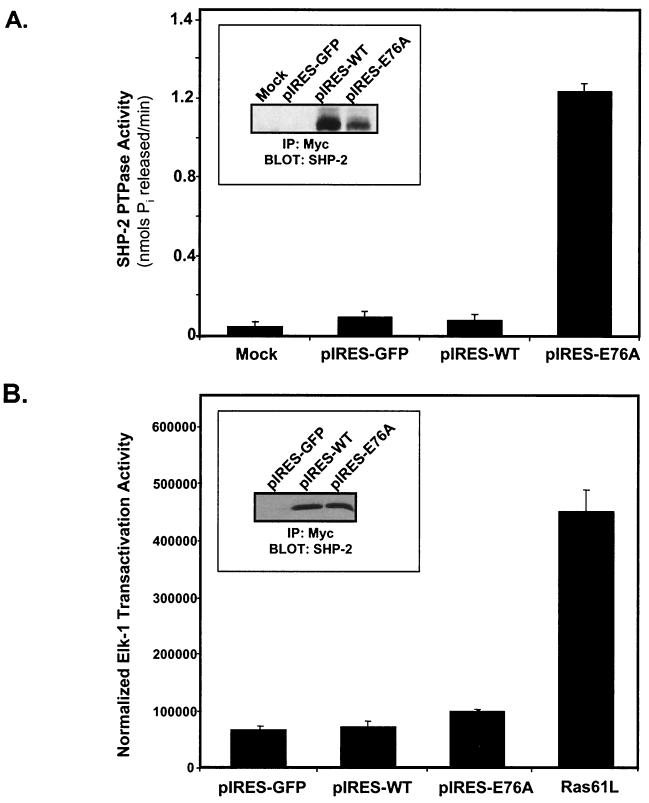
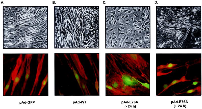
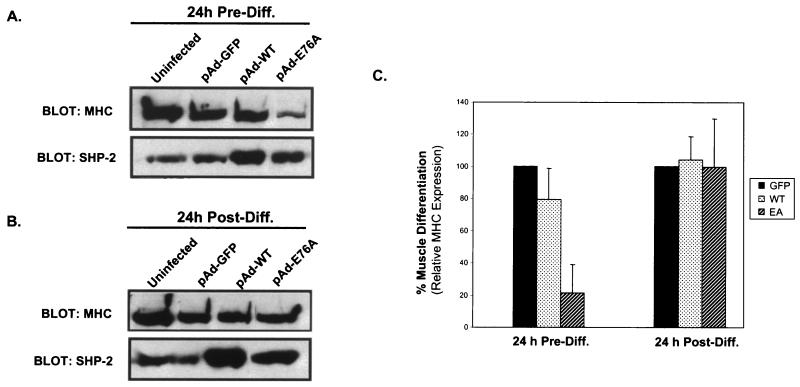
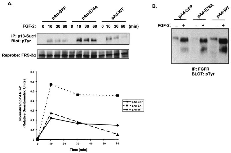
Ligand activation of the fibroblast growth factor receptor (FGFR) represses myogenesis and promotes activation of extracellular signal-regulated kinases 1 and 2 (Erks). The precise mechanism through which the FGFR transmits both of these signals in myoblasts remains unclear. The SH2 domain-containing protein tyrosine phosphatase, SHP-2, has been shown to participate in the regulation of FGFR signaling. However, no role for SHP-2 in FGFR myogenic signaling is known. In this study, we show that stimulation of C2C12 myoblasts with FGF-2 induces SHP-2 complex formation with tyrosyl-phosphorylated FGFR substrate 2 alpha (FRS-2 alpha). Both the catalytic activity and, to a much lesser extent, the Grb2 binding-tyrosyl phosphorylation sites of SHP-2 are required for maximal FGF-2-induced Erk activity and Elk-1 transactivation. When overexpressed in C2C12 myoblasts, wild-type SHP-2, but not a catalytically inactive SHP-2 mutant, potentiates the suppressive effects of FGF-2 on muscle-specific gene expression. In addition, expression of a constitutively active mutant of SHP-2 is sufficient to prevent myogenesis. The constitutively active mutant of SHP-2 induces hyper-tyrosyl phosphorylation of FRS-2 alpha but fails to stimulate or potentiate either FGF-2-induced Erk activation or Elk-1 transactivation. These data suggest that in myoblasts, SHP-2 represses myogenesis via a pathway that is independent of the Erks. We propose that SHP-2 plays a pivotal role in FGFR signaling in myoblasts via both Erk-dependent and Erk-independent pathways.


Figures

FIG. 1.
FIG. 1.
FGF-2 induces FRS-2α tyrosyl phosphorylation and complex formation with SHP-2 in C2C12 myoblasts. (A) C2C12 myoblasts were serum starved overnight and then either left untreated (−) or treated (+) with FGF-2 (10 ng/ml) for 10 min. Lysates from unstimulated and stimulated myoblasts were immunoprecipitated either with NRS or with anti-SHP-2 antibodies. Immune complexes and total-cell lysates (TCL) from unstimulated and FGF-2-stimulated C2C12 myoblasts were resolved by SDS-8% PAGE and analyzed by immunoblotting with antiphosphotyrosine (4G10) antibodies (pTyr). The lower panel represents a reprobe of the membrane with anti-SHP-2 antibodies. (B) Lysates prepared from either unstimulated (−) or FGF-2-stimulated (+) C2C12 myoblasts were prepared as described above. Lysates were subjected either to affinity precipitation with p13-Suc-1 agarose beads (Suc1) or to immunoprecipitation with anti-SHP-2 antibodies. p13-Suc-1 agarose affinity complexes or anti-SHP-2 antibody immune complexes were resolved by SDS-PAGE, and tyrosyl-phosphorylated proteins were visualized by immunoblotting with 4G10 antibodies. (C) Lysates prepared as described in the legend to panel B were subjected to immunoprecipitation (IP) either with NRS or with anti-FRS-2α antibodies (1st IP). The supernatants from the first IP were either left alone or subsequently immunoprecipitated with anti-SHP-2 antibodies or anti-FRS-2α antibodies (2nd IP). Immune complexes were analyzed by immunoblotting with 4G10 antibodies. (D) Lysates were prepared from C2C12 myoblasts that were either left untreated (−) or treated (+) with FGF-2 (10 ng/ml) for 10 min. Lysates were immunoprecipitated either with NRS or with anti-FRS-2α antibodies, and immune complexes were resolved and immunoblotted with anti-SHP-2 antibodies. The lower panel represents a reprobe of the membrane with anti-FRS-2α antibodies.
FIG. 1.
FIG. 1.
FGF-2 induces FRS-2α tyrosyl phosphorylation and complex formation with SHP-2 in C2C12 myoblasts. (A) C2C12 myoblasts were serum starved overnight and then either left untreated (−) or treated (+) with FGF-2 (10 ng/ml) for 10 min. Lysates from unstimulated and stimulated myoblasts were immunoprecipitated either with NRS or with anti-SHP-2 antibodies. Immune complexes and total-cell lysates (TCL) from unstimulated and FGF-2-stimulated C2C12 myoblasts were resolved by SDS-8% PAGE and analyzed by immunoblotting with antiphosphotyrosine (4G10) antibodies (pTyr). The lower panel represents a reprobe of the membrane with anti-SHP-2 antibodies. (B) Lysates prepared from either unstimulated (−) or FGF-2-stimulated (+) C2C12 myoblasts were prepared as described above. Lysates were subjected either to affinity precipitation with p13-Suc-1 agarose beads (Suc1) or to immunoprecipitation with anti-SHP-2 antibodies. p13-Suc-1 agarose affinity complexes or anti-SHP-2 antibody immune complexes were resolved by SDS-PAGE, and tyrosyl-phosphorylated proteins were visualized by immunoblotting with 4G10 antibodies. (C) Lysates prepared as described in the legend to panel B were subjected to immunoprecipitation (IP) either with NRS or with anti-FRS-2α antibodies (1st IP). The supernatants from the first IP were either left alone or subsequently immunoprecipitated with anti-SHP-2 antibodies or anti-FRS-2α antibodies (2nd IP). Immune complexes were analyzed by immunoblotting with 4G10 antibodies. (D) Lysates were prepared from C2C12 myoblasts that were either left untreated (−) or treated (+) with FGF-2 (10 ng/ml) for 10 min. Lysates were immunoprecipitated either with NRS or with anti-FRS-2α antibodies, and immune complexes were resolved and immunoblotted with anti-SHP-2 antibodies. The lower panel represents a reprobe of the membrane with anti-FRS-2α antibodies.
FIG. 2.
FIG. 2.
FGF-2 induces tyrosyl phosphorylation of FRS-2α but not IRS-1 in C2C12 myoblasts. C2C12 myoblasts were serum deprived for 18 h and then either left unstimulated (−) or stimulated (+) with either FGF-2 (10 ng/ml), insulin (100 nM), or IGF-I (100 ng/ml) for 10 min. These cell lysates were immunoprecipitated (IP) with either anti-FRS-2α or anti-IRS-1 antibodies; immune complexes were resolved by SDS-PAGE and immunoblotted by using 4G10 antiphosphotyrosine antibodies. The same membranes were then reprobed with either anti-FRS-2α or anti-IRS-1 antibodies.
FIG. 3.
FIG. 3.
SHP-2's catalytic activity is required for FGF-2-mediated activation of Elk-1 and Erk2 in C2C12 myoblasts. (A) C2C12 myoblasts were transfected with 0.5 μg of 5XGAL4-luciferase, 0.5 μg of Elkc-GAL4, 0.5 μg of β-galactosidase, and 2.5 μg of either vector alone, WT SHP-2, the catalytically inactive cysteine-to-serine (CS) mutant of SHP-2, or the Y542F/Y580F (Y2F) double tyrosyl phosphorylation site mutant of SHP-2. Twenty-four hours after transfection, myoblasts were transferred to serum deprivation medium for a further 24 h and were either left unstimulated (−) or stimulated (+) with FGF-2 (10 ng/ml) for 4 h. Luciferase and β-galactosidase activity assays were performed as described in Materials and Methods, and the raw luciferase values were normalized to β-galactosidase activity. Transfections were performed in triplicate for each condition, and the normalized (relative to the unstimulated vector control) data are means ± standard errors of the means from at least three separate experiments. Immunoblotting of C2C12 myoblasts with anti-Myc antibodies confirmed similar expression levels of transfected SHP-2 (data not shown). (B) C2C12 myoblasts were transfected with 2 μg of HA-Erk2 and 10 μg of either vector alone, WT SHP-2, or the CS or Y2F mutant of SHP-2 into serum deprivation medium. Twenty-four hours after transfection, the cells were either left unstimulated or stimulated with FGF-2 (10 ng/ml) for 10, 30, or 60 min. Myoblasts were then harvested, and HA-tagged Erk2 was immunoprecipitated (IP) with anti-HA antibodies. These HA-Erk2 immune complexes were assayed for Erk2 activity by using MBP as a substrate. Lower panels show results of representative control experiments with the above lysates in which the HA-Erk2 immune complexes were resolved by SDS-PAGE and immunoblotted with anti-Erk2 antibodies, and the cell lysates were examined for SHP-2 expression levels by immunoblotting.
FIG. 4.
FIG. 4.
SHP-2 potentiates the inhibition of FGF-2 on myogenic gene expression. (A) C2C12 myoblasts were cultured either in the presence of serum (10% FBS) or in the presence of FGF-2 (10 ng/ml), and SHP-2 was immunoprecipitated (IP) as described in the text. Anti-SHP-2 immune complexes were resolved by SDS-PAGE and immunoblotted with antiphosphotyrosine (4G10) antibodies (pTyr). The lower panel represents a reprobe of this membrane with anti-SHP-2 antibodies. (B) C2C12 myoblasts cultured in the presence of either serum or FGF-2 were transfected with 0.5 μg of MCK-luciferase (MCK-Luc), 0.5 μg of β-galactosidase, and 2.5 μg of either the pJ3 vector control, WT SHP-2, or the CS mutant of SHP-2. Twenty-four hours after transfection, lysates were prepared for analysis of MCK-luciferase and β-galactosidase activities. Transfections were performed in triplicate and normalized to the vector control. Results are means ± standard errors of the means from three separate experiments, and data were analyzed by using a one-tailed paired t test. ∗, P = 0.01 for WT SHP-2 in serum versus WT SHP-2 in FGF-2; ∗∗, P = 0.03 for WT SHP-2 in serum versus the SHP-2 CS mutant in serum; ∗∗∗, P = 0.03 for WT SHP-2 in FGF-2 versus the SHP-2 CS mutant in FGF-2.
FIG. 5.
FIG. 5.
Constitutive activation and association of SHP-2 E76A with FRS-2α and p120 tyrosyl-phosphorylated proteins in C2C12 myoblasts. (A) Schematic representation of the adenovirus construct expressing GFP and the constitutively active mutant of SHP-2 in which E76 within theN-SH2 domain was mutated to A76. (B) C2C12 myoblasts were either left uninfected or infected with either pAd-GFP or pAd-GFP-SHP-2-E76A (pAd-E76A) for 24 h. Representative photomicrographs (magnification, ×100) are phase-contrast images (left) and corresponding indirect-immunofluorescence images (right) and show ∼100% of the adenovirus-infected myoblasts expressing GFP. (C) C2C12 myoblasts were either left uninfected or infected with pAd-GFP or pAd-E76A for 24 h. Myoblasts were then serum starved for an additional 18 h and either left unstimulated or restimulated with FGF-2 (10 ng/ml) for 10 min. Lysates from these C2C12 myoblasts were subjected to immunoprecipitation (IP) with either anti-SHP-2 antibodies (for uninfected myoblasts) or anti-Myc (9E10) antibodies (for infected myoblasts). These immune complexes were resolved by SDS-PAGE and immunoblotted with antiphosphotyrosine (pTyr) antibodies. The lower panel represents a reprobe of the membrane with anti-SHP-2 antibodies. (D) 293 cells were left untransduced or were transduced with pAd-GFP, pAd-WT, or pAd-E76A for 24 h. SHP-2 was immunoprecipitated with anti-Myc antibodies, and immune complexes were used to perform a phosphatase assay using pNPP as a substrate. Results shown are representative of two independent experiments, where SHP-2 E76A gives an ∼25-fold-higher level of pNPP hydrolysis than WT SHP-2. The inset shows that equivalent levels of SHP-2 protein were expressed in these immune complex phosphatase assays.
FIG. 5.
FIG. 5.
Constitutive activation and association of SHP-2 E76A with FRS-2α and p120 tyrosyl-phosphorylated proteins in C2C12 myoblasts. (A) Schematic representation of the adenovirus construct expressing GFP and the constitutively active mutant of SHP-2 in which E76 within theN-SH2 domain was mutated to A76. (B) C2C12 myoblasts were either left uninfected or infected with either pAd-GFP or pAd-GFP-SHP-2-E76A (pAd-E76A) for 24 h. Representative photomicrographs (magnification, ×100) are phase-contrast images (left) and corresponding indirect-immunofluorescence images (right) and show ∼100% of the adenovirus-infected myoblasts expressing GFP. (C) C2C12 myoblasts were either left uninfected or infected with pAd-GFP or pAd-E76A for 24 h. Myoblasts were then serum starved for an additional 18 h and either left unstimulated or restimulated with FGF-2 (10 ng/ml) for 10 min. Lysates from these C2C12 myoblasts were subjected to immunoprecipitation (IP) with either anti-SHP-2 antibodies (for uninfected myoblasts) or anti-Myc (9E10) antibodies (for infected myoblasts). These immune complexes were resolved by SDS-PAGE and immunoblotted with antiphosphotyrosine (pTyr) antibodies. The lower panel represents a reprobe of the membrane with anti-SHP-2 antibodies. (D) 293 cells were left untransduced or were transduced with pAd-GFP, pAd-WT, or pAd-E76A for 24 h. SHP-2 was immunoprecipitated with anti-Myc antibodies, and immune complexes were used to perform a phosphatase assay using pNPP as a substrate. Results shown are representative of two independent experiments, where SHP-2 E76A gives an ∼25-fold-higher level of pNPP hydrolysis than WT SHP-2. The inset shows that equivalent levels of SHP-2 protein were expressed in these immune complex phosphatase assays.
FIG. 6.
FIG. 6.
A constitutively active mutant of SHP-2 fails to either activate or potentiate FGF-2-induced Erk. (A) C2C12 myoblasts that were left uninfected or infected with pAd-GFP, pAd-WT, or pAd-E76A were serum starved and either left untreated (−) or treated (+) with FGF-2 (10 ng/ml) for the indicated lengths of time. Activation of the Erks was assessed by immunoblotting of lysates prepared from the indicated conditions with anti-phospho-Erk antibodies (pErk). Immunoblots were reprobed for Erk and SHP-2 expression levels. Arrows to the right indicate the positions of Erk1 and Erk2. The graph shows results of densitometric analysis of pErk1/2 levels from experiments performed as described above. Normalized pErk1/2 represents the ratio of the densitometric units of pErk1/2 to the corresponding densitometric units of Erk1/2. Data shown are means ± standard errors of the means from four to five independent experiments. (B) C2C12 myoblasts either were left uninfected or were infected with pAd-GFP, pAd-WT, or pAd-E76A. Twenty-four hours after infection, the cells were serum starved for an additional 24 h and then were either left unstimulated or restimulated with FGF-2 (10 ng/ml) for 10 min. Myoblasts were then harvested, and lysates were immunoprecipitated (IP) with anti-Erk2 antibodies. These immune complexes were assayed for Erk2 activity by using MBP as a substrate. Lower panels show results of representative control experiments from the same lysates in which the Erk2 immune complexes were resolved by SDS-PAGE and immunoblotted with anti-Erk2 antibodies, and cell lysates were examined for SHP-2 expression levels by immunoblotting.
FIG. 7.
FIG. 7.
Effect of the constitutively active SHP-2 mutant on Elk-1 transactivation. (A) Myc-tagged WT SHP-2 and SHP-2 E76A were subcloned into the pIRES-GFP vector and were transfected, along with the pIRES-GFP control, into 293 cells. Anti-Myc-SHP-2 immune complexes were measured for phosphatase activity as described in Materials and Methods. Results shown are representative of two independent experiments, where E76A gives an ∼15-fold-higher level of pNPP hydrolysis than WT SHP-2. (Inset) Levels of SHP-2 protein assayed for phosphatase activity in these immune complexes were determined by immunoblotting of the resolved immune complexes with anti-SHP-2 antibodies. (B) C2C12 myoblasts were transfected with either pIRES-GFP, WT SHP-2 (pIRES-WT), or the constitutively active mutant of SHP-2 (pIRES-E76A). As a positive control, activated Ras [Ras61(Leu)] was also transfected into C2C12 myoblasts. After 24 h, the cultures were transferred to serum-free medium for a further 24 h, after which Elk-1 luciferase and β-galactosidase activities were determined. The experiments were performed in triplicate, and results are means ± standard errors of the means from three separate experiments, with Elk-1 luciferase activity normalized to β-galactosidase activity. (Inset) Equivalent expression levels of WT SHP-2 and SHP-2 E76A were confirmed following transfection of C2C12 myoblasts with pIRES-GFP, pIRES-WT, or pIRES-E76A. Transfectants were cultured for 48 h, and lysates were prepared and immunoprecipitated with anti-Myc (9E10) antibodies. IP, immunoprecipitation.
FIG. 8.
FIG. 8.
The constitutively active mutant of SHP-2 is sufficient to block myogenesis. C2C12 myoblasts were infected either with pAd-GFP (A), pAd-WT (B), or pAd-E76A (C) 24 h prior to the initiation of differentiation or with pAd-E76A 24 h postdifferentiation (D). Shown are representative photomicrographs of Wright-Giemsa-stained C2C12 myoblasts (top row) and coimmunofluorescence images of adenovirus-infected myoblasts expressing GFP that were stained with Texas red phalloidin (bottom row). Infection of C2C12 myoblasts with the pAd-E76A adenovirus 24 h prior to the initiation of differentiation prevents multinucleated myotube formation as compared to pAd-GFP- or pAd-WT-infected myoblasts. When C2C12 myoblasts are infected with pAd-E76A 24 h after the initiation of differentiation, multinucleated myotube formation proceeds normally.
FIG. 9.
FIG. 9.
The constitutively active mutant of SHP-2 is sufficient to block muscle-specific gene expression. Lysates were prepared from C2C12 myoblasts that were either uninfected or infected 24 h prior to (A) or 24 h after (B) differentiation with pAd-GFP, pAd-WT, or pAd-E76A. Lysates were immunoblotted for the expression of SHP-2 and MHC. (C) Densitometric analysis of MHC expression in C2C12 myoblasts infected with either pAd-GFP, pAd-WT, or pAd-E76A 24 h prior to or 24 h following the initiation of differentiation. Data are means ± standard errors of the means from four to six separate experiments.
FIG. 10.
FIG. 10.
The activated mutant of SHP-2 induces hyper- and sustained tyrosyl phosphorylation of FRS-2α. C2C12 myoblasts were infected with pAd-GFP, pAd-E76A, or pAd-WT for 24 h prior to serum deprivation. Twenty-four hours later, the cells were stimulated with 10 ng of FGF-2/ml for the indicated times. (A) Cells were harvested, and lysates were subjected to affinity precipitation with p13-Suc-1 agarose beads. This complex was resolved by SDS-PAGE and immunoblotted with antiphosphotyrosine (4G10) antibodies (pTyr) or anti-FRS-2α antibodies for the reprobe. The graph shows the results of densitometric analysis of the above experiment represented as arbitrary phosphotyrosyl FRS-2α levels normalized to total FRS-2α levels. (B) C2C12 myoblasts were infected with either pAd-GFP, pAd-E76A, or pAd-WT for 24 h prior to serum deprivation. Twenty-four hours later, the cells were stimulated with 10 ng of FGF-2/ml for 10 min. Cells were harvested, and lysates were subjected to immunoprecipitation with anti-FGFR antibodies, resolved by SDS-PAGE, and immunoblotted with anti-pTyr (4G10) antibodies. IP, immunoprecipitation.

Similar articles

Cited by

References

    1. Adachi, M., M. Sekiya, T. Miyachi, K. Matsuno, Y. Hinoda, K. Imai, and A. Yachi. 1992. Molecular cloning of a novel protein-tyrosine phosphatase, SH-PTP3, with sequence similarity to the src-homology region 2. FEBS Lett. 314:335-339. - PubMed
    1. Ahmad, S., D. Banville, Z. Zhao, E. H. Fischer, and S.-H. Shen. 1993. A widely expressed human protein tyrosine phosphatase contains src homology 2 domains. Proc. Natl. Acad. Sci. USA 90:2197-2201. - PMC - PubMed
    1. Bennett, A. M., S. F. Hausdorff, A. M. O'Reilly, R. M. Freeman, and B. G. Neel. 1996. Multiple requirements for SHPTP2 in epidermal growth factor-mediated cell cycle progression. Mol. Cell. Biol. 16:1189-1202. - PMC - PubMed
    1. Bennett, A. M., T. L. Tang, S. Sugimoto, C. T. Walsh, and B. G. Neel. 1994. Protein-tyrosine-phosphatase SHPTP2 couples platelet-derived growth factor receptor β to Ras. Proc. Natl. Acad. Sci. USA 91:7335-7339. - PMC - PubMed
    1. Bennett, A. M., and N. K. Tonks. 1997. Regulation of distinct stages of skeletal muscle differentiation by mitogen-activated protein kinases. Science 278:1288-1291. - PubMed

Publication types

MeSH terms

Substances