. 2002 Jun;76(11):5835-45.
doi: 10.1128/jvi.76.11.5835-5845.2002.

Induction of the bovine papillomavirus origin "onion skin"-type DNA replication at high E1 protein concentrations in vivo

Affiliations

Induction of the bovine papillomavirus origin "onion skin"-type DNA replication at high E1 protein concentrations in vivo

Andres Männik et al. J Virol. 2002 Jun.

Abstract

We have studied the replication of plasmids composed of bovine papillomavirus type 1 (BPV1) origin of replication and expression cartridges for viral proteins E1 and E2 in hamster and mouse cells. We found that the replication mode changed dramatically at different expression levels of the E1 protein. At high levels of the E1 protein, overreplication of the origin region of the plasmid was observed. Analysis of the replication products by one-dimensional and two-dimensional gel electrophoresis suggested that initially "onion skin"-type replication intermediates were generated, presumably resulting from initiation of the new replication forks before the leading fork completed the synthesis of the DNA on the episomal plasmid. These replication intermediates served as templates for generation of a heterogeneous set of origin region-containing linear fragments by displacement synthesis at the partially replicated plasmid. Additionally, the linear fragments may have been generated by DNA break-up of the onion skin-type intermediates. Analysis of replication products indicated that generated linear fragments recombined and formed concatemers or circular molecules, which presumably were able to replicate in an E1- and E2-dependent fashion. At moderate and low levels of E1, generated by transcription of the E1 open reading frame using weaker promoters, DNA replication was initiated at much lower levels, which allowed elongation of the replication fork starting from the origin to be more balanced and resulted in the generation of full-sized replication products.


Figures
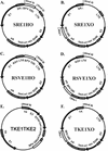
FIG. 1.
FIG. 1.
Schematic representation of designed plasmids. The arrows represent the location and direction of expression of genes (E1 gene, E2 gene, neomycin-kanamycin resistance marker gene neo), and the shaded boxes represent transcriptional elements like promoters, polyadenylation sites (pA), and bacterial origin. The open boxes represent the origin regions (BPV URR), which comprise the minimal origins together with the MME. Promoters of different strengths are driving the transcription of E1 and E2 in the constructs SRα, RSV LTR, TK, and MoMuLV LTR.
FIG. 2.
FIG. 2.
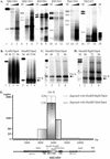
The replication properties of the designed plasmids are dependent on E1 protein expression. (A and D) A transient-replication assay was carried out with plasmids with promoters of different strengths for E1 in CHO cells. Increasing amounts of plasmid DNA (1, 2, and 5 μg) were transfected by electroporation into CHO cells, and episomal DNA was extracted 72 h later. (A) Purified episomal DNA was digested using linearizing enzymes EcoRI (lanes 2 to 4) or HindIII (lanes 5 to 10 [also panel D]) together with DpnI. Two hundred picograms of linear DNA (SRE1HO) was used as a marker on the blot (lane 1). X-ray film was exposed for 24 h (A) or 48 h (D). The position of unit-sized plasmid is indicated by the arrow (D). (B) Western blot analysis for E1 protein expression from transfected CHO cells. In this experiment, the strong SRα promoter was driving E1 expression in the case of plasmids SRE1HO and SRE1XO, which differ from each other in ori location in the plasmid (lanes 2 to 4 and 5 to 7, respectively). The weaker RSV promoter was driving E1 expression in the plasmid RSVE1HO (lanes 8 to 10). As a marker, the lysate from the COS7 cells, transfected with 0.5 μg of E1 expression construct pCGEag, was used as a positive control (lane 1), and only carrier DNA-transfected CHO cells were used as the negative control (lane 11). (C) Western blot analysis for E2 expression directed by the MoMuLV LTR from the plasmids SRE1HO (lanes 2 to 4), SRE1XO (lanes 5 to 7), and RSVE1HO (lanes 8 to 10).
FIG. 3.
FIG. 3.
Restriction analysis of replication products. (A) Transient-replication assay of vector constructs with promoters of different strengths driving E1 expression (SRα, RSV, TK) and with two ori locations (in HindIII or in XhoI sites). After transfection of 1 and 2 μg of vector DNA into CHO cells, episomal replication products were isolated, purified, and analyzed 72 h posttransfection using DpnI and different enzyme combinations: HindIII (lanes 2 and 3), MunI/Eco47III (lanes 5, 6, 11, 12, 14, 15, 17, and 18), and MunI/Eco47III/HindIII (lanes 8 and 9). Lanes 1, 4, 7, 10, 13, and 16 contain markers for the input plasmids, cleaved with respective enzymes. (B to D) Analysis of replication products in plasmids with high-level E1 expression (construct SRE1HO) in the CHO cell line, using 1.0 μg of transfected DNA. The cells were harvested 48, 72, and 96 h after transfection, and low-molecular-weight DNA was analyzed on 0.8% agarose gel by Southern blotting. DNA was digested with DpnI and additionally with the linearizing enzyme EcoRI (panel B, lanes 2 to 4); with enzyme HindIII (panel B, lanes 6 to 8), which cleaves out the origin-containing fragment (arrow); or with the enzyme combinations HindIII/XbaI (C) and HindIII/BglII (D). ori-sequence containing bands are shown by arrows. Marker DNAs (400 pg), digested with respective enzymes, are also shown. The blots were exposed for 24 h. (E) Schematic representation of amplification of SRE1HO vector. 32P-labeled dCTP incorporation was normalized to that based on the length of restriction fragments. The number of copies of origin sequence-containing fragments synthesized was calculated by comparing the incorporation of labeled nucleotides in the origin sequence-containing fragment to the incorporation of label into other fragments generated by the enzyme cleavage. A schematic representation of the linearized SRE1HO construct is shown below the graph, where viral origin, coding sequences, promoters, and used restriction enzyme cleavage sites are presented. Ori fr., origin sequence-containing fragment.
FIG. 4.
FIG. 4.
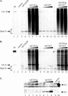
Admixture of replication proteins together with TKE1TKE2 vector DNA into CHO cells. (A and B) One microgram of the vector was transfected alone (lanes 4, 8, and 12) or with 0.1, 0.25, or 0.75 μg of the appropriate expression vector—pCGEag (for E1 expression) (lanes 5 to 7) or pCGE2 (for E2 expression) (lanes 9 to 11)—or with two expression vectors together (lanes 13 to 15). Mock-transfected cells (lane 2) and cotransfection with green fluorescent protein expression vector pCGGFP (0.75 μg) (lane 3) were used as negative controls. Cells were harvested 48 h after transfection, and episomal DNA was analyzed by Southern blotting with appropriate size markers (lane 1). Full-length vector DNA (B) and the 1-kb origin fragment (A) were used as probes, respectively. DpnI-resistant replicated DNA fragments as well nonreplicated DpnI-digested material are indicated on both blots. (A) The linearizing enzyme HindIII was used together with DpnI. (B) Enzyme KspAI (cleaves out an ∼1.0-kbp origin fragment) together with DpnI was used for restriction of the replication products. (C) Western blot analyses of expression levels of replication proteins E1 and E2 in samples at 48 h after incubation. Purified E1 and E2 proteins were loaded as positive controls (indicated by arrows, respectively). Abbreviations: Lin. fr., linear fragment; Ori fr., origin sequence-containing fragment.
FIG. 5.
FIG. 5.
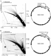
Neutral-neutral 2D agarose gel electrophoresis analysis of replication products of SRE1HO plasmid in CHO cells. (A) Plasmid DNA (1 μg) was transfected into CHO cells by electroporation, and episomal DNA was extracted by alkaline lysis. Extracted DNA was digested with KspAI, which cleaves the plasmid at the minimal origin region and within the SRα promoter, resulting in the fragment carrying most of the URR. The digested material was separated on the 2D gel, transferred to the nylon filter, and probed with the URR region. The arc of linear molecules, along with positions of double-stranded monomers (1n) (size, 1,650 bp) and dimers (2n), is indicated. In addition, replication intermediates (simple and branched Y structures) and termination structures (tts) were clearly seen on the blot. Appropriate molecular size marker positions in both dimensions are indicated also. The schematic map of the plasmid with recognition sites for KspAI is presented to the right of the blot. The replication initiation site is indicated by an arrow. (B) Analysis of replication intermediates within the 1.1-kb URR fragment. A 2-μg aliquot of SRE1HO plasmid was transfected by electroporation into CHO cells. Episomal DNA was extracted by Hirt lysis 48 h posttransfection, digested with HindIII, and separated by 2D gel electrophoresis. Migration of the monomer (1n)- and dimer (2n)-sized double-stranded DNA fragments on the arc of linear molecules and location of the simple Y arc and branched Y fragments as well as the arc for the asymmetric bubble (ab) are indicated. Recognition sites for HindIII and E1 binding sites (initiation site) are indicated by an arrow on the schematic map of the plasmid. Both blots have been probed with a radiolabeled BPV-1 URR probe (nt 6959 to 40), identical to the BPV Ori region indicated on the vector map.
FIG. 6.
FIG. 6.
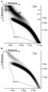
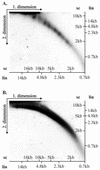
Neutral-neutral 2D electrophoresis of uncut plasmid molecules. CHO cells were transfected with 2 μg of SRE1HO plasmid DNA (size ≈ 11 kb). The uncut episomal DNA was extracted at 24 h (A) or at 72 h (B) after transfection and analyzed simultaneously by 2D gel electrophoresis with the appropriate molecular size markers for linear DNA fragments in both dimensions as indicated on the blots. (A) The arcs of the linear molecules are indicated as well the migrating positions of the covalently closed circular (ccc), linear (lin), and relaxed circular (oc) topological forms of the SRE1HO in the second dimension. The blots were hybridized with the BPV1 URR probe (nt 6959 to 40) and exposed for 48 h.
FIG. 7.
FIG. 7.
Neutral-neutral 2D gel electrophoresis of replication products fractionated by CsCl gradient centrifugation. Extrachromosomal Hirt method-extracted DNA was harvested 48 h after transfection of CHO cells with 2 μg of SRαHO plasmid DNA (∼11 kb) and fractionated by the CsCl-ethidium bromide density gradient. The fractions containing covalently closed circular form and open circular and linear DNA replication products were isolated. The fraction of supercoiled molecules (A) and the linear molecules and the relaxed circles (B) were analyzed simultaneously under the same 2D gel conditions. Positions of marker bands for linear DNA (lin) and for supercoiled DNA (sc) are indicated for both dimensions. Both blots were hybridized with the BPV1 URR probe (nt 6959 to 40).

Similar articles

Cited by

References

    1. Amin, A. A., S. Titolo, A. Pelletier, D. Fink, M. G. Cordingley, and J. Archambault. 2000. Identification of domains of the HPV11 E1 protein required for DNA replication in vitro. Virology 272:137-150. - PubMed
    1. Barksdale, S., and C. Baker. 1993. Differentiation-specific expression from the bovine papillomavirus type 1 P2443 and late promoters. J. Virol. 67:5605-5616. - PMC - PubMed
    1. Bedell, M. A., J. B. Hudson, T. R. Golub, M. E. Turyk, M. Hosken, G. D. Wilbanks, and L. A. Laimins. 1991. Amplification of human papillomavirus genomes in vitro is dependent on epithelial differentiation. J. Virol. 65:2254-2260. - PMC - PubMed
    1. Belyavskyi, M., M. Westerman, L. DiMichele, and V. Wilson. 1996. Perturbation of the host cell cycle and DNA replication by the bovine papillomavirus replication protein E1. Virology 219:206-219. - PubMed
    1. Bonne-Andrea, C., S. Santucci, and P. Clertant. 1995. Bovine papillomavirus E1 protein can, by itself, efficiently drive multiple rounds of DNA synthesis in vitro. J. Virol. 69:3201-3205. - PMC - PubMed

Publication types

Substances

LinkOut - more resources