. 2002 May;22(10):3460-73.
doi: 10.1128/MCB.22.10.3460-3473.2002.

Mutational analysis of all conserved basic amino acids in RAG-1 reveals catalytic, step arrest, and joining-deficient mutants in the V(D)J recombinase

Affiliations

Mutational analysis of all conserved basic amino acids in RAG-1 reveals catalytic, step arrest, and joining-deficient mutants in the V(D)J recombinase

Leslie E Huye et al. Mol Cell Biol. 2002 May.

Abstract

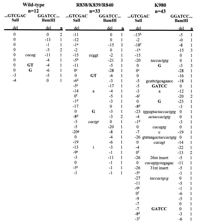
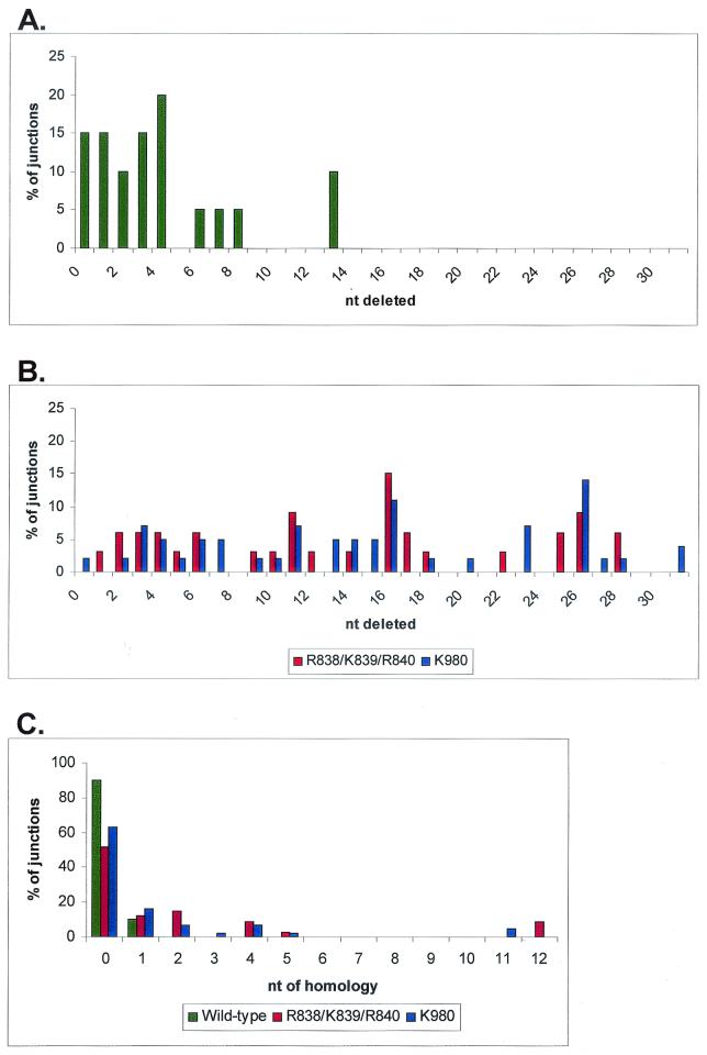
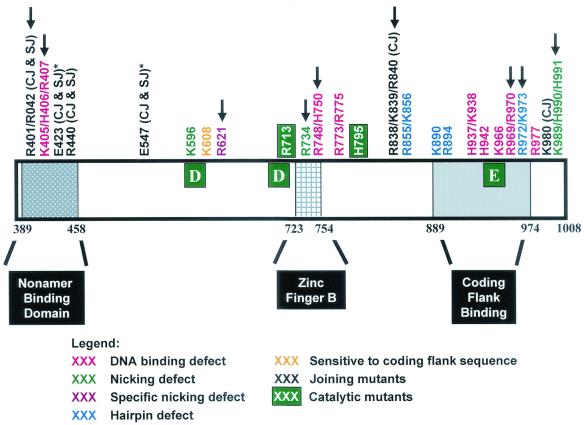
Although both RAG-1 and RAG-2 are required for all steps of V(D)J recombination, little is known about the specific contribution of either protein to these steps. RAG-1 contains three acidic active-site amino acids that are thought to coordinate catalytic metal ions. To search for additional catalytic amino acids and to better define the functional anatomy of RAG-1, we mutated all 86 conserved basic amino acids to alanine and evaluated the mutant proteins for DNA binding, nicking, hairpin formation, and joining. We found several amino acids outside of the canonical nonamer-binding domain that are critical for DNA binding, several step arrest mutants with defects in nicking or hairpin formation, and four RAG-1 mutants defective specifically for joining. Analysis of coding joints formed by some of these mutants revealed excessive deletions, frequent use of short sequence homologies, and unusually long palindromic junctional inserts, known as P nucleotides, that result from aberrant hairpin opening. These features characterize junctions found in scid mice, which are deficient for the catalytic subunit of DNA-dependent protein kinase (DNA-PKcs), suggesting that the RAG proteins and DNA-PKcs perform overlapping functions in coding joint formation. Interestingly, the amino acids that are altered in 12 of our mutants are also mutated in human inherited immunodeficiency syndromes. Our analysis of these mutants provides insights into the molecular mechanisms underlying these disorders.


Figures

FIG. 1.
FIG. 1.
Basic amino acids in RAG-1 targeted for mutagenesis. Eighty-six conserved basic residues (71 absolutely conserved [blue] and 15 with charge conservations [red]) were identified by aligning the RAG-1 sequences from nine species (human, rabbit, mouse, opossum, chicken, Xenopus, bull shark, rainbow trout, and zebra fish). The active-site residues D600, D708, and E962 are shown as white letters on a black background.
FIG. 2.
FIG. 2.
Mutations outside of the nonamer-binding domain in RAG-1 affect DNA binding. DNA binding was evaluated by electrophoretic mobility shift assays in which GST-tagged RAG-1 mutants coexpressed with GST-tagged RAG-2 were incubated with a radiolabeled 12-RSS oligonucleotide substrate. The positions of the free substrate, the protein-DNA complex, and the origin are indicated. Mutants with severe DNA-binding defects are in boldface type and are indicated by an asterisk. All lanes within each panel are from the same gel. Quantification was done by PhosphorImager analysis of the results of binding experiments with two independent protein preparations. WT, wild type. (Mutant K966 was previously shown to be proficient for RSS binding [30].)
FIG. 3.
FIG. 3.
Analysis of coupled cleavage of oligonucleotide substrates in Mg2+ allows classification of RAG-1 mutants. GST-tagged RAG-1 mutants coexpressed with GST-tagged RAG-2 were incubated with a radiolabeled 12-RSS oligonucleotide substrate, an unlabeled 23-RSS substrate, and HMG-1 in Mg2+. (A) Coupled cleavage with oligonucleotide substrates. Mutants that were identified as having a DNA-binding defect (panel I; DNA binding) were also severely defective for both nicking and hairpin formation (lanes 3 to 10). The second mutant class (panel II; nicking) was defective for nicking (lanes 11 to 16). The third mutant class (panel III; hairpin) was specifically defective for hairpin formation (lanes 17 to 20). The fourth class of mutants (panel IV; coding flank) contained one mutant, K608, which exhibited wild-type (Wt) levels of nicking and hairpin formation on substrates with permissive coding flanks (5′-TCTTA-heptamer) (lane 21). The fifth class of mutants (panel V; joining) was capable of both nicking and hairpin formation (lanes 22 to 25). Representative data are shown. All lanes are from the same exposure of the same gel but have been rearranged for clarity. The positions of the uncleaved substrate and the nick and hairpin products are indicated by diagrams on the left. The star indicates the position of the radioactive label. (B) Coupled cleavage with nonpermissive coding flank (5′-TCGAC-heptamer) oligonucleotide substrates. Compared to the wild type, mutant K608 exhibited a decrease in hairpin formation with a concurrent accumulation of nicks on substrates with nonpermissive coding flanks (compare lanes 2 and 3), indicating that this mutant is sensitive to the sequence of the coding flank.
FIG. 4.
FIG. 4.
Analysis of nick-defective and hairpin-defective mutants under relaxed RSS cleavage conditions in Mn2+. The 10 mutants showing defective nicking and/or hairpin formation in the coupled cleavage assay were tested for cleavage of a radiolabeled 12-RSS substrate under relaxed cleavage conditions (in Mn2+). Mutants are grouped and labeled according to their phenotypes in Mg2+. Three mutants (R621, R713, and H795) remained defective for nicking (lanes 2 to 4), and three mutants remained specifically defective for hairpin formation (lanes 8 to 10). Interestingly, these hairpin-defective mutants made low levels of aberrant hairpins, indicated by arrows. Representative data are shown. All lanes are from the same exposure of the same gel but have been rearranged for clarity. The positions of the uncleaved substrate, the nick product, and the normal hairpin product are indicated by diagrams on the left. The star indicates the position of the radiolabel. WT, wild type.
FIG. 5.
FIG. 5.
Prenicked oligonucleotide substrates specifically assay for the hairpin formation step. Mutants that exhibited a nicking defect (lanes 2 to 4) or aberrant hairpin formation (lanes 5 to 7) under relaxed cleavage conditions were tested for hairpin formation with a prenicked substrate in Mn2+, a strategy which bypasses the requirement for the generation of a nick at the correct position. The position of the aberrant hairpin is indicated by an arrow. Representative data are shown. All lanes are from the same exposure of the same gel but have been rearranged for clarity. Lane 5 is overloaded, but quantification reveals ratios of products and substrate similar to those in other lanes. See the legend to Fig. 3 for explanations of symbols. WT, wild type.
FIG. 6.
FIG. 6.
Identification of joining-deficient RAG-1 mutants. (A) Diagram illustrating the three recombination substrates used for in vivo analysis of the mutants. pJH299 assays for coding joints and signal joints formed by inversion on the plasmid. pJH289 assays for signal and coding joints formed by deletion, with the formation of signal joints on the plasmid product and coding joints on the excised product. pJH290 assays for coding joints and signal joints formed by deletion, with the formation of coding joints on the plasmid product and signal joints on the excised product. Coding flanks are represented by open and filled rectangles. The 12- and 23-RSS are indicated by open and filled triangles, respectively. The arrow on the plasmid products indicates the promoter for the chloramphenicol acetyltransferase (cat) gene, represented by the stippled rectangle. cat is expressed upon recombination, removing the transcription terminator (represented by S). Joints formed on the plasmid products can be identified by a bacterial transformation assay (23). (B to G) PCR assays (24 cycles) to detect coding joints (CJ) (B, E, and G), signal joints (SJ) (C and E), and signal ends (SE) (F). Diagrams of the PCR substrates are shown on the right. Wild-type (WT)-transfected DNA was assayed at 1× and 1:10 and 1:100 dilutions. The mutants were assayed at 1× concentrations. M, radiolabeled markers. (B) PCR for inversional coding joints on pJH299. (C) PCR for inversional signal joints on pJH299. (D) PCR for coding joints on the excised product of pJH289. (E) PCR for signal joints on the excised product of pJH290. (F) Signal ends (12 RSS) from the pJH290 substrate were detected by ligation-mediated PCR. Analysis of signal ends from the pJH299 substrate gave similar results (data not shown). (G) PCR for coding joints on the plasmid product of pJH290. (H and I) Analysis of coding joints on the plasmid product of pJH290 (H) and signal joints on the plasmid product of pJH289 (I) by a transformation assay. The wild-type result is normalized to 1, and recombination by the mutants is shown relative to that of the wild type. Data from two independent experiments are shown (white and black bars).
FIG. 7.
FIG. 7.
Nucleotide sequence analysis of coding joints from R838/K839/R840 and K980. PCR products containing coding joints were cloned and sequenced. The total numbers of unique sequences obtained for the wild type, R838/K839/R840, and K980 are indicated. The first 6 nt on either side of a perfect junction are shown. A perfect coding joint is GGTCGTTGATCCCCCATCGATGAGAGTCGAC-GGATCCTCTCATCGATGAGAGGATCGACGACGACATGGC. n, number of unique junctions with the indicated sequence. The number of deleted (del) nucleotides from each end is indicated beneath the corresponding end sequences. Letters in boldface type between the two ends indicate presumptive P nucleotides. Lowercase letters between the two ends indicate nucleotide insertions. Italics denote nucleotides derived from imprecise cleavage events. The sequence of the 26-nt insert is CTAGAGTCGATCCGTCCCCGGGCGAG. The sequence of the 31-nt insert is CAAAACCCTCTGTAACTCTAGATCCAGGAAT. Superscripts designate junctions that exhibit short sequence homologies, as follows: a, TCGA; b, CATCGATGAGAG; c, TCGAC; d, GA; e, G; f, C; g, TC; h, ATC; I, CATCGATGAGA.
FIG. 8.
FIG. 8.
Coding joint deletions and short sequence homologies. Distribution of the number of nucleotides deleted from coding joints from the wild type (A) (includes sequences from this study and reference 47) and R838/K839/R840 and K980 (B). The x axis represents the total number of nucleotides lost from both coding ends. Data are from Fig. 7. (C) Mutant coding joints exhibit short sequence homologies at the coding junction. The x axis represents the number of homologous nucleotides at the junction. Data are from Fig. 7.
FIG. 9.
FIG. 9.
Schematic diagram of the core domain of RAG-1 (amino acids 384 to 1008) mapping the phenotypic classes of mutants identified in this study. The active-site residues, D600, D708, and E962, are shown as large white letters in green boxes. The nonamer-binding domain, zinc finger B, and coding flank region are also shown. Asterisks indicate two acidic residues previously identified as joining mutants (54). Arrows indicate mutants with corresponding mutants found in T B Scid and Omenn syndrome patients. The human mutations are R396C, R396L, or R396H; R404W; R410Q; R474H; R507W; R624H or R624C; R737H; H753L; R841W; R973H; R975Q; and K992E (7, 55, 66, 67).

Similar articles

Cited by

References

    1. Agrawal, A., and D. G. Schatz. 1997. RAG1 and RAG2 form a stable postcleavage synaptic complex with DNA containing signal ends in V(D)J recombination. Cell 89:43-53. - PubMed
    1. Aidinis, V., D. C. Dias, C. A. Gomez, D. Bhattacharyya, E. Spanopoulou, and S. Santagata. 2000. Definition of minimal domains of interaction within the recombination-activating genes 1 and 2 recombinase complex. J. Immunol. 164:5826-5832. - PubMed
    1. Akamatsu, Y., and M. A. Oettinger. 1998. Distinct roles of RAG1 and RAG2 in binding the V(D)J recombination signal sequences. Mol. Cell. Biol. 18:4670-4678. - PMC - PubMed
    1. Bailin, T., X. Mo, and M. J. Sadofsky. 1999. A RAG1 and RAG2 tetramer complex is active in cleavage in V(D)J recombination. Mol. Cell. Biol. 19:4664-4671. - PMC - PubMed
    1. Bogue, M. A., C. Wang, C. Zhu, and D. B. Roth. 1997. V(D)J recombination in Ku86-deficient mice: distinct effects on coding, signal, and hybrid joint formation. Immunity 7:37-47. - PubMed

Publication types

MeSH terms

LinkOut - more resources