. 2002 May;22(10):3425-36.
doi: 10.1128/MCB.22.10.3425-3436.2002.

AMP-activated kinase regulates cytoplasmic HuR

Affiliations

AMP-activated kinase regulates cytoplasmic HuR

Wengong Wang et al. Mol Cell Biol. 2002 May.

Abstract

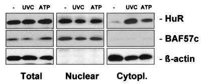
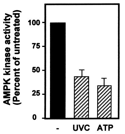
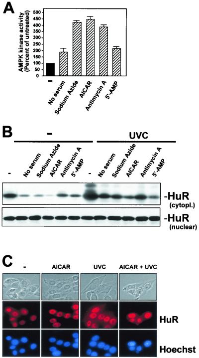
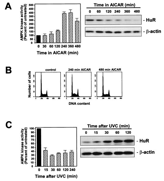
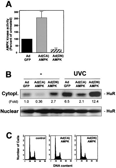
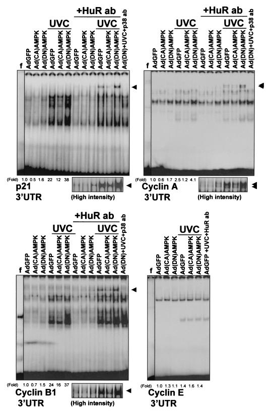
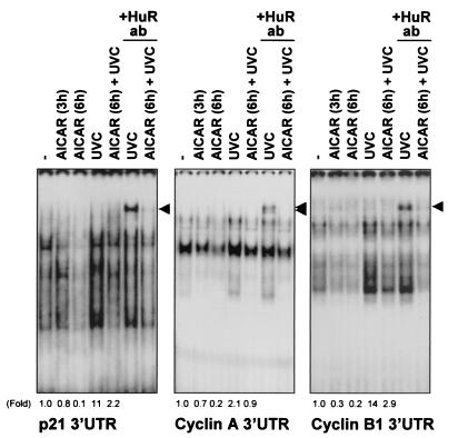
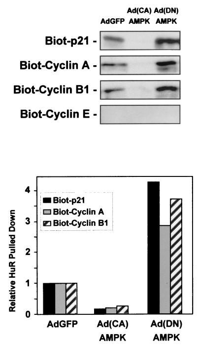
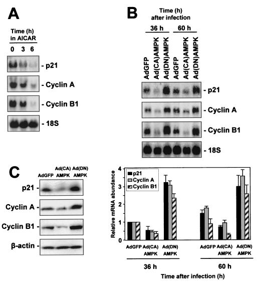
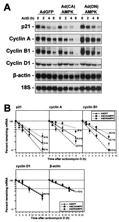
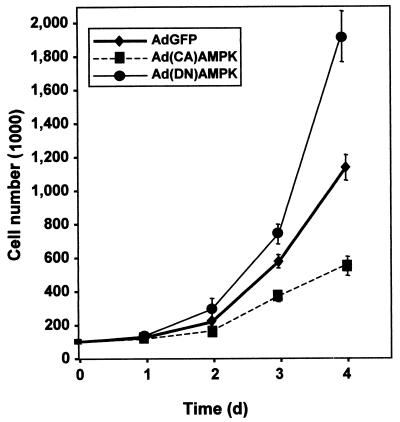
While transport of RNA-binding protein HuR from nucleus to cytoplasm is emerging as a key regulatory step for HuR function, the mechanisms underlying this process remain poorly understood. Here, we report that the AMP-activated kinase (AMPK), an enzyme involved in responding to metabolic stresses, potently regulates the levels of cytoplasmic HuR. Inhibition of AMPK, accomplished either through cell treatment or by adenovirus infection to express dominant-negative AMPK, was found to increase the level of HuR in the cytoplasm and to enhance the binding of HuR to p21, cyclin B1, and cyclin A mRNA transcripts and elevate their expression and half-lives. Conversely, AMPK activation, achieved by means including infection to express constitutively active AMPK, resulted in reduced cytoplasmic HuR; decreased levels and half-lives of mRNAs encoding p21, cyclin A, and cyclin B1; and diminished HuR association with the corresponding transcripts. We therefore propose a novel function for AMPK as a regulator of cytoplasmic HuR levels, which in turn influences the mRNA-stabilizing function of HuR and the expression of HuR target transcripts.


Figures

FIG. 1.
FIG. 1.
Subcellular localization of HuR in RKO cells. Three hours after exposure of RKO cells to either 20-J/m2 UVC (lane UVC) or 1 mM ATP (lane ATP), whole-cell (20 μg), nuclear (10 μg), and cytoplasmic (40 μg) lysates were subjected to Western blot analysis to monitor the expression of HuR. Sequential hybridizations using antibodies against BAF57c and β-actin were carried out to assess the quality of the fractionation process and the uniformity in loading and transfer of nuclear and cytoplasmic samples, respectively. Lane −, untreated lysates.
FIG. 2.
FIG. 2.
Methodology used to assess the effect of the various treatments on the cytoplasmic HuR levels. Shown is a Western blot analysis of HuR abundance in whole-cell, nuclear, and cytoplasmic lysates prepared as described in the legend for Fig. 1 from RKO cells that were either left untreated or treated with AICAR. Lanes −, unirradiated cells (control); lanes UVC, treatment with UVC.
FIG. 3.
FIG. 3.
Inhibition of AMPK kinase activity in RKO cells by treatments that enhance HuR presence in the cytoplasm. Three hours after exposure of RKO cells to either 20-J/m2 UVC (lane UVC) or 1 mM ATP (lane ATP), whole-cell lysates were prepared and AMPK kinase activity was tested after IP with polyclonal antibodies recognizing the AMPK α1 and α2 subunits and with synthetic peptide SAMS as the substrate. −, untreated lysates.
FIG. 4.
FIG. 4.
Effect of AMPK activators on the subcellular localization of HuR. Shown is an assessment of AMPK activity (A) and HuR levels (B) in cytoplasmic (40 μg) and nuclear (10 μg) lysates prepared from RKO cells that were serum starved for 36 h or were treated for 4 h with either 2 mM sodium azide, 2 mM AICAR, 1μM antimycin A, or 1 mM ATP. For Western blot analysis, cells were either mock irradiated but otherwise exposed to AMPK-activating treatments continuously for 4 h (−) or were exposed to 20-J/m2 UVC 6 h after addition of AMPK activators and then cultured for an additional 3 h. (C) Immunofluorescence detection of HuR in RKO cells that were treated with AICAR, UVC, or both AICAR and UVC as explained in the legend for panel B. Top, phase-contrast images; middle, HuR immunofluorescence; bottom, Hoechst staining to visualize nuclei.
FIG. 5.
FIG. 5.
Kinetics of AMPK kinase activity and HuR subcellular localization. (A) Assessment of AMPK activity (left) and HuR presence in the cytoplasm (right) at the indicated times following treatment of RKO cells with 2 mM AICAR. (B) FACS analysis after treatment of RKO cells with AICAR for the times indicated. (C) Assessment of AMPK activity (left) and HuR presence in the cytoplasm (right) at the indicated times following treatment of RKO cells with 20-J/m2 UVC. Western blots (A and C) were stripped and rehybridized to assess β-actin levels in order to control for sample loading and transfer.
FIG. 6.
FIG. 6.
Influence of adenoviruses expressing mutant AMPK catalytic subunits on AMPK kinase activity and cytoplasmic HuR levels. Forty-eight hours after infection of RKO cells with 100 PFU of either AdGFP, Ad(CA)AMPK, or Ad(DN)AMPK/cell, AMPK activity (A) and HuR levels (B) were measured in the cytoplasmic (40 μg) and nuclear (10 μg) fractions of cells that were either left unirradiated or exposed to UVC (20 J/m2) and collected 3 h later. Fold, difference in HuR signal between indicated population and AdGFP-infected, unirradiated population. In panel A, data represent the means ± standard errors of the means from four independent experiments. (C) FACS analysis of RKO populations 48 h after infection.
FIG. 7.
FIG. 7.
Effect of AMPK-expressing adenoviruses on the association of HuR with target transcripts. EMSA were performed using radiolabeled RNAs encoding the 3′ UTRs of p21, cyclin A, and cyclin B1 genes (see Materials and Methods) and proteins present in cytoplasmic lysates of RKO cells that were infected with either AdGFP, Ad(CA)AMPK, or Ad(DN)AMPK and then were either exposed to 20-J/m2 UVC or left unirradiated and collected 3 h later. The presence of HuR in RNA-protein complexes was assayed by monitoring the formation of supershifted bands in the presence of anti-HuR antibodies (+HuR ab) or control antibodies (+p38 ab). Arrowheads, supershifted complexes. Fold, difference in total signals of radiolabeled complexes between indicated cells and AdGFP-infected, unirradiated cells. Fold differences were not calculated for supershift lanes. f, free probe, not incubated with cytoplasmic lysate; high intensity, supershifted bands developed at greater intensity.
FIG. 8.
FIG. 8.
Influence of AMPK modulators on the association of HuR with target transcripts. EMSA was performed using radiolabeled transcripts encoding the 3′ UTRs of p21, cyclin A, and cyclin B1 genes and proteins present in cytoplasmic lysates of RKO cells that had either been treated with 2 mM AICAR for the times indicated and then left unirradiated or exposed to 20-J/m2 UVC (+UVC). The presence of HuR in the RNA-protein complexes was assayed by monitoring the formation of supershifted bands in the presence of anti-HuR antibodies (+HuR ab). Arrowheads, supershifted complexes. Fold, relative signals of radiolabeled complexes compared with those measured in untreated (−), unirradiated cells. Fold differences were not calculated for supershift lanes.
FIG. 9.
FIG. 9.
HuR forms complexes with biotinylated target transcripts. Cytoplasmic lysates prepared 48 h after infection of RKO cells with either AdGFP, Ad(CA)AMPK, or Ad(DN)AMPK were incubated with biotinylated transcripts corresponding to the 3′ UTRs of the genes encoding the proteins indicated. Biotinylated (Biot) RNA-protein complexes were pulled down with streptavidin-conjugated magnetic beads and analyzed by Western blotting to assess HuR levels. The pull-down shown is representative of two independent experiments. The graph depicts quantitation of the signals on the Western blots.
FIG. 10.
FIG. 10.
AMPK-modulatory interventions influence the expression of targets of HuR-mediated mRNA stabilization. (A) RKO cells were either left untreated or were treated with AICAR (2 mM) for 3 or 6 h, whereupon RNA was prepared and subjected to Northern blot analysis to assess the expression levels of mRNAs encoding p21, cyclin A, and cyclin B1. (B) Top, measurement of the levels of mRNAs encoding p21, cyclin A, and cyclin B1 in RKO cells at the indicated times following adenovirus infection. Bottom, quantitation of mRNA levels, with data representing the means ± standard errors of the means of three independent experiments. 18S rRNA signals served to monitor the equality of loading and transfer among samples. (C) Western blot analysis of protein expression 48 h after infection.
FIG. 11.
FIG. 11.
AMPK-modulatory interventions influence the half-lives of mRNAs that are stabilized by HuR. (A) Assessment of the stability of mRNAs encoding p21, cyclin A, cyclin B1, cyclin D1, and β-actin in RKO cells 48 h after infection with the adenoviruses indicated. (B) mRNA half-lives were calculated after treating cells with 2 μg of ActD/ml, preparing RNA at the times indicated, and measuring p21, cyclin A, and cyclin B1 mRNA Northern blot signals, normalizing them to 18S rRNA signals, and plotting them on a logarithmic scale (bottom). Note that the time scales for the cyclin D1 and β-actin mRNA plots are different. Horizontal dashed lines, 50% of level for untreated cells. Data represent the means ± standard errors of the means for three independent experiments.
FIG. 12.
FIG. 12.
Influence of AMPK on cell proliferation. RKO cells (100,000 cells per dish) were infected with 100 PFU/cell, and cell numbers were monitored every 24 h thereafter with a hemacytometer. The experiment was done in triplicate, and data are means ± standard errors of the means.

Similar articles

Cited by

References

    1. Atasoy, U., J. Watson, D. Patel, and J. D. Keene. 1998. ELAV protein HuA (HuR) can redistribute between nucleus and cytoplasm and is upregulated during serum stimulation and T cell activation. J. Cell Sci. 111:3145-3156. - PubMed
    1. Bohjanen, P. R., B. Petryniak, C. H. June, C. B. Thompson, and T. Lindsten. 1992. AU RNA-binding factors differ in their binding specificities and affinities. J. Biol. Chem. 267:6302-6309. - PubMed
    1. Bost, F., N. Dean, R. McKay, and D. Mercola. 1997. The JUN kinase/stress-activated protein kinase pathway is required for epidermal growth factor stimulation of growth of human A549 lung carcinoma cells. J. Biol. Chem. 272:33422-33429. - PubMed
    1. Brennan, C. M., and J. A. Steitz. 2001. HuR and mRNA stability. Cell. Mol. Life Sci. 58:266-277. - PMC - PubMed
    1. Carballo, E., W. S. Lai, and P. J. Blackshear. 1998. Feedback inhibition of macrophage tumor necrosis factor-α production by tristetraprolin. Science 281:1001-1005. - PubMed

MeSH terms

LinkOut - more resources