. 2002 Apr;3(4):378-83.
doi: 10.1093/embo-reports/kvf072.

The Drosophila homolog of NTF-2, the nuclear transport factor-2, is essential for immune response

Affiliations

The Drosophila homolog of NTF-2, the nuclear transport factor-2, is essential for immune response

Ananya Bhattacharya et al. EMBO Rep. 2002 Apr.

Abstract

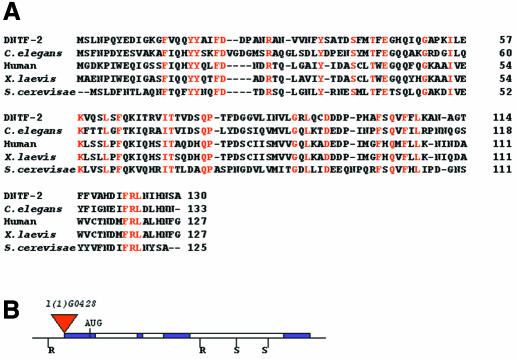
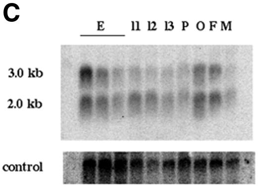
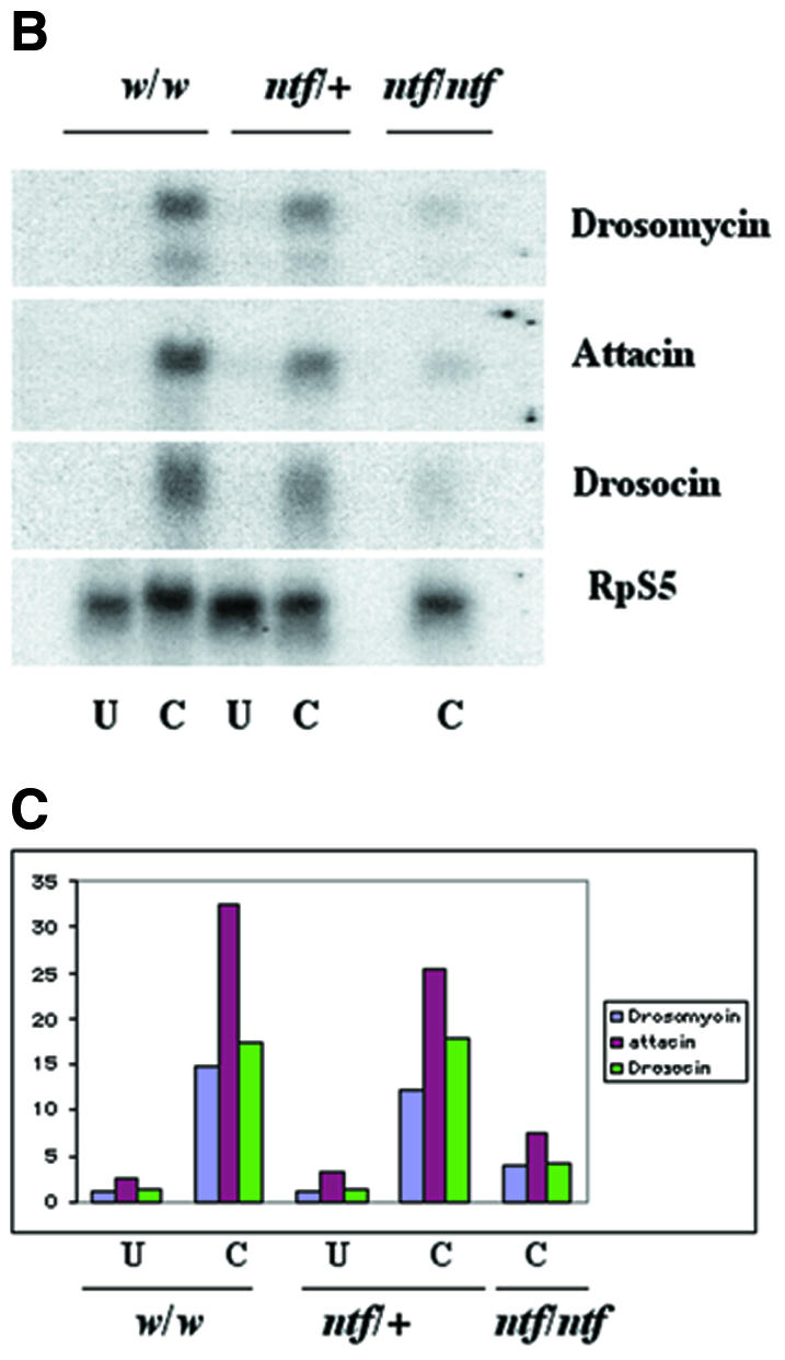
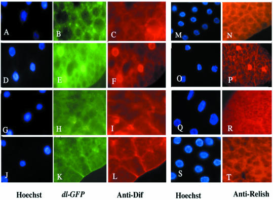
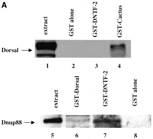
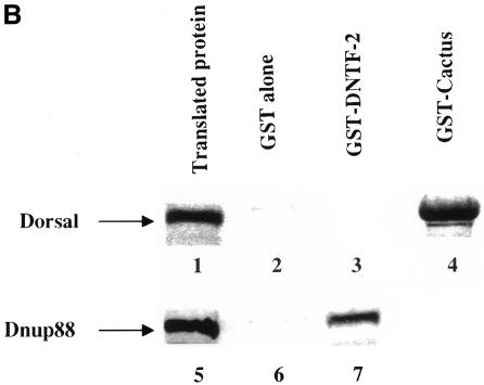
Nuclear transport factor-2 (NTF-2) functions in yeast and mammalian cell culture in targeting proteins into the nucleus. The Drosophila homolog, DNTF-2, is an essential component of the nuclear import machinery, since ntf mutants are lethal. Interestingly, hypomorphic alleles show specific phenotypes. Some are viable, but the number of omatidia in the eye is severely reduced. The immune response in the Drosophila larval fat body is also affected; the three NF-kappaB/Rel proteins Dorsal, Dif and Relish do not target to the nucleus after infection, and, consequently, the expression of the anti-microbial peptide genes drosomycin, attacin and drosocin is severely impaired. Hence, in spite of its general requirement in many developmental processes, DNTF-2 has a higher specific requirement in the development of the eye and in the immune response. We also found that DNTF-2 interacts directly with Mbo/DNup88, which does not contain phenylalanine-glycine-rich repeats, but has been shown to function in the import of Rel proteins.


Figures

None
Fig. 1. (A) Amino-acid sequence of DNTF-2. The sequence is aligned with human, C. elegans, Xenopus laevis and Saccharomyces cerevisiae NTF-2 sequences. Residues that are identical in all known NTF-2 proteins are shown in red. (B) Organization of the DNTF-2 genomic region. The solid blue boxes represent exons, empty boxes represent introns. The position of the P element l(1)G0428, a few bases upstream from the start of the cDNA, is shown as a red triangle. (C) RNA expression profile of the ntf-2 gene. In the upper panel, a blot of poly(A)+ RNA is probed with the ntf-2 cDNA. E refers to the RNA from the three embryonic stages, 0–4 h, 4–8 h and 8–16 h. L1–3 represent the three larval stages; P, pupal stage; O, ovaries; F, females and M, male RNA. The same blot was probed with RpS5, encoding the ribosomal protein (lower panel). (D) Hypomorphic mutants in the ntf-2 gene show abnormalities in eye development. (a) Mutants have small eyes with a strongly reduced number of ommatidia. (b) The wild-type eye.
None
Fig. 2. P[ntf] mutant larvae show low induction of the anti-microbial peptides when challenged with a bacterial solution. (A) Comparison of the induction of the GFP-tagged drosomycin reporter gene between wild-type and l(1)G0428/l(1)G0428 larvae. U represents bacterially unchallenged larvae; C, challenged larvae. (B) Northern blots of total RNA samples from wild-type, l(1)G0428/+ and l(1)G0428/l(1)G0428 larvae probed with cDNA probes corresponding to drosomycin, attacin and drosocin genes. The RpS5 loading control is shown at the bottom. (C) Profiles of anti-microbial gene induction. The data from the northern analyses shown in (B) were quantitated by phosphorimager analysis, and the amount of induction is calculated as a percentage of RpS5 RNA.
None
Fig. 3. The Rel proteins Dorsal, Dif and Relish do not target to the nuclei of fat body cells in response to bacterial challenge in ntf mutants. The distribution of Dorsal, Dif and Relish proteins in unchallenged wild-type (AC, M and N), challenged wild-type (DF, O and P), unchallenged larvae from l(1)G0428/l(1)G0428 (GI, Q and R) and challenged larvae from l(1)G0428/l(1)G0428 (JL, S and T) are shown. The nuclei stained with Hoechst are shown in (A, D, G, J, M, O, Q and S). In the homozygous mutant fat bodies, none of the Rel proteins are seen in the nuclei after bacterial infection (K, L and T), in contrast to the wild type, where all three Rel proteins are translocated to nuclei after bacterial infection (E, F and P).
None
Fig. 4. GST–DNTF-2 associates with the Drosophila homolog of Nup88 (DNup88)/Mbo. (A) Western blot probed with anti-Mbo antibody show binding of DNup88 from embryonic extracts with GST–DNTF-2 (lane 7) and weaker interaction with Dorsal (lane 6) but not with GST alone (lane 8). In contrast, Dorsal does not associate with GST–DNTF-2 (lane 3) and GST alone (lane 2) but associates with GST–Cactus (lane 4), the positive control. (B) DNup88 directly binds to DNTF-2. cDNA clones of DNup88 and Dorsal were in vitro translated with [35S]met and tested for binding with GST–DNTF-2, GST–Cactus and GST alone. Only DNup88 shows direct binding with GST–DNTF-2 (lane 7), whereas Dorsal (lane 3) shows no association with DNTF-2. As a positive control, Dorsal strongly binds to GST–Cactus (lane 4). As a negative control, all the translated proteins were tested for their binding with GST alone (lanes 2 and 6), which shows no binding.

Similar articles

Cited by

References

    1. Arai Y., Hosoda, F., Kobayashi, H., Arai, K., Hayashi, Y., Kamada, N., Kaneko, Y. and Okhi, M. (1997) The inv(11)(p15q22) chromosome translocation of de novo and therapy-related myeloid malignancies result in fusion of the nucleoporin gene, NUP98, with the putative RNA helicase gene DDX10. Blood, 89, 3936–3944. - PubMed
    1. Bayliss R., Ribbeck, K., Akin, D., Kent, H.M., Feldherr, C., Görlich, D. and Stewart, M. (1999) Interaction between NTF2 and xFxFG-containing nucleoporins is required to mediate nuclear import of RanGDP. J. Mol. Biol., 293, 579–593. - PubMed
    1. Clarkson W.D., Kent, H.M. and Stewart, M. (1996) Separate binding sites on nuclear transport factor 2 for GDP–Ran and the phenylalanine-rich repeat regions of nucleoporins p62 and Nsp1p. J. Mol. Biol., 263, 517–524. - PubMed
    1. Corbett A.H. and Silver, P.A. (1996) The NTF2 gene encodes an essential, highly conserved protein that functions in nuclear transport in vivo. J. Biol. Chem., 271, 18477–18484. - PubMed
    1. Drier E.A., Huang, L.H. and Steward, R. (1999) Nuclear import of the Drosophila Rel protein Dorsal is regulated by phosphorylation. Genes Dev., 13, 556–568. - PMC - PubMed

Publication types

MeSH terms

LinkOut - more resources