. 2002 Apr;22(8):2487-97.
doi: 10.1128/MCB.22.8.2487-2497.2002.

Critical but distinct roles for the pleckstrin homology and cysteine-rich domains as positive modulators of Vav2 signaling and transformation

Affiliations

Critical but distinct roles for the pleckstrin homology and cysteine-rich domains as positive modulators of Vav2 signaling and transformation

Michelle A Booden et al. Mol Cell Biol. 2002 Apr.

Abstract

Vav2, like all Dbl family proteins, possesses tandem Dbl homology (DH) and pleckstrin homology (PH) domains and functions as a guanine nucleotide exchange factor for Rho family GTPases. Whereas the PH domain is a critical positive regulator of DH domain function for a majority of Dbl family proteins, the PH domains of the related Vav and Vav3 proteins are dispensable for DH domain activity. Instead, Vav proteins contain a cysteine-rich domain (CRD) critical for DH domain function. We evaluated the contribution of the PH domain and the CRD to Vav2 guanine nucleotide exchange, signaling, and transforming activity. Unexpectedly, we found that mutations of the PH domain impaired Vav2 signaling, transforming activity, and membrane association. However, these mutations do not influence exchange activity on Rac and only slightly affect exchange on RhoA and Cdc42. We also found that the CRD was critical for the exchange activity in vitro and contributed to Vav2 membrane localization. Finally, we found that phosphoinositol 3-kinase activation synergistically enhanced Vav2 transforming and signaling activity by stimulating exchange activity but not membrane association. In conclusion, the PH domain and CRD are mechanistically distinct, positive modulators of Vav2 DH domain function in vivo.


Figures

FIG. 1.
FIG. 1.
Primary structure and protein expression levels of the human Vav2 truncation and point mutants. (A) Schematic structure of full-length human Vav2, NH2-terminal and COOH-terminal truncation mutants, and PH domain and CRD point mutants. Horizontal lines below full-length Vav2 show the predicted translational product of each truncation or point mutant initiated at the indicated amino acid. NH2-terminally truncated Vav2 (Δ1-191) is a strongly transforming variant of Vav2, and further COOH-terminal truncation to generate the Vav2-DPC fragment (DPC) did not reduce this activity. Therefore, to avoid potential complications in interpretation, all PH and CRD point mutants were generated in the background of the highly transforming Vav2-DPC fragment. Vertical lines, mutations in the PH domain (K407A and W503L) and the CRD (K533A, K538A, K563A, and V568E). Truncation mutants containing the DH and PH domains (DP) or DH domain (D) were also generated. The Δ1-191 and wild-type and mutant Vav2-DPC, -DP, and -D sequences were fused in frame to an HA epitope tag at the NH2 terminus. (B) Expression of the Vav2-DPC, truncation, and point mutants in stably and transiently transfected NIH 3T3 cells.
FIG. 2.
FIG. 2.
Mutation of the Vav2 PH domain decreases Vav2 transforming and signaling activity. (A) NIH 3T3 cells were transfected with the empty pCGN-hygro plasmid (Vector) or pCGN-hygro plasmids encoding the various PH domain mutants (Fig. 1; 1 μg per 60-mm-diameter dish). The appearance of transformed foci was quantitated on day 14. The values represent the averages ± standard errors of three dishes and are representative of at least three independent assays. WT, wild type. (B) NIH 3T3 cells were transiently transfected with the empty pCGN-hygro plasmid or pCGN-hygro plasmids encoding the various PH domain mutants (100 ng per 60-mm-diameter dish), together with a mutant serum response element luciferase reporter plasmid to determine stimulation of SRF. Fold activation was determined by the number of relative luciferase units relative to the number of units seen with the empty vector control. Data are representative of at least three independent assays performed on duplicate plates.
FIG. 3.
FIG. 3.
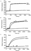
Stimulation of mant-GDP incorporation into Rac1, Cdc42, and RhoA by the Vav2 PH domain mutants in vitro. The abilities of bacterially expressed wild-type Vav2-DPC and Vav2-DPC PH domain mutants (50 nM) to stimulate the incorporation of mant-GDP into bacterially expressed (2 μM) Rac1 (A), Cdc42 (B), and RhoA (C) were measured by fluorescence spectroscopy (λex = 360 nm; λem = 440 nm). Arrows, time point (300 s) at which the Vav2 PH or CRD polypeptides were added to the exchange reaction mixture. Results are representative of two independent assays.
FIG. 4.
FIG. 4.
The Vav2 PH domain, but not the CRD, influences subcellular distribution. NIH 3T3 cells were transiently transfected with the empty pCGN mammalian expression vector and pCGN plasmids encoding the various PH domain and CRD mutants (Fig. 1; 1 μg per 60-mm-diameter dish). Subcellular fractions were prepared 48 h after transfection by lysis of cells in hypotonic buffer (0.1 M Tris [pH 7.4], 0.5 M MgCl2, 1 mM Pefabloc, 1 μM leupeptin, 2 μM pepstatin, 0.1% aprotinin) and addition of NaCl to adjust the ionic strength to 0.15 M, followed by ultracentrifugation for 30 min at 100,000 × g as described previously (5). Proteins in the crude S100 (S) and P100 (P) fractions were precipitated with 5 ml of acetone for 1 h at 4°C, collected by centrifugation at 2,000 × g for 30 min, and resuspended in 100 μl of electrophoresis sample buffer. Equal volumes of each fraction were separated by sodium dodecyl sulfate-polyacrylamide gel electrophoresis, and protein expression was determined by Western blot analysis using the anti-HA epitope antibody.
FIG. 5.
FIG. 5.
Alignment of representative CRDs. The conserved cysteine and histidine residues in the different Raf serine/threonine kinases and Vav protein CRDs were aligned. Black shading, identical residues; gray shading, highly conserved residues, dashes, gaps in the alignment with PKCδ C1A. Residues in PKCδ C1A involved in diacylglycerol (DAG) and membrane binding (membrane) are indicated (19). Residues in human c-Raf-1 involved in Ras and 14-3-3 binding are also indicated (19). Arrows, residues that were mutated in human Vav2.
FIG. 6.
FIG. 6.
Mutation of the Vav2 CRD decreases Vav2 transforming and signaling activity. (A) NIH 3T3 cells were transfected with the empty pCGN-hygro plasmid or pCGN-hygro plasmids encoding the various CRD mutants (Fig. 1; 1 μg per 60-mm-diameter dish). The appearance of transformed foci was quantitated on day 14. The values represent the averages ± standard errors of three dishes and are representative of at least three independent assays. WT, wild type. (B) NIH 3T3 cells were transiently transfected with empty pCGN plasmid or pCGN plasmids encoding the various CRD mutants (100 ng per 60-mm-diameter dish), together with the SRF luciferase reporter plasmid. Fold activation was determined by the number of relative luciferase units relative to the number of units seen with the empty vector control. Data are representative of at least three independent assays performed on duplicate plates.
FIG. 7.
FIG. 7.
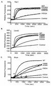
Stimulation of mant-GDP incorporation into Rac1, Cdc42, and RhoA by the Vav2 CRD mutants in vitro. The ability of bacterially expressed wild-type (WT) Vav2-DPC (50 nM) and Vav2-DPC with the listed substitutions within the CRD (50 nM) to stimulate the incorporation of mant-GDP into bacterially expressed (2 μM) Rac1 (A), Cdc42 (B), and RhoA (C) was measured by fluorescence spectroscopy (λex = 360 nm; λem = 440 nm). Arrows, time point (300 s) at which the Vav2 CRD polypeptides were added to the exchange reaction mixture. Results are representative of two independent assays.
FIG. 8.
FIG. 8.
Coexpression of activated PI3K causes enhancement of the transforming and signaling activities of Vav2 DPC but has minimal effect on the Vav2 PH or CRD mutants. Assays were performed as described in the legends for Fig. 2 and 5 except that 500 ng of the pZIP-NeoSV(x)1 empty vector (black bars) or pZIP-NeoSV(x)1 encoding activated PI3K (p110-CAAX) (open bars) was cotransfected with 100 ng of pCGN-hygro plasmid DNA encoding the indicated Vav2 protein. (A) NIH 3T3 cells were cotransfected with control plasmid pCGN-hygro (vector) encoding the indicated Vav2 PH domain mutant proteins (Fig. 1; 100 ng per 60-mm-diameter dish), together with pZIP-NeoSV(x)1 (vector) or pZIP-p110-CAAX (500 ng per 60-mm-diameter dish). Values are the averages ± standard errors for two dishes and are representative of three independent assays. WT, wild type. (B) NIH 3T3 cells were transiently transfected with the above plasmids along with a luciferase reporter construct for SRF transcriptional activity. Data are representative of at least three independent assays performed on duplicate plates. (C) The expression of p110-CAAX in the SRF reporter assay (B) was analyzed by Western blotting 20 μg of total cell lysates with an anti-Akt and an anti-phospho-Akt (P-Akt) antibody.
FIG. 9.
FIG. 9.
Coexpression of activated PI3K and Vav2-DPC caused a synergistic increase in the level of Rac1-GTP but did not increase Vav2-DPC membrane association. (A) NIH 3T3 cells stably expressing Vav2-DPC, p110CAAX, or both were cultured for 24 h in low serum (0.1%) and lysed, and the lysates were used in GST pull-down assays using GST-PAK Rac binding domain (RBD) immobilized on glutathione agarose beads. Bound proteins and total cell lysates were analyzed by Western blotting with anti-Rac1 antibodies. The expression of Vav2-DPC was analyzed by Western blotting total cell lysates with an anti-HA antibody. The expression of p110-CAAX was analyzed by Western blotting total cell lysates with an anti-phospho-Akt antibody (data not shown). (B) PI3K activation does not enhance Vav2-DPC association with membranes. NIH 3T3 cells were cotransfected with the control plasmid pCGN (vector) encoding Vav2-DPC (1 μg per 60-mm-diameter dish), together with pZIP-NeoSV(x)1 (vector) or pZIP-p110CAAX (500 ng per 60-mm-diameter dish). Subcellular fractionation was performed as described for Fig. 4.

Similar articles

Cited by

References

    1. Abe, K., K. L. Rossman, B. Liu, K. D. Ritola, D. Chiang, S. L. Campbell, K. Burridge, and C. J. Der. 2000. Vav2 is an activator of Cdc42, Rac1, and RhoA. J. Biol. Chem. 275:10141-10149. - PubMed
    1. Abe, K., I. P. Whitehead, J. P. O'Bryan, and C. J. Der. 1999. Involvement of N-terminal sequences in the negative regulation of vav signaling and transforming activity. J. Biol. Chem. 274:30410-30418. - PubMed
    1. Bishop, A. L., and A. Hall. 2000. Rho GTPases and their effector proteins. Biochem. J. 348:241-255. - PMC - PubMed
    1. Brtva, T. R., J. K. Drugan, S. Ghosh, R. S. Terrell, S. Campbell-Burk, R. M. Bell, and C. J. Der. 1995. Two distinct Raf domains mediate interaction with Ras. J. Biol. Chem. 270:9809-9812. - PubMed
    1. Buss, J. E., P. A. Solski, J. P. Schaeffer, M. J. MacDonald, and C. J. Der. 1989. Activation of the cellular proto-oncogene product p21Ras by addition of a myristylation signal. Science 243:1600-1603. - PubMed

Publication types

MeSH terms

LinkOut - more resources