. 2002 Apr;22(8):2463-71.
doi: 10.1128/MCB.22.8.2463-2471.2002.

Histone H1 represses estrogen receptor alpha transcriptional activity by selectively inhibiting receptor-mediated transcription initiation

Affiliations

Histone H1 represses estrogen receptor alpha transcriptional activity by selectively inhibiting receptor-mediated transcription initiation

Edwin Cheung et al. Mol Cell Biol. 2002 Apr.

Abstract

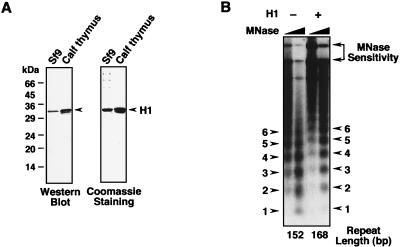
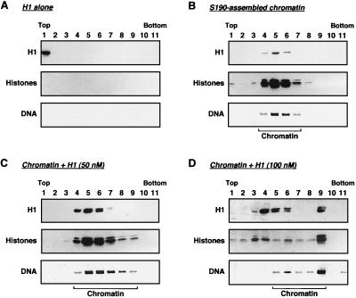
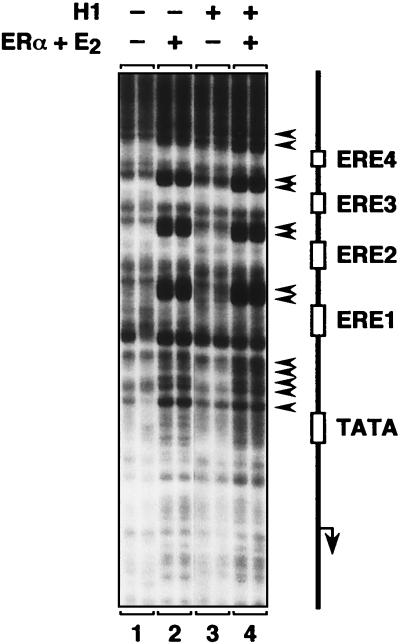
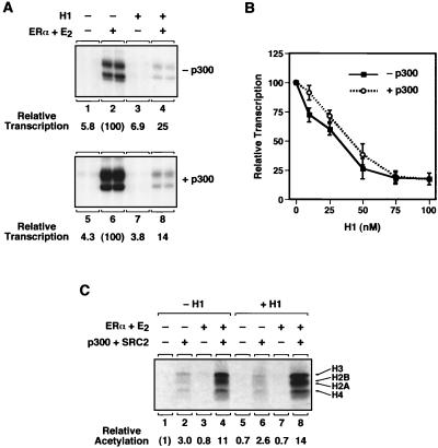
Chromatin is the physiological template for many nuclear processes in eukaryotes, including transcription by RNA polymerase II. In vivo, chromatin is assembled from genomic DNA, core histones, linker histones such as histone H1, and nonhistone chromatin-associated proteins. Histone H1 is thought to act as a general repressor of transcription by promoting the compaction of chromatin into higher-order structures. We have used a biochemical approach, including an in vitro chromatin assembly and transcription system, to examine the effects of histone H1 on estrogen receptor alpha (ER alpha)-mediated transcription with chromatin templates. We show that histone H1 acts as a potent repressor of ligand- and coactivator-regulated transcription by ER alpha. Histone H1 exerts its repressive effect without inhibiting the sequence-specific binding of ER alpha to chromatin or the overall extent of targeted acetylation of nucleosomal histones by the coactivator p300. Instead, histone H1 acts by blocking a specific step in the ER alpha-dependent transcription process, namely, transcription initiation, without affecting transcription reinitiation. Together, our data indicate that histone H1 acts selectively to reduce the overall level of productive transcription initiation by restricting promoter accessibility and preventing the ER alpha-dependent formation of a stable transcription pre-initiation complex.


Figures

FIG. 1.
FIG. 1.
Assembly of chromatin containing histone H1 by using a Drosophila chromatin assembly extract. (A) SDS-polyacrylamide gel electrophoresis and Western blotting analyses of histone H1 purified from Sf9 cells and calf thymus. The purified proteins were subjected to SDS-10% polyacrylamide gel electrophoresis with subsequent Western blotting with an anti-histone H1 antibody (left) or staining with Coomassie brilliant blue R-250 (right). (B) Addition of histone H1 to chromatin increases resistance to micrococcal nuclease and the nucleosomal repeat length. Chromatin was assembled by using the Drosophila S190 extract in the presence or absence of purified Sf9 cell histone H1 (100 nM) and then subjected to digestion with micrococcal nuclease (MNase). Note the increased resistance to micrococcal nuclease cleavage in the presence of histone H1 as indicated by increased amounts of higher-molecular-weight DNA species. The positions of the mono, di, and tri, etc., nucleosomal DNA fragments are indicated. Similar results were obtained with calf thymus histone H1 (not shown).
FIG. 2.
FIG. 2.
Histone H1 is efficiently incorporated into chromatin assembled in vitro. S190-assembled chromatin was incubated with calf thymus histone H1 at final concentrations of 0, 50, and 100 nM for 15 min at 27°C. After incubation, each sample was run on a 15 to 40% glycerol gradient. Fractions from the gradients were analyzed for histone H1 and core histones by Western blotting and for DNA by agarose gel electrophoresis with ethidium bromide staining. Note that the anti-core histone serum preferentially detects histones H2A and H2B (H2A > H2B > H3 > H4).
FIG. 3.
FIG. 3.
Histone H1 is a potent inhibitor of ERα-mediated transcription with chromatin templates. (A) Histone H1 inhibits ERα-mediated transcription with chromatin templates in a concentration-dependent manner. The plasmid template p2ERE-AdE4 was assembled into chromatin in the presence or absence of increasing amounts of purified Sf9 cell or calf thymus histone H1 (H1Sf9 and H1ct, respectively), as well as ERα (10 nM) and E2 (100 nM), as indicated. The concentrations of histone H1 in the chromatin assembly reaction mixtures were 12.5, 25, 50, and 100 nM. The chromatin samples were subjected to in vitro transcription analysis, and the resulting RNA products were analyzed by primer extension. The signals in each lane were quantified with a PhosphorImager. Similar results were obtained by using as a template pERE, which contains four EREs (not shown). (B) Histone H1 inhibits ERα-mediated transcription with chromatin templates when added either during or after chromatin assembly. The plasmid template p2ERE-AdE4 was assembled into chromatin in the presence or absence of purified Sf9 cell histone H1, ERα (10 nM), and E2 (100 nM), as indicated. Histone H1 was added at a concentration of 100 nM either during chromatin assembly or after chromatin assembly was complete. The chromatin samples were subjected to in vitro transcription analysis, and the resulting RNA products were analyzed by primer extension. The signals in each lane were quantified with a PhosphorImager. Similar results were obtained with calf thymus histone H1 (not shown).
FIG. 4.
FIG. 4.
Histone H1 does not inhibit sequence-specific binding of ERα to chromatin. The effect of histone H1 on the binding of liganded ERα to the EREs in the chromatin-assembled pERE template was analyzed by DNase I primer extension footprinting. The experiment was performed under the conditions described for Fig. 3B (i.e., under conditions in which ERα-dependent transcription is inhibited by histone H1 but in the absence of the HeLa cell nuclear extract used for the transcription assays). The schematic diagram denotes the locations of the EREs, TATA box, and transcription start site for the pERE template (arrow). Binding of ERα to the chromatin template results in regions of DNase I hypersensitivity, which are indicated by arrowheads. Each sample was run in duplicate.
FIG. 5.
FIG. 5.
Histone H1 inhibits ERα-mediated transcription in the presence of exogenously added p300 but not the targeted acetylation of nucleosomal histones by p300. (A) Histone H1 inhibits ERα-mediated transcription in the presence of added p300. In vitro chromatin assembly and transcription experiments were performed with the plasmid template p2ERE-AdE4 as described in the legend to Fig. 3B. Purified p300 was added to the assembled chromatin at a concentration of 60 nM. (B) Histone H1 inhibits ERα-mediated transcription in the presence of added p300 in a concentration-dependent manner. In vitro chromatin assembly and transcription experiments were performed with the plasmid template p2ERE-AdE4 and increasing concentrations of histone H1 as described in the legend to Fig. 3A. Purified p300 was added to the assembled chromatin at a concentration of 60 nM. Each point represents the mean ± the standard error of the mean of three or more separate determinations. (C) Histone H1 does not inhibit the targeted acetylation of nucleosomal histones by p300. The plasmid template pERE was assembled into chromatin with or without histone H1 and subsequently purified by column chromatography with Sepharose CL-4B to remove traces of unincorporated (i.e., free) histones. ERα, E2, p300, and SRC2(RID/PID) were added, and histone acetylation reactions were performed in the presence of [3H]acetyl coenzyme A as described in Methods and Materials. The reaction mixtures were separated on an SDS-15% polyacrylamide gel and subjected to fluorography. The 3H-labeled histone bands were excised from the gel and quantified by liquid scintillation counting. The results are expressed as relative acetylation compared to the control sample.
FIG. 6.
FIG. 6.
Histone H1 inhibits localized increases in restriction enzyme accessibility induced by the binding of ERα. Specific structural changes at the promoter induced by histone H1 were analyzed by restriction enzyme accessibility assays using chromatin-assembled pERE. The experiment was performed under the conditions described for Fig. 3B (i.e., under conditions in which ERα-mediated transcription is inhibited by histone H1 but in the absence of the HeLa cell nuclear extract used for the transcription assays). (A) Restriction enzyme accessibility assay. (Top) Aliquots of chromatin, with or without added ERα or histone H1, were digested with increasing amounts of the restriction endonuclease XbaI, deproteinized, double digested with the restriction endonucleases HindIII and EcoRI, and subjected to Southern blot analysis with an end-labeled oligonucleotide probe that hybridizes to the XbaI/EcoRI fragment. The HindIII/EcoRI fragments (Uncut) and the XbaI-digested fragments (XbaI cut) are indicated by arrows. (Bottom) The schematic diagram denotes the locations of the EREs, TATA box, transcription start site, oligonucleotide probe, and restriction enzymes used in the assay. (B) Quantification of restriction enzyme accessibility assays. The signals in each lane in panel A were quantified with a PhosphorImager. The extent of digestion by XbaI under each condition was determined by dividing the signal from the XbaI-cut bands by the sum of the signals from the XbaI-cut and uncut bands. Each point represents the mean ± the standard error of the mean of three or more separate determinations.
FIG. 7.
FIG. 7.
Histone H1 inhibits transcription initiation, but not reinitiation, by liganded ERα. (A) Histone H1 inhibits ERα-mediated transcription in a single round of transcription. In vitro chromatin assembly and transcription experiments were performed with the plasmid template p2ERE-AdE4 as described for Fig. 3B. For the single-round transcription experiments, Sarkosyl was added after initiation of transcription by addition of rNTPs. Under these conditions, Sarkosyl inhibits transcription reinitiation but not elongation and, thus, a single round of transcription is obtained. The chromatin samples were subjected to in vitro transcription analysis, and the resulting RNA products were analyzed by primer extension. The signals in each lane were quantified with a PhosphorImager. (B) Histone H1 does not inhibit ERα-mediated transcription reinitiation. The number of rounds of transcription in experiments like the one shown in panel A was determined by dividing the amount of transcription in the absence of Sarkosyl by the amount of transcription in the corresponding sample in the presence of Sarkosyl. Each bar represents the mean ± the range of two separate determinations.
FIG. 8.
FIG. 8.
Histone H1 represses ERα-dependent transcription by selectively inhibiting the formation of a stable PIC. (A) Schematic representation of the order-of-addition, single-round transcription experiments with chromatin templates shown in panel B. The chromatin assembly and single-round transcription assays were performed under the conditions described in the legend to Fig. 7. Histone H1 was added at the following times: 1, directly to the chromatin-assembled template prior to the addition of the other reagents; 2, with the HeLa cell nuclear extract (HNE); 3, before the initiation of transcription (i.e., 1 min prior to addition of the rNTPs); 4, just after the initiation of transcription but prior to the addition of Sarkosyl to limit transcription to a single round. (B) Results of the order-of-addition, single-round transcription experiments. The chromatin samples were subjected to in vitro transcription analysis, and the resulting RNA products were analyzed by primer extension. The signals in each lane were quantified with a PhosphorImager.

Similar articles

Cited by

References

    1. Bates, D. L., and J. O. Thomas. 1981. Histones H1 and H5: one or two molecules per nucleosome? Nucleic Acids Res. 9:5883-5894. - PMC - PubMed
    1. Belikov, S., and V. Karpov. 1998. Linker histones: paradigm lost but questions remain. FEBS Lett. 441:161-164. - PubMed
    1. Bouvet, P., S. Dimitrov, and A. P. Wolffe. 1994. Specific regulation of Xenopus chromosomal 5S rRNA gene transcription in vivo by histone H1. Genes Dev. 8:1147-1159. - PubMed
    1. Bresnick, E. H., M. Bustin, V. Marsaud, H. Richard-Foy, and G. L. Hager. 1992. The transcriptionally-active MMTV promoter is depleted of histone H1. Nucleic Acids Res. 20:273-278. - PMC - PubMed
    1. Brown, D. T., A. Gunjan, B. T. Alexander, and D. B. Sittman. 1997. Differential effect of H1 variant overproduction on gene expression is due to differences in the central globular domain. Nucleic Acids Res. 25:5003-5009. - PMC - PubMed

Publication types

MeSH terms

LinkOut - more resources