. 2002 Mar 15;22(6):2206-14.
doi: 10.1523/JNEUROSCI.22-06-02206.2002.

Neuregulin expression at neuromuscular synapses is modulated by synaptic activity and neurotrophic factors

Affiliations

Neuregulin expression at neuromuscular synapses is modulated by synaptic activity and neurotrophic factors

Jeffrey A Loeb et al. J Neurosci. .

Abstract

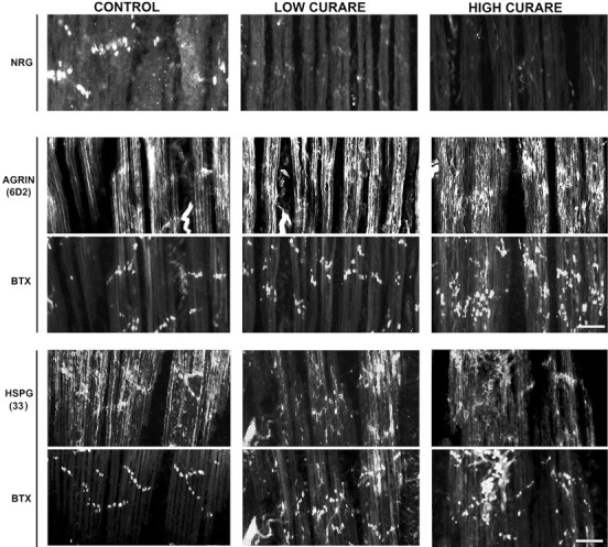
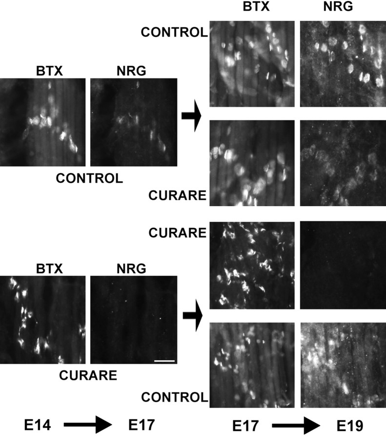
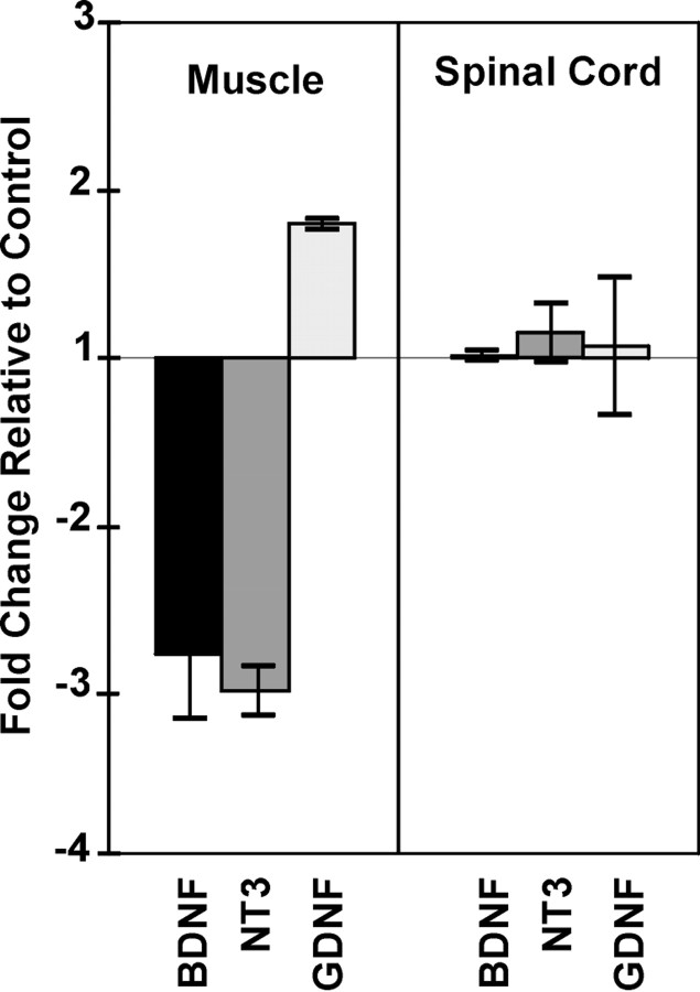
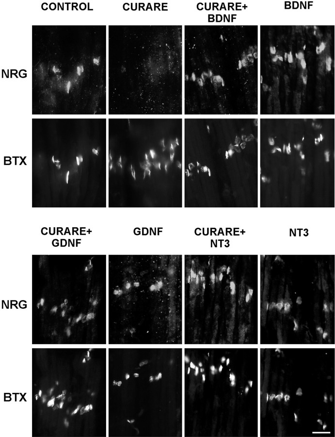
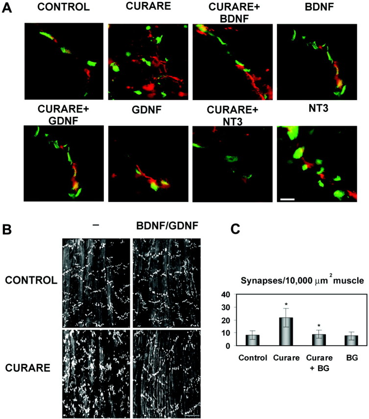
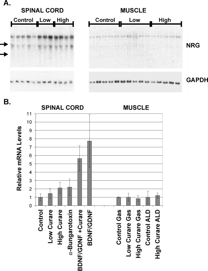
The proper formation of neuromuscular synapses requires ongoing synaptic activity that is translated into complex structural changes to produce functional synapses. One mechanism by which activity could be converted into these structural changes is through the regulated expression of specific synaptic regulatory factors. Here we demonstrate that blocking synaptic activity with curare reduces synaptic neuregulin expression in a dose-dependent manner yet has little effect on synaptic agrin or a muscle-derived heparan sulfate proteoglycan. These changes are associated with a fourfold increase in number and a twofold reduction in average size of synaptic acetylcholine receptor clusters that appears to be caused by excessive axonal sprouting with the formation of new, smaller acetylcholine receptor clusters. Activity blockade also leads to threefold reductions in brain-derived neurotrophic factor and neurotrophin 3 expression in muscle without appreciably changing the expression of these same factors in spinal cord. Adding back these or other neurotrophic factors restores synaptic neuregulin expression and maintains normal end plate band architecture in the presence of activity blockade. The expression of neuregulin protein at synapses is independent of spinal cord and muscle neuregulin mRNA levels, suggesting that neuregulin accumulation at synapses is independent of transcription. These findings suggest a local, positive feedback loop between synaptic regulatory factors that translates activity into structural changes at neuromuscular synapses.


Figures

Fig. 1.
Fig. 1.
Activity blockade with curare results in a disorganized end plate band architecture with an increase in number and a decrease in average synaptic size. A, Chick embryos were treated daily with saline (CONTROL), a nonparalytic concentration (0.3 mg/d) of d-tubocurarine (LOW CURARE), or a paralytic dose (3 mg/d) ofd-tubocurarine (HIGH CURARE) for 4 d from E14 to E18. ALD muscles at E18 were double-labeled with rhodamine α-bungarotoxin (BTX), which labels AChRs, and SV2, which labels synaptic vesicles in the nerve terminals.Insets, Higher-magnification views of AChR clusters and nerve terminals showing that with curare treatment, a mixture of AChR clusters of variable size is seen, and most of these are associated with nerve terminals. Scale bar, 50 μm. Quantitation of the number (B) and size (C) of postsynaptic AChRs stained with α-bungarotoxin revealed a fourfold to fivefold increase in the number of postsynaptic AChR clusters and a twofold reduction in the average size of the AChR clusters. *Significantly different from the control; p = 0.0018 (B, HIGH CURARE), 0.048 (C, LOW CURARE), and 0.016 (C, HIGH CURARE).
Fig. 2.
Fig. 2.
Activity blockade blocks synaptic NRG expression in a dose-dependent manner but has little effect on synaptic HSPGs. NRG immunoreactivity (top panel) at neuromuscular synapses was assessed with affinity-purified 183N antisera on the same ALD muscle sections shown in Figure 1 treated with saline or low or high doses of curare. A dose-dependent reduction in NRG immunoreactivity was noted so that, at paralytic doses of curare, little to no NRG immunoreactivity was detected. Agrin immunoreactivity at neuromuscular synapses was measured using the monoclonal antibody 6D2 double-labeled with Bodipy-α-bungarotoxin (BTX;middle two panels), and a muscle HSPG antibody (33) was double-labeled with Bodipy-α-bungarotoxin (bottom two panels). Scale bars, 50 μm.
Fig. 3.
Fig. 3.
Synaptic NRG expression resumes after reversal of activity blockade. Chicken embryos were treated with either paralytic concentrations of curare (3 mg/d) or the same volume of saline from E14 to E17. Some of the embryos were examined on E17, showing that activity blockade blocked the appearance of NRG immunoreactivity compared with untreated embryos that expressed submaximal, synaptic NRG levels. All muscle sections were double-labeled at the same time with 183N antibodies against NRG and α-bungarotoxin (BTX) for localization of synapses. Each group was either continued with the same treatment or switched to the opposite treatment from E17 to E19. Those that were in saline for the entire period from E14 to E19 had maximal NRG staining, whereas curare treatment from E17 to E19 prevented any additional increase in NRG staining. Those animals maintained in curare for the entire period had no NRG staining at synapses, whereas those switched from curare to saline recovered their ability to release NRG at synapses. Scale bar, 20 μm.
Fig. 4.
Fig. 4.
Activity blockade results in altered neurotrophic factor expression in muscle but not in spinal cord. Using real-time RT-PCR methods normalized to chicken GAPDH, both BDNF and NT-3 mRNAs were reduced by approximately threefold with paralytic concentrations of curare from E14 to E18 relative to saline-treated ALD muscle. In contrast, GDNF expression increased slightly in muscle. None of these neurotrophic factors was changed in spinal cords from the same animals, suggesting that curare is working postsynaptically to induce its effects on these factors. Each point represents the average ± 1 SD from RT-PCR measurements from three independent animals, and each RT-PCR from each animal was performed in quadruplicate.
Fig. 5.
Fig. 5.
Neurotrophic factors restore synaptic NRG expression in the presence of activity blockade. Chick embryos were treated daily with saline (CONTROL), a paralytic dose of curare (3 mg/d; CURARE) or the same dose of curare with 1 μg (∼20 ng/ml) of BDNF, NT-3, or GDNF for 4 d from E14 to E18. ALD muscles at E18 were double-labeled for NRG (183N) and acetylcholine receptors (α-bungarotoxin; BTX) to localize synapses. Although the curare alone blocked NRG expression, each of these neurotrophic factors restored NRG expression in the presence of activity blockade. The neurotrophic factors alone had no effect. Scale bar, 20 μm.
Fig. 6.
Fig. 6.
Neurotrophic factors maintain normal end plate band architecture in the face of activity blockade. A, High-magnification views of synapses treated with saline (CONTROL), high-dose curare (CURARE), or high-dose curare plus BDNF, NT-3, or GDNF (1 μg/d) double-labeled with α-bungarotoxin (green) and neurofilaments and synaptic terminals (red) using a mixture of RT-97 and SV2 antibodies. This reveals that the increased number of smaller synapses seen with curare is attributable to excessive sprouting of the terminal axons and that this sprouting is suppressed by the coaddition of BDNF, NT-3, or GDNF. Scale bar, 10 μm. B, Examination of AChR patterns in ALD with α-bungarotoxin at lower magnification reveals that BDNF (2 ng/ml) plus GDNF (20 ng/ml) in the presence of paralytic doses of curare helps maintain a normal architecture. These factors given without curare had no effects. Scale bar, 100 μm. C, Quantitation of the number of AChR clusters per 10,000 μm2 revealed increased numbers with curare alone (as in Fig. 1) that normalized to control levels with BDNF and GDNF (BG). The density with curare treatment was significantly increased from controls (p= 0.010) and the curare with BDNF and GDNF treatment was significantly lower than with curare alone (p = 0.014).
Fig. 7.
Fig. 7.
NRG mRNA levels are increased in spinal cord with activity blockade and neurotrophic factors but are unchanged in muscle.A, Total RNA from lumbar spinal cords and gastrocnemius muscles taken from the embryos described previously were analyzed for NRG mRNA by Northern blot analysis using a full-length pro-NRG probe revealing two transcripts. Note that considerably less NRG mRNA is expressed in muscle compared with spinal cord. Although there were significant increases in NRG mRNA for both low and high doses of curare, no differences were noted in muscle. Positions of the ribosomal RNA bands are indicated by arrows on theleft. B, The top, 7 kb transcript shown in A together with data from similar Northern blots were quantified and normalized to the level of chicken GAPDH mRNA (shown in the bottom of A) and summarized as the mean ± SD for spinal cord samples on theleft and gastrocnemius and ALD muscles on theright. Although curare alone produced a twofold induction of NRG mRNA levels, BDNF and GDNF alone or together with curare produced a fivefold to eightfold induction of NRG mRNA. No difference was noted between gastrocnemius and ALD muscles. The numbers of animals in each condition were as follows: Control,n = 32; Low Curare,n = 8; High Curare,n = 13; α-Bungarotoxin,n = 2; BDNF/GDNF +Curare, n = 6;BDNF/GDNF, n = 2; Control Gas, n = 2; Low Curare Gas,n = 3; High Curare Gas,n = 5; Control ALD,n = 4; and High Curare ALD,n = 2.

Similar articles

Cited by

References

    1. Alcantara S, Frisen J, del Rio JA, Soriano E, Barbacid M, Silos-Santiago I. TrkB signaling is required for postnatal survival of CNS neurons and protects hippocampal and motor neurons from axotomy-induced cell death. J Neurosci. 1997;17:3623–3633. - PMC - PubMed
    1. Anderson MJ, Klier FG, Tanguay KE. Acetylcholine receptor aggregation parallels the deposition of a basal lamina proteoglycan during development of the neuromuscular junction. J Cell Biol. 1984;99:1769–1784. - PMC - PubMed
    1. Bayne EK, Anderson MJ, Fambrough DM. Extracellular matrix organization in developing muscle: correlation with acetylcholine receptor aggregates. J Cell Biol. 1984;99:1486–1501. - PMC - PubMed
    1. Brown MC, Holland RL, Hopkins WG. Motor nerve sprouting. Annu Rev Neurosci. 1981;4:17–42. - PubMed
    1. Burden S. Development of the neuromuscular junction in the chick embryo: the number, distribution, and stability of acetylcholine receptors. Dev Biol. 1977;57:317–329. - PubMed

Publication types

MeSH terms

LinkOut - more resources