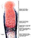
The life cycle of chondrocytes in the developing skeleton
- PMID: 11879545
- PMCID: PMC128921
- DOI: 10.1186/ar396
The life cycle of chondrocytes in the developing skeleton
Abstract
Cartilage serves multiple functions in the developing embryo and in postnatal life. Genetic mutations affecting cartilage development are relatively common and lead to skeletal malformations, dysfunction or increased susceptibility to disease or injury. Characterization of these mutations and investigation of the molecular pathways in which these genes function have contributed to an understanding of the mechanisms regulating skeletal patterning, chondrogenesis, endochondral ossification and joint formation. Extracellular growth and differentiation factors including bone morphogenetic proteins, fibroblast growth factors, parathyroid hormone-related peptide, extracellular matrix components, and members of the hedgehog and Wnt families provide important signals for the regulation of cell proliferation, differentiation and apoptosis. Transduction of these signals within the developing mesenchymal cells and chondrocytes results in changes in gene expression mediated by transcription factors including Smads, Msx2, Sox9, signal transducer and activator of transcription (STAT), and core-binding factor alpha 1. Further investigation of the interactions of these signaling pathways will contribute to an understanding of cartilage growth and development, and will allow for the development of strategies for the early detection, prevention and treatment of diseases and disorders affecting the skeleton.
Figures

Similar articles
-
Endochondral ossification: how cartilage is converted into bone in the developing skeleton.Int J Biochem Cell Biol. 2008;40(1):46-62. doi: 10.1016/j.biocel.2007.06.009. Epub 2007 Jun 29. Int J Biochem Cell Biol. 2008. PMID: 17659995 Review.
-
The chondrocytic journey in endochondral bone growth and skeletal dysplasia.Birth Defects Res C Embryo Today. 2014 Mar;102(1):52-73. doi: 10.1002/bdrc.21060. Birth Defects Res C Embryo Today. 2014. PMID: 24677723 Review.
-
ALK2 functions as a BMP type I receptor and induces Indian hedgehog in chondrocytes during skeletal development.J Bone Miner Res. 2003 Sep;18(9):1593-604. doi: 10.1359/jbmr.2003.18.9.1593. J Bone Miner Res. 2003. PMID: 12968668
-
Dentin matrix protein 1, a target molecule for Cbfa1 in bone, is a unique bone marker gene.J Bone Miner Res. 2002 Oct;17(10):1822-31. doi: 10.1359/jbmr.2002.17.10.1822. J Bone Miner Res. 2002. PMID: 12369786
-
Molecular mechanisms of endochondral bone development.Biochem Biophys Res Commun. 2005 Mar 18;328(3):658-65. doi: 10.1016/j.bbrc.2004.11.068. Biochem Biophys Res Commun. 2005. PMID: 15694399 Review.
Cited by
-
NFI-C Is Required for Epiphyseal Chondrocyte Proliferation during Postnatal Cartilage Development.Mol Cells. 2020 Aug 31;43(8):739-748. doi: 10.14348/molcells.2020.2272. Mol Cells. 2020. PMID: 32759468 Free PMC article.
-
Enhancing Cartilage Repair: Surgical Approaches, Orthobiologics, and the Promise of Exosomes.Life (Basel). 2024 Sep 11;14(9):1149. doi: 10.3390/life14091149. Life (Basel). 2024. PMID: 39337932 Free PMC article. Review.
-
Naproxen affects osteogenesis of human mesenchymal stem cells via regulation of Indian hedgehog signaling molecules.Arthritis Res Ther. 2014 Jul 17;16(4):R152. doi: 10.1186/ar4614. Arthritis Res Ther. 2014. PMID: 25034046 Free PMC article.
-
Relating the chondrocyte gene network to growth plate morphology: from genes to phenotype.PLoS One. 2012;7(4):e34729. doi: 10.1371/journal.pone.0034729. Epub 2012 Apr 30. PLoS One. 2012. PMID: 22558096 Free PMC article.
-
Microarray analyses of gene expression during chondrocyte differentiation identifies novel regulators of hypertrophy.Mol Biol Cell. 2005 Nov;16(11):5316-33. doi: 10.1091/mbc.e05-01-0084. Epub 2005 Aug 31. Mol Biol Cell. 2005. PMID: 16135533 Free PMC article.
References
-
- Nuckolls GH, Shum L, Slavkin HC. Progress toward understanding craniofacial malformations. Cleft Palate Craniofac J. 1999;36:12–26. - PubMed
-
- Felson DT, Lawrence RC, Dieppe PA, Hirsch R, Helmick CG, Jordan JM, Kington RS, Lane NE, Nevitt MC, Zhang Y, Sowers M, McAlindon T, Spector TD, Poole AR, Yanovski SZ, Ateshian G, Sharma L, Buckwalter JA, Brandt KD, Fries JF. Osteoarthritis: new insights. Part 1: the disease and its risk factors. Ann Intern Med. 2000;133:635–646. - PubMed
-
- Hill DJ, Logan A. Peptide growth factors and their interactions during chondrogenesis. Prog Growth Factor Res. 1992;4:45–68. - PubMed
-
- Underhill TM, Sampaio AV, Weston AD. Retinoid signalling and skeletal development. Novartis Found Symp. 2001;232:171–185. - PubMed
Publication types
MeSH terms
Substances
Grants and funding
LinkOut - more resources
Full Text Sources
Other Literature Sources
Research Materials

