

Review
doi: 10.1172/JCI15217.
Mucus clearance as a primary innate defense mechanism for mammalian airways
Affiliations
- PMID: 11877463
- PMCID: PMC150901
- DOI: 10.1172/JCI15217
Item in Clipboard
Review
Mucus clearance as a primary innate defense mechanism for mammalian airways
J Clin Invest.
2002 Mar.
No abstract available
Figures
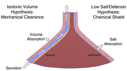
Pulmonary defense mechanisms preventing chronic bacterial infection. The lung is depicted as an inverted funnel, reflecting the relative surface area of distal versus proximal airways. The mechanical-clearance-of-mucus hypothesis is shown on the left. The schema depicts discrete mucus and periciliary liquid layers and ascribes to the epithelium a volume-absorbing function. The chemical shield hypothesis is shown on the right, with the epithelium depicted as having a salt- but not a volume-absorbing function to produce the hypotonic ASL required for defensin activity.
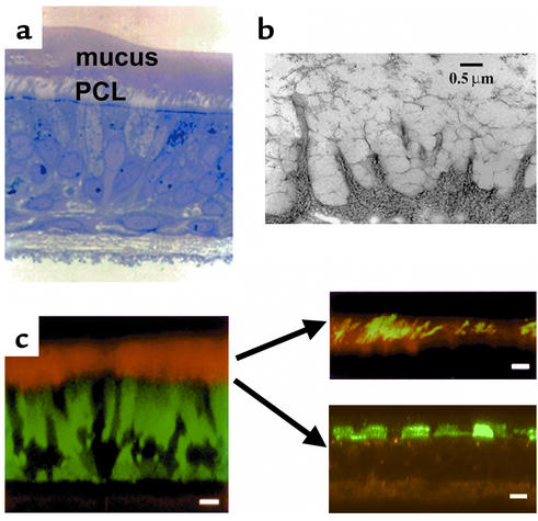
Microanatomy of human ASL. (a) WD human airway epithelial culture exhibiting rotational mucus transport (see Figure 3), fixed with perfluorocarbon/osmium. Note a distinct mucus layer atop a distinct PCL. (b) Visualization of glycocalyx on WD human airway epithelia by the freeze substitution technique. Note the high degree of organization of this barrier. (c) Left: X-Z confocal image of living WD human airway epithelial culture. The cells were stained with calcein, AM, (green), and the ASL was visualized with Texas red dextran. Scale bar = 10 μm. Top right: Fluorescent “dissection” of mucus layer and PCL in living WD airway epithelia by confocal microscopy. The mucus layer is visualized as green fluorescent beads and the PCL as the “bead-free zone” interposed between the mucus layer and cell surface (black). Scale bar = 10 μm. Bottom right: Detection of glycocalyx by fluorescence/confocal microscopy. The keratan sulfate component of the glycocalyx is visualized by Texas red–labeled anti–keratan sulfate (anti-KS) antibodies. Scale bar = 5 μm.
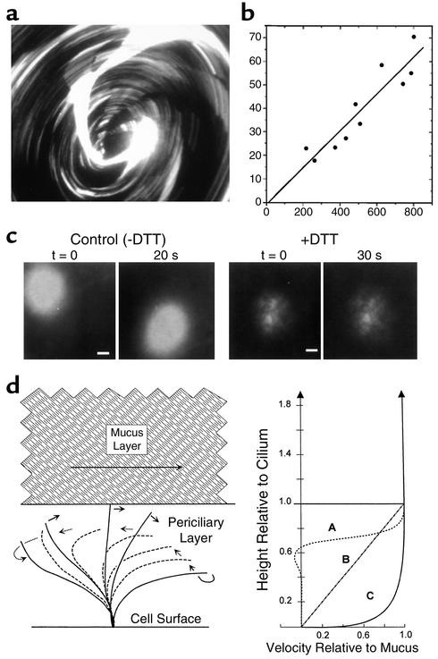
Mucus layer and PCL transport by human airway epithelia. (a) Mucus transport in WD airway epithelial cultures was identified by the rotational movement of 1-μm fluorescent microspheres in the mucus layer. The image shown was acquired as a single 5-second exposure; the streaks represent the paths of individual microspheres (field diameter ∼2 mm). (b) Mucus transport rates were calculated as the linear velocities of fluorescent microspheres (a) and plotted against the distance from the center of the rotation (radial). (c) Transport of PCL revealed by photoactivation of caged fluorescein-conjugated dextrans. The ASL was labeled with caged fluorescein-conjugated dextran (10,000 Da) and the fate of photoreleased dextran fluorescence was determined. Conventional fluorescence microscopy at low power was used (bars = 0.2 mm). Left: Migration of the released fluorescent dextran during the 20-second period of observation. Right: After removing the mucus layer (+DTT), the migration of released fluorescent dextran was minimal. Both the mucus layer and the PCL are labeled by photoactivation. The absence of a “smear” during movement implies that both the mucus layer and the PCL move at the same velocity. The absence of PCL movement after mucus removal suggests that mucus movement is critical for PCL movement. (d) Models of ASL transport. Lateral fluid velocity profiles in ASL predicted for three different considerations, with the ordinate aligned to the diagram of the ciliary beat cycle at the left. Curve A approximates velocity profiles in the PCL predicted from theoretical considerations of ciliary propulsion of water (60, 61). Note nominally zero velocities at 70–75% of the ciliary length, below the level of the return stroke. Curve B depicts the velocity profile predicted for PCL flow driven solely by frictional interactions with the mucus layer. Curve C depicts the velocity profile for the PCL from the observations in this work that the flows of the PCL and mucus layer are essentially indistinguishable.
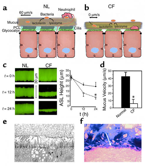
PCL is required for effective mucociliary and cough clearance. (a) Schema of microanatomy of normal ASL. Note mixing of bacteria in “turbulent” mucus. (b) Schema depicting hypothetical volume depletion of ASL covering CF airway epithelial surfaces. Note that the volume depletion is reflected in both the generation of a more concentrated mucus layer and the depletion of the PCL. PCL depletion allows interactions to occur between the tethered mucins of the glycocalyx and the mucus layer. Note motile bacteria penetrating into thickened, stationary mucus. (c) Evidence for ASL volume depletion in CF airway epithelia. ASL height was measured immediately, 12 hours, and 24 hours after deposition of PBS containing Texas red dextran on the epithelial surface of the cell (pseudocolored green). Left: Representative confocal microscopy images. Right: Mean data for normal (circles) and CF (squares) ASL heights. *CF ASL is significantly shallower than normal (P < 0.05; n = 6 per group). (d) Mucus (bead) rotational velocity 24 hours after administration of PBS containing fluorescent markers. At t = 0 hours, both normal and CF cultures exhibited rotational velocities of about 45 μm/s. (*P < 0.05, CF vs. normal; n = 6 per group). (e) Low-power electron micrograph of perfluorocarbon/osmium–fixed CF airway culture 24 hours after volume addition and with rotational mucus transport abolished. Note close apposition of mucus layer and the glycocalyx covering flattened cilia and the cell surface. (f) Light micrograph of freshly excised CF bronchus stained with Alcin blue periodic acid-Schiffs for mucus. As in the in vitro model, note close apposition (annealing) between secreted mucins and the cell surface (indicated by white arrow). NL, normal.
Similar articles
-
Innate immune functions of the airway epithelium.Contrib Microbiol. 2008;15:147-163. doi: 10.1159/000136349. Contrib Microbiol. 2008. PMID: 18511860 Review.
-
Mucociliary clearance--a critical upper airway host defense mechanism and methods of assessment.Curr Opin Allergy Clin Immunol. 2007 Feb;7(1):5-10. doi: 10.1097/ACI.0b013e3280114eef. Curr Opin Allergy Clin Immunol. 2007. PMID: 17218804 Review.
-
Effective mucus clearance is essential for respiratory health.Am J Respir Cell Mol Biol. 2006 Jul;35(1):20-8. doi: 10.1165/rcmb.2006-0082SF. Epub 2006 Mar 9. Am J Respir Cell Mol Biol. 2006. PMID: 16528010 Free PMC article. Review. No abstract available.
-
Model of mucociliary clearance in cystic fibrosis lungs.J Theor Biol. 2015 May 7;372:81-8. doi: 10.1016/j.jtbi.2015.02.023. Epub 2015 Mar 5. J Theor Biol. 2015. PMID: 25746843
-
Airway mucus function and dysfunction.N Engl J Med. 2010 Dec 2;363(23):2233-47. doi: 10.1056/NEJMra0910061. N Engl J Med. 2010. PMID: 21121836 Free PMC article. Review. No abstract available.
Cited by
-
Detrimental role of the airway mucin Muc5ac during ventilator-induced lung injury.Mucosal Immunol. 2013 Jul;6(4):762-75. doi: 10.1038/mi.2012.114. Epub 2012 Nov 28. Mucosal Immunol. 2013. PMID: 23187315 Free PMC article.
-
Computational Modeling Insights into Extreme Heterogeneity in COVID-19 Nasal Swab Data.Viruses. 2023 Dec 30;16(1):69. doi: 10.3390/v16010069. Viruses. 2023. PMID: 38257769 Free PMC article.
-
Acquired cilia dysfunction in chronic rhinosinusitis.Am J Rhinol Allergy. 2012 Jan-Feb;26(1):1-6. doi: 10.2500/ajra.2012.26.3716. Am J Rhinol Allergy. 2012. PMID: 22391065 Free PMC article. Review.
-
Epithelial tethering of MUC5AC-rich mucus impairs mucociliary transport in asthma.J Clin Invest. 2016 Jun 1;126(6):2367-71. doi: 10.1172/JCI84910. Epub 2016 May 16. J Clin Invest. 2016. PMID: 27183390 Free PMC article.
-
Glucocorticoid receptor and histone deacetylase-2 mediate dexamethasone-induced repression of MUC5AC gene expression.Am J Respir Cell Mol Biol. 2012 Nov;47(5):637-44. doi: 10.1165/rcmb.2012-0009OC. Epub 2012 Jul 12. Am J Respir Cell Mol Biol. 2012. PMID: 22798432 Free PMC article.
References
-
- Kilburn KH. A hypothesis for pulmonary clearance and its implications. Am Rev Respir Dis. 1968;98:449–463. - PubMed
-
- Guggino WB. Cystic fibrosis and the salt controversy. Cell. 1999;96:607–610. - PubMed
-
- Wanner A, Salathe M, O’Riordan TG. Mucociliary clearance in the airways. Am J Respir Crit Care Med. 1996;154:1868–1902. - PubMed
-
- Boucher RC. Human airway ion transport. Part 1. Am J Respir Crit Care Med. 1994;150:271–281. - PubMed
Publication types
MeSH terms
Grants and funding
LinkOut - more resources
Full Text Sources
Other Literature Sources
