

Cloning and characterization of the rat homologues of the Inhibitor of Apoptosis protein 1, 2, and 3 genes
- PMID: 11860601
- PMCID: PMC65541
- DOI: 10.1186/1471-2164-3-5
Cloning and characterization of the rat homologues of the Inhibitor of Apoptosis protein 1, 2, and 3 genes
Abstract
Background: Inhibitor of Apoptosis (IAP) proteins are key intrinsic regulators of apoptosis induced by a variety of triggers. We isolated the rat Inhibitor of Apoptosis genes 1, 2 and 3 and characterized their tissue distribution and expression.
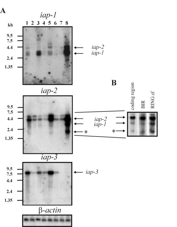
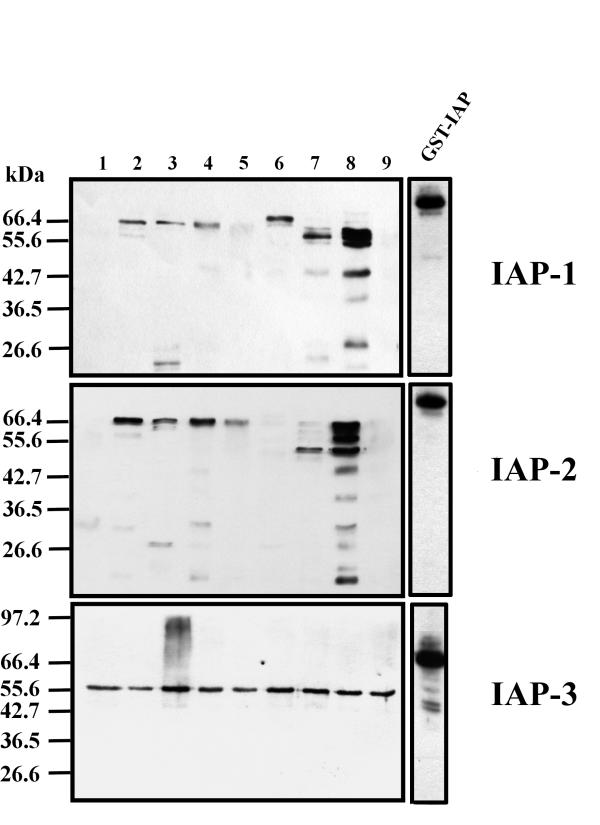
Results: Rat iap-1 encodes a protein of 67.1 kDa with 73 % and 89.2 % homology to human and mouse iap-1 respectively. Rat iap-2 encodes a protein of 66.7 kDa with 81.6 % and 89.3 % homology to human and mouse iap-2 respectively. Rat iap-3 encodes a protein of 56.1 kDa with 89.5 % and 93.1 % homology to human and mouse iap-3 respectively. We have generated rabbit polyclonal antibodies against all three rat IAP genes. Northern and Western blot analysis detected rat IAP transcripts and proteins in majority of the tissues examined. In addition, a shorter, alternatively spliced transcript corresponding to iap-2 was found in testes.
Conclusions: We have identified three rat homologues of the IAP genes. The elevated expression of rat iap-1 and iap2 in testes suggests that these two genes play an important antiapoptotic role in spermatogenesis.
Figures
Similar articles
-
Genomic organization and primary characterization of miap-3: the murine homologue of human X-linked IAP.Genomics. 1997 Jun 15;42(3):514-8. doi: 10.1006/geno.1997.4742. Genomics. 1997. PMID: 9205126
-
Gene promoter of apoptosis inhibitory protein IAP2: identification of enhancer elements and activation by severe hypoxia.Biochem J. 2002 Jun 1;364(Pt 2):413-21. doi: 10.1042/BJ20011431. Biochem J. 2002. PMID: 12023884 Free PMC article.
-
A baculovirus anti-apoptosis gene homolog of the Trichoplusia ni granulovirus.Virus Genes. 1999;19(2):95-101. doi: 10.1023/a:1008148922330. Virus Genes. 1999. PMID: 10541013
-
The inhibitor of apoptosis (IAP) proteins are critical regulators of signaling pathways and targets for anti-cancer therapy.Exp Oncol. 2012 Oct;34(3):200-11. Exp Oncol. 2012. PMID: 23070005 Review.
-
IAP family of cell death and signaling regulators.Methods Enzymol. 2014;545:35-65. doi: 10.1016/B978-0-12-801430-1.00002-0. Methods Enzymol. 2014. PMID: 25065885 Review.
Cited by
-
IGF2BP1 controls cell death and drug resistance in rhabdomyosarcomas by regulating translation of cIAP1.Oncogene. 2015 Mar 19;34(12):1532-41. doi: 10.1038/onc.2014.90. Epub 2014 Apr 7. Oncogene. 2015. PMID: 24704827
-
Posttranscriptional downregulation of c-IAP2 by the ubiquitin protein ligase c-IAP1 in vivo.Mol Cell Biol. 2005 Apr;25(8):3348-56. doi: 10.1128/MCB.25.8.3348-3356.2005. Mol Cell Biol. 2005. PMID: 15798218 Free PMC article.
-
Chronic crude garlic-feeding modified adult male rat testicular markers: mechanisms of action.Reprod Biol Endocrinol. 2009 Jun 24;7:65. doi: 10.1186/1477-7827-7-65. Reprod Biol Endocrinol. 2009. PMID: 19552815 Free PMC article.
-
Intermediate domain of receptor-interacting protein kinase 1 (RIPK1) determines switch between necroptosis and RIPK1 kinase-dependent apoptosis.J Biol Chem. 2012 Apr 27;287(18):14863-72. doi: 10.1074/jbc.M111.288670. Epub 2012 Feb 23. J Biol Chem. 2012. PMID: 22362767 Free PMC article.
-
High level expression of apoptosis inhibitor in hepatoma cell line expressing Hepatitis B virus.Int J Med Sci. 2005;2(1):30-35. doi: 10.7150/ijms.2.30. Epub 2005 Jan 5. Int J Med Sci. 2005. PMID: 15968337 Free PMC article.
References
LinkOut - more resources
Full Text Sources
Molecular Biology Databases
Research Materials
Miscellaneous
