. 2002 Mar;76(5):2529-42.
doi: 10.1128/jvi.76.5.2529-2542.2002.

Translation of polioviral mRNA is inhibited by cleavage of polypyrimidine tract-binding proteins executed by polioviral 3C(pro)

Affiliations

Translation of polioviral mRNA is inhibited by cleavage of polypyrimidine tract-binding proteins executed by polioviral 3C(pro)

Sung Hoon Back et al. J Virol. 2002 Mar.

Abstract

The translation of polioviral mRNA occurs through an internal ribosomal entry site (IRES). Several RNA-binding proteins, such as polypyrimidine tract-binding protein (PTB) and poly(rC)-binding protein (PCBP), are required for the poliovirus IRES-dependent translation. Here we report that a poliovirus protein, 3C(pro) (and/or 3CD(pro)), cleaves PTB isoforms (PTB1, PTB2, and PTB4). Three 3C(pro) target sites (one major target site and two minor target sites) exist in PTBs. PTB fragments generated by poliovirus infection are redistributed to the cytoplasm from the nucleus, where most of the intact PTBs are localized. Moreover, these PTB fragments inhibit polioviral IRES-dependent translation in a cell-based assay system. We speculate that the proteolytic cleavage of PTBs may contribute to the molecular switching from translation to replication of polioviral RNA.


Figures
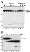
FIG. 1.
FIG. 1.
Cleavage of PTBs in poliovirus-infected cells. (A) HeLa cells (lanes 1 to 7) and H1-HeLa cells (lanes 8 and 9) were infected with poliovirus (lanes 2 to 7) or HRV-14 (lane 9). Samples were then harvested at the indicated times, Aliquots were subjected to SDS-PAGE and immunoblotted with monoclonal antibody against PTB. The numbers on top of panel A indicate the number of hours postinfection (h.p.i). The PTB fragments generated by poliovirus infection are indicated by solid and open arrowheads. Only the C-terminal parts of PTBs are revealed by Western blot analysis. (B) HA-tagged PTB4 was expressed in the stable HeLa cell line HeLa12/HA-PTB4 by withdrawal of tetracycline from culture medium. Forty-eight hours after induction, the cells were infected with poliovirus and analyzed by immunoblotting with anti-HA monoclonal antibody. The positions of the major and the minor cleavage products are indicated by solid and open arrows, respectively (Fig. 3A). Only the N-terminal parts of HA-PTB4 are revealed by Western blot analysis.
FIG. 2.
FIG. 2.
In vitro cleavage of PTB proteins by recombinant 3Cpro. (A) Different amounts of purified recombinant 3Cpro were incubated with HeLa cell extracts (20 μg) at 37°C for 3 h. (B and C) Purified recombinant PTB1 or PTB4 (1.0 μg) was incubated with the indicated quantities of protease at 37°C for 4 h. Reactions were stopped by the addition of 2× Laemmli sample buffer and were analyzed by SDS-PAGE followed by immunoblotting with anti-PTB monoclonal antibody (A, B, and C). (D) Five microliters of [35S]methionine-labeled PTB isoforms was incubated with 1.0 μg of 3Cpro for 1 h (lanes 2, 5, and 8) or for 3 h (lanes 3, 6, and 9). Negative control samples incubated for 3 h without addition of 3Cpro are shown in lanes 1, 4, and 7. Samples were resolved by SDS-PAGE, and the protein bands were detected by autoradiography. The positions of major cleavage products of PTB are indicated by a solid arrow and solid arrowhead, and the minor cleavage products are indicated by an open arrow and open arrowhead.
FIG. 3.
FIG. 3.
Identification of 3Cpro cleavage sites in PTBs. (A) Schematic diagram of 3Cpro cleavage sites in PTBs. The amino acid sequences around the major and minor cleavage sites in PTBs are shown in the single-letter motif. The NLS and RRMs in PTB are indicated by open and solid boxes, respectively, and the sizes of the cleaved products are shown in kilodaltons. Ab, antibody. (B) [35S]methionine-labeled PTB4 and its mutants were incubated with (+) or without (−) 3Cpro at 37°C for 3 h. Protease reactions were stopped by the addition of 2× Laemmli sample buffer, and the samples were analyzed by SDS-PAGE followed by autoradiography.
FIG. 3.
FIG. 3.
Identification of 3Cpro cleavage sites in PTBs. (A) Schematic diagram of 3Cpro cleavage sites in PTBs. The amino acid sequences around the major and minor cleavage sites in PTBs are shown in the single-letter motif. The NLS and RRMs in PTB are indicated by open and solid boxes, respectively, and the sizes of the cleaved products are shown in kilodaltons. Ab, antibody. (B) [35S]methionine-labeled PTB4 and its mutants were incubated with (+) or without (−) 3Cpro at 37°C for 3 h. Protease reactions were stopped by the addition of 2× Laemmli sample buffer, and the samples were analyzed by SDS-PAGE followed by autoradiography.
FIG. 4.
FIG. 4.
Subcellular localization of PTB and its derivatives. (A) Effect of poliovirus infection on PTB localization. HeLa cells were mock infected or infected with poliovirus for 6 h and then stained with antibody directed against PTB. The top panels show cells examined with a TRITC filter, the middle panels show Hoechst 33258 staining of the same field with a 4",6"-diamidino-2-phenylindole (DAPI) filter, and the bottom panels show the merged image of the TRITC and Hoechst images. (B) Effect of transcription blockage by actinomycin D on PTB localization. Uninfected HeLa cells were incubated without or with actinomycin D (5 μg/ml) for 6 h and then fixed and stained with anti-PTB monoclonal antibody. (C) HeLa cells were transfected with plasmids expressing GFP/PTB4/RFP and then mock infected or infected with poliovirus for 6 h from 40 h posttransfection. GFP fluorescence and RFP fluorescence were visualized with a fluorescein isothiocyanate (FITC) filter and a TRITC filter, respectively. DNA was examined with a DAPI filter after Hoechst 33258 staining. Panels 4 and 8 are merged images of FITC, TRITC, and Hoechst images. (D) Subcellular localization of PTB fragments. HeLa cells were transiently transfected with plasmids expressing GFP (panels 1, 6, and 11), GFP fused with PTB1-RRM2-3-4 (panels 2, 7, and 12), PTB4-RRM1-2 (panels 3, 8, and 13), PTB4-RRM1-2 lacking NLS (panels 4, 9, and 14), and PTB4-RRM3-4 (panels 5, 10, 15). GFP fluorescence was visualized with an FITC filter. DNA was stained with Hoechst 33258 and examined with a DAPI filter.
FIG. 4.
FIG. 4.
Subcellular localization of PTB and its derivatives. (A) Effect of poliovirus infection on PTB localization. HeLa cells were mock infected or infected with poliovirus for 6 h and then stained with antibody directed against PTB. The top panels show cells examined with a TRITC filter, the middle panels show Hoechst 33258 staining of the same field with a 4",6"-diamidino-2-phenylindole (DAPI) filter, and the bottom panels show the merged image of the TRITC and Hoechst images. (B) Effect of transcription blockage by actinomycin D on PTB localization. Uninfected HeLa cells were incubated without or with actinomycin D (5 μg/ml) for 6 h and then fixed and stained with anti-PTB monoclonal antibody. (C) HeLa cells were transfected with plasmids expressing GFP/PTB4/RFP and then mock infected or infected with poliovirus for 6 h from 40 h posttransfection. GFP fluorescence and RFP fluorescence were visualized with a fluorescein isothiocyanate (FITC) filter and a TRITC filter, respectively. DNA was examined with a DAPI filter after Hoechst 33258 staining. Panels 4 and 8 are merged images of FITC, TRITC, and Hoechst images. (D) Subcellular localization of PTB fragments. HeLa cells were transiently transfected with plasmids expressing GFP (panels 1, 6, and 11), GFP fused with PTB1-RRM2-3-4 (panels 2, 7, and 12), PTB4-RRM1-2 (panels 3, 8, and 13), PTB4-RRM1-2 lacking NLS (panels 4, 9, and 14), and PTB4-RRM3-4 (panels 5, 10, 15). GFP fluorescence was visualized with an FITC filter. DNA was stained with Hoechst 33258 and examined with a DAPI filter.
FIG.5.
FIG.5.
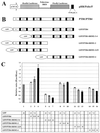
Effect of PTB and its derivatives on poliovirus IRES activity. (A) Schematic diagram of the reporter plasmid. This plasmid expresses a dicistronic mRNA consisting of the Renilla luciferase (RLuc) gene at the first cistron and the polioviral IRES-firefly luciferase (FLuc) gene at the second cistron. The RLuc and FLuc are directed by scanning and IRES-dependent translation, respectively. (B) Schematic diagram of effector plasmids. These plasmids produce full-length PTB4 or PTB1 and PTB4 derivatives fused with GFP. (C) Effect of PTB or its derivatives on poliovirus IRES activity. 293T cells were cotransfected with 0.75 μg of dicistronic plasmid and effector plasmids as indicated in the chart at the bottom. The total amount of effector plasmids was maintained at a constant level by adding control plasmid pEGFP-C1 when necessary. Forty-eight hours after transfection, Renilla luciferase and firefly luciferase activities were measured as described in Materials and Methods, and the relative ratio of firefly luciferase to Renilla luciferase activity in each cell lysate was calculated. The columns and bars represent the means and standard deviations of four independent transfection experiments. The numbers in the chart represent micrograms of DNA.
FIG. 6.
FIG. 6.
Model of molecular switching from translation to replication of poliovirus RNA. Proteins identified as interacting with the 5"NTR of poliovirus are depicted by ovals. Different proteins are shaded differently. For simplicity of the model, only one PTB binding is drawn, even though several PTB-binding sites are present in polioviral IRES. The events required for translation at the early stage of viral infection are depicted with thin lines. The effect of the interaction between PCBP and PTB on translation is hypothetical, even though the protein-protein interaction between these proteins was demonstrated experimentally. Changes after the production of 3CDpro are depicted by thick lines. PTB is cleaved by 3CDpro as indicated by the folded line on PTB. The C-terminal PTB may stay on the IRES element blocking translation from this mRNA. At the same time, the removal of the N-terminal end of PTB prevents its interaction with other proteins, such as PCBP, hnRNP K, hnRNP L, and PTB itself (48). The PCBP, previously bound to the IRES element, may be transferred to the CL element in the presence of 3CDpro. With these changes, replication will commence.

Similar articles

Cited by

References

    1. Agol, V. I., A. V. Paul, and E. Wimmer. 1999. Paradoxes of the replication of picornaviral genomes. Virus Res. 62:129-147. - PubMed
    1. Andino, R., N. Böddeker, D. Silvera, and A. V. Gamarnik. 1999. Intracellular determinants of picornavirus replication. Trends Microbiol. 7:76-82. - PubMed
    1. Andino, R., G. E. Rieckhof, and D. Baltimore. 1990. A functional ribonucleoprotein complex forms around the 5" end of poliovirus RNA. Cell 63:369-380. - PubMed
    1. Arnold, E., M. Luo, G. Vriend, M. G. Rossmann, A. C. Palmenberg, G. D. Parks, M. J. Nicklin, and E. Wimmer. 1987. Implications of the picornavirus capsid structure for polyprotein processing. Proc. Natl. Acad. Sci. USA 84:21-25. - PMC - PubMed
    1. Baltimore, D. 1968. Structure of the poliovirus replicative intermediate RNA. J. Mol. Biol. 32:359-368. - PubMed

Publication types

MeSH terms

LinkOut - more resources