. 2002 Mar;76(5):2350-62.
doi: 10.1128/jvi.76.5.2350-2362.2002.

An endoplasmic reticulum protein, p180, is highly expressed in human cytomegalovirus-permissive cells and interacts with the tegument protein encoded by UL48

Affiliations

An endoplasmic reticulum protein, p180, is highly expressed in human cytomegalovirus-permissive cells and interacts with the tegument protein encoded by UL48

K Ogawa-Goto et al. J Virol. 2002 Mar.

Abstract

We have used a virus overlay assay to detect cellular proteins associated with human cytomegalovirus (HCMV) particles. The radiolabeled HCMV particles specifically bound to two host proteins with molecular sizes of 150 and 180 kDa. By a micro-amino-acid sequencing technique, the 180-kDa protein was identified as a human homologue of the ES130/p180 ribosome receptor (p180), which is an integral endoplasmic reticulum (ER) membrane protein possessing a very unique tandem repeat domain at its N-terminal region. The virus overlay assay using truncated p180 polypeptides revealed that HCMV binding to human p180 occurred through the N-terminal region. In HCMV-permissive cells the high level of expression of the human p180 protein was clearly observed regardless of cell type. Furthermore, we showed that p180 binds to the UL48 gene product, which is one of the predominant tegument proteins of HCMV and which is considered to be tightly associated with the capsid. The interaction between the two proteins was assumed to be specific and was observed both in vitro and in vivo. During the late phase of infection, the unique relocation of human p180 was observed, that is, to the juxtanuclear region, which appeared to be in the vicinity of the area where naked virions were frequently observed in an electron-microscopic study. Thus our data suggest that p180 interacts with the HCMV tegument, at least through pUL48, during the HCMV replication process. We discuss the possible role of the interaction between p180 and pUL48 in the intracellular transport of HCMV virions.


Figures

FIG. 1.
FIG. 1.
The radiolabeled HCMV probe specifically bound to two host proteins with molecular masses of 150 and 180 kDa. (a) The membrane fractions of MRC-5 (lanes 2 and 4) and authentic annexin II (lanes 1 and 3) were separated in SDS-12% PAGE gels and subjected to the virus overlay assay as described in Materials and Methods in the absence (lanes 1 and 2) and the presence (lanes 3 and 4) of heparin (100 μg/ml). (b to e) The membrane fraction was separated in SDS-5% PAGE gels, transferred to a PVDF membrane under strong conditions, and a virus overlay assay was carried out as for panel a. (b) Assay in the absence (lane 1) and the presence (lane 2) of heparin. (c) Assay in the absence (lane 1) and the presence of cold virions at a 10-fold excess (lane 2) and a 20-fold excess (lane 3) or in the presence of myelin basic protein (MBP; lane 5). Lane 4, protein pattern of the membrane fraction stained by amido black. (d) Virus overlay assay performed in the absence (lane 1) and the presence (lane 2) of Triton X-100 (0.5%, final concentration). (e) The membrane fraction (mem) was solubilized with 1% Triton X-100 in PBS for 30 min on ice and centrifuged to separate the soluble (sol) and insoluble (insol) fractions. Then immunoprecipitation with a MAb against CD13 (WM15) was carried out, followed by either a virus overlay assay (lanes 1 to 4) or immunostaining by WM15 (lanes 5 to 8). Shown are the membrane fraction of MRC-5 cells prior to solubilization (lanes 1 and 5), the Triton X-100-soluble fraction (lanes 2 and 6), the immunoprecipitates (IP) with WM15 (lanes 3 and 7), and the Triton X-100-insoluble fraction (lanes 4 and 8). bi-HCMV, biotinylated HCMV.
FIG. 2.
FIG. 2.
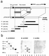
The domain structure of human p180 isoforms and HCMV binding to recombinant p180 polypeptides containing various regions. (a) Schematic representation of the domain structure of the human p180 protein and the relationship between isoforms possessing 54 and 24 tandem repeats, which were deduced from the primary amino acid sequences encoded by pTSX3.8F and pTSX4.8F. They each have a predicted transmembrane domain (TM) close to the N terminus, a highly basic N-terminal domain consisting of lysine clusters, a proline-rich domain (solid), a basic tandem repeat domain (10aa repeats), and a C-terminal acidic coiled-coil domain (19). ∗, insertion of three amino acids in this clone at amino acid 488. Various truncated human p180-GST fusion polypeptides were designed to address the domain responsible for the binding to HCMV. Open portion of the bar for each truncated mutant, N-terminal GST. Numbers in parentheses denote the amino acid residues encoded by pTSX3.8F. A summary of the binding experiments in the virus overlay assay is shown on the right. (b) Various truncated human p180/GST-fusion polypeptides were purified and used for the overlay assay. (A) Protein staining with amido black; (B) overlay assay with a biotinylated HCMV (bi-HCMV) probe; (C) overlay assay with biotinylated BSA.
FIG. 3.
FIG. 3.
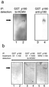
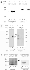
HCMV binding to the cellular p180 protein. The binding of the HCMV probe to the cellular p180 protein was tested using immunoprecipitated (IP) antigens. MRC-5 cell lysates were subjected to immunoprecipitation and either to a virus overlay assay or Western blotting. (a) Proteins immunoprecipitated by the anti-GST antibody (lanes 1 and 3) or the anti-p180 antibody (lanes 2 and 4) were used for either virus overlay assays (left) or Western blotting (right). In lane 2, a positive band with an apparent molecular size of 180 kDa was detected (arrow). bi-HCMV, biotinylated HCMV. (b) The biotinylated HCMV probe was pretreated with antiserum against UL48/S1 at dilution of 1:100 (left) or 1:1,000 (middle) or normal rabbit serum at a 1:100 dilution (right). After 2 h of incubation at 4°C, a virus overlay assay was carried out for panel a. Proteins immunoprecipitated by the anti-GST antibody (lanes 1, 3, and 5) or the anti-p180 antibody (lanes 2, 4, and 6) were used as antigens. p180 is shown by an arrow.
FIG. 4.
FIG. 4.
Human p180 and related proteins were highly expressed in the HCMV-permissive cell lines. Total cell lysates from various cell lines were subjected to Western blotting and analyzed with the affinity-purified anti-p180 C1 antibody (a) or a MAb against calnexin, an ER resident membrane protein (b). p180 was most highly expressed in MRC-5, K-1034, and Caco-2 cells, which were known as fully permissive for HCMV infection. Lanes: 1, MRC-5; 2, K-1034; 3, U937; 4, THP-1; 5, HSB-2; 6, Ramos 3; 7, HeLa; 8, 293T; 9, NIH 3T3; 10, Vero; 11, CHO; 12, Caco-2.
FIG. 5.
FIG. 5.
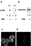
Induction of p180 protein expression in THP-1 cells upon treatment with TPA. (a) Western blotting analysis of THP-1 cell lysates. The p180 protein was detected by affinity-purified antibodies against N1 or C1, as indicated. (b) A MAb against calnexin was used. Lanes (a and b): 1, no TPA treatment; 2, TPA treatment. (c and d) IF analyses on THP-1 cells using the affinity-purified antibody against p180 with TPA treatment (c) and without TPA treatment (d).
FIG. 6.
FIG. 6.
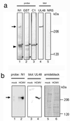
The recombinant p180 polypeptide specifically bound to the UL48 protein. (a) The purified virus proteins were separated in SDS-5% PAGE gels and subjected to either an in vitro overlay assay (lanes 1 to 3) or Western blotting (lanes 4 and 5). The N1 probe (lane 1), GST probe (lane 2), and C1 probe (lane 3) were used for the in vitro overlay assay, and the anti-UL48 antibody (lane 4) or normal rabbit serum (NRS) (lane 5) were used for Western blotting. Arrow, 230-kDa positive band; arrowhead, 140-kDa band; ∗, protein bands assumed to be nonspecific. (b) Mock-infected (lanes 1, 3, and 5) and HCMV-infected (lanes 2, 4, and 6) fibroblasts at 4 days p.i. were used instead of the virion preparation and analyzed as for panel a. Lanes 1 and 2, N1 probe; lanes 3 and 4, anti-UL48 antibody; lanes 5 and 6, protein staining by amido black.
FIG. 7.
FIG. 7.
The UL48 protein specifically interacted with endogenous p180. (a) 293T cells were transfected with pcUL48 (lanes 1, 2, 4, and 5 [the left lane is considered lane 1]) or vehicle only (lanes 3 and 6). Immunoprecipitation (IP) was carried out using either the anti-p180 antibody (lanes 2, 3, 5, and 6) or the anti-GST antibody (lanes 1 and 4), followed by blotting with anti-Xpress or anti-p180 antibodies. (b) An HCMV IE1-expressing plasmid was used instead of pcUL48 and analyzed as for panel a. (c) At 4 days p.i. mock-infected and HCMV-infected cells were lysed and centrifuged at 50,000 × g for 30 min to remove capsids and analyzed as for panel a. The UL48 protein (arrow) was detected by the anti-UL48 antibody.
FIG. 8.
FIG. 8.
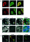
Immunofluorescence analyses of the p180 protein in cultured cells. (A) MRC-5 (a to c) or K-1034 (d to f) cells were cultured on a coverslip and processed for IF assay. (a and d) Antibody against p180; (b) anti-calreticulin antibody; (e) MAb against calnexin; (c and f), merged signal. The perinuclear reticular staining patterns for human p180 in MRC-5 (a) and K-1034 cells (d) were observed; the patterns resemble those for ER markers calreticulin and calnexin. (B) HCMV-infected (a to d) or mock-infected (e to h) MRC-5 cells at 3 days p.i. were processed for triple staining. (a and e) Anti-p180 antibody; (b and f) MAb against IE protein; (c and g) anti-calreticulin antibody; (d and h) merged signals. The infected cells exhibiting the enlargement of IE-protein positive nucleus showed a unique relocation of p180 signal to the perinuclear area, which is segregated from that of calreticulin, an ER protein. (C) HCMV-infected (a and b) and mock-infected (c and d) MRC-5 cells at 3 days p.i. were stained with anti-UL48/S1 antibody and a MAb against IE protein. (a and c) Anti-UL48/S1 antibody; (b and d) merged signal. UL48 signal (green) was observed as a juxtanuclear vacuolar pattern in the IE protein (red)-positive cells.
FIG. 9.
FIG. 9.
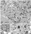
Ultrastructural features of HCMV-infected fibroblasts at late phase of infection. HCMV-infected MRC-5 cells (multiplicity of infection, 1 to 3) were harvested 3 or 4 days p.i. and processed for electron microscopy. (a) A number of cytoplasmic naked virions without envelopes were observed (small arrows) in the area between the nucleus (N) and centriole (C). Note that enveloped virions (large arrows) and dense bodies with mature shapes are mostly found in the distal area. The subcellular organelles exhibited altered membrane morphology such as dilation of RER and morphologically aberrant Golgi apparatus. Normal organelles were segregated to the cell periphery. (b to d). Higher-magnification micrographs revealed a thin filamentous network between the fine filamentous tegument of the naked virions (arrows) and the cellular vesicles (V) or membranes (M). Mt, mitochondrion. Bars, 2 μm (a) and 0.2 μm (b to d).
FIG. 10.
FIG. 10.
Schematic representation of the interaction between p180 and tegument protein during the course of HCMV infection. A model of the interaction between p180 and HCMV through the tegument protein is presented. The cytoplasmic disposition of p180 accounts for a dual possibility of interaction with the tegument before and after nuclear events. First (A), upon entry of HCMV into the host cell, released capsids which are transported to the nucleus may bind to the N-terminal domain of p180 (Nt) located on the ER or ER-derived vesicles. Second (B), after the nuclear assembly process, transported capsids that egressed from the nucleus to the final envelopment compartments may also interact with p180. The coiled-coil structure in the p180 carboxyl domain (Ct) is the most probable candidate for the interaction with other proteins involved in the cellular transport system which results in facilitating capsid transport in the cytoplasm. TGN, trans-Golgi network.

Similar articles

Cited by

References

    1. Aebersold, R. H., J. Leavitt, R. A. Saavedra, L. E. Hood, and S. B. Kent. 1987. Internal amino acid sequence analysis of proteins separated by one- or two-dimensional gel electrophoresis after in situ protease digestion. Proc. Natl. Acad. Sci. USA 84:6970-6974. - PMC - PubMed
    1. Ando, Y., T. Iwasaki, T. Sata, S. Soushi, T. Kurata, and Y. Arao. 1997. Enhanced cytopathic effect of human cytomegalovirus on a retinal pigment epithelium cell line, K-1034, by serum-free medium. Arch. Virol. 142:1645-1658. - PubMed
    1. Baldick, C. J., Jr., and T. Shenk. 1996. Proteins associated with purified human cytomegalovirus particles. J. Virol. 70:6097-6105. - PMC - PubMed
    1. Batterson, W., D. Furlong, and B. Roizman. 1983. Molecular genetics of herpes simplex virus. VIII. Further characterization of a temperature-sensitive mutant defective in release of viral DNA and in other stages of the viral reproductive cycle. J. Virol. 45:397-407. - PMC - PubMed
    1. Becker, F., L. Block-Alper, G. Nakamura, J. Harada, K. D. Wittrup, and D. I. Meyer. 1999. Expression of the 180-kD ribosome receptor induces membrane proliferation and increased secretory activity in yeast. J. Cell Biol. 146:273-284. - PMC - PubMed

MeSH terms

Substances

LinkOut - more resources