. 2002 Feb 1;30(3):810-7.
doi: 10.1093/nar/30.3.810.

Efficient polyadenylation of Rous sarcoma virus RNA requires the negative regulator of splicing element

Affiliations

Efficient polyadenylation of Rous sarcoma virus RNA requires the negative regulator of splicing element

Brent L Fogel et al. Nucleic Acids Res. .

Abstract

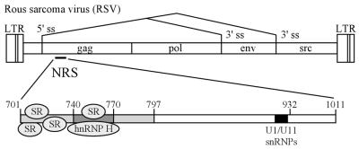
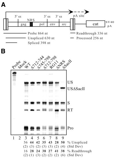
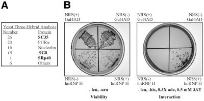
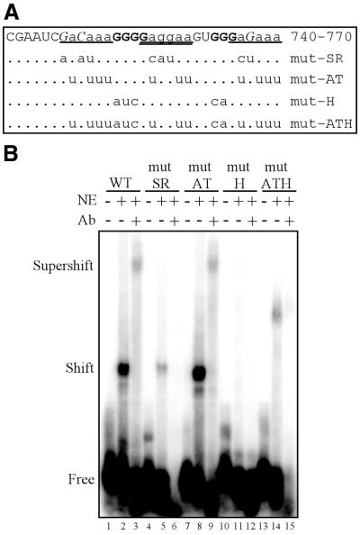
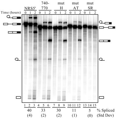
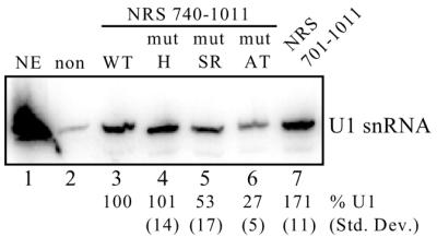
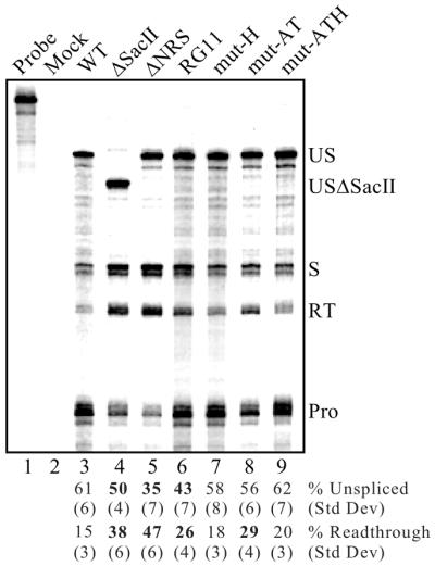
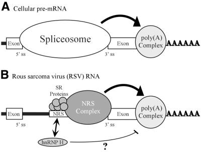
Rous sarcoma virus pre-mRNA contains an element known as the negative regulator of splicing (NRS) that acts to inhibit viral RNA splicing. The NRS binds serine/arginine-rich (SR) proteins, hnRNP H and the U1/U11 snRNPs, and appears to inhibit splicing by acting as a decoy 5' splice site. Deletions within the gag gene that encompass the NRS also lead to increased read-through past the viral polyadenylation site, suggesting a role for the NRS in promoting polyadenylation. Using NRS-specific deletions and mutations, we show here that a polyadenylation stimulatory activity maps directly to the NRS and is most likely dependent upon SR proteins and U1 and/or U11 snRNP. hnRNP H does not appear to mediate splicing control or stimulate RSV polyadenylation, since viral RNAs containing hnRNP H-specific mutations were spliced and polyadenylated normally. However, the ability of hnRNP H mutations to suppress the read-through caused by an SR protein mutation suggests the potential for hnRNP H to antagonize polyadenylation. Interestingly, disruption of splicing control closely correlated with increased read-through, indicating that a functional NRS is necessary for efficient RSV polyadenylation rather than binding of an individual factor. We propose a model in which the NRS serves to enhance polyadenylation of RSV unspliced RNA in a process analogous to the stimulation of cellular pre-mRNA polyadenylation by splicing complexes.


Figures

Figure 1
Figure 1
RSV and the NRS. The RSV genome is shown with the relative positions of the long terminal repeats (LTRs) and the gag, pol, env and src genes shown (not to scale). Alternative splicing is indicated by lines between the 5′ and 3′ splice sites (ss). The location of the NRS is shown within the gag gene and is expanded below with the positions of key sequence features noted. The overlapping binding site for the U1 and U11 snRNPs is located at nt 913–926 and is denoted by a black box. The 5′ portion of the NRS (nt 701–797) is lightly shaded and the region where hnRNP H binds (nt 740–770) is darkly shaded. The relative positions of SR protein and hnRNP H binding are indicated.
Figure 2
Figure 2
A functional NRS is required for efficient RSV polyadenylation. (A) Schematic diagram of the constructs used in splicing and polyadenylation control assays. These constructs contain the RSV Prague A strain genome (not drawn to scale) fused to the cat gene which is followed by the SV40 early polyadenylation signal. The relative positions of the four viral genes are shown and the NRS is indicated by a black box. Viral transcription begins in the viral 5′ LTR (dark line) and continues to the polyadenylation site within the 3′ LTR, where it can either undergo cleavage or read-through (dashed line). The region encompassed by the riboprobe used in these studies is shown below the diagram and the sizes of the probe and the protected fragments are indicated. This probe can be utilized for analyzing both splicing and polyadenylation, since it spans both the 5′ splice site and the 5′ LTR (which is duplicated at the 3′ end). (B) RNase protection assay using RNA from transfected CEFs. Total RNA from CEFs transfected with the indicated proviral DNA was hybridized to the riboprobe described in (A). Protected fragments were separated on a denaturing gel and a phosphorimager was used for visualization and quantitation. Probe, a sample of unprocessed probe; Mock, assay performed using RNA from mock-transfected CEFs. The positions of unspliced (US), spliced (S), read-through (RT) and processed (Pro) RNAs are shown. Because the ΔSacII construct lacks nt 543–630, the unspliced protected band is slightly smaller and is indicated (USΔSacII). Bands were quantitated and normalized for uridine content. The percentages of unspliced and read-through RNA were calculated from the total of spliced plus unspliced RNA and read-through plus processed RNA, respectively. The average percentages of unspliced and read-through RNA from at least three separate transfections and protection assays are shown below the lanes with corresponding standard deviations. Significant deviations (P < 0.02) from wild-type (WT) values are indicated in bold.
Figure 3
Figure 3
SR proteins and hnRNP H interact with the NRS in vivo in a yeast three-hybrid assay. (A) Summary of results from a three-hybrid screen using NRS 740–797 RNA in tandem (740–797×2) as bait for proteins expressed from a HeLa cDNA library. Positive clones are ordered relative to the number of times they were isolated. SR proteins are indicated in bold. (B) Three-hybrid results using the Gal4 activation domain (Gal4AD) alone or fused to hnRNP H (hnRNP H) and expressed with NRS 740–797×2 in either the sense (+) or antisense (–) orientation. Individual colonies were streaked on medium lacking leucine and uracil to confirm viability of the transformants or on medium lacking histidine to assess RNA-mediated activation of the HIS3 reporter gene. Results shown are representative of four independent colonies selected from each transformation.
Figure 4
Figure 4
Mapping of the hnRNP H binding site and representative hnRNP H mutants. (A) Summary of hnRNP H binding studies using NRS nt 740–770. Residues that are essential for hnRNP H binding are indicated in bold, non-essential residues are shown in lower case and unconfirmed residues are in italic. Bases that were not tested are in normal type. The positions of three repeated sequence elements that resemble SR consensus binding sites are underlined. The sequences of the four 740–770 mutants described in this study are shown with the corresponding mutations indicated. (B) Electrophoretic mobility shift assays were performed using 2.5 fmol individual NRS 740–770 RNAs and 5 µg HeLa nuclear extract (NE) in the presence or absence of hnRNP H antiserum (Ab). The positions of free, shifted and supershifted RNAs are indicated.
Figure 5
Figure 5
Splicing enhancer activity of NRS5′ mutants. Splicing enhancer assays were performed using the indicated in vitro transcribed doublesex–NRS RNAs incubated in HeLa nuclear extract under splicing conditions for either 1 or 2 h. The positions of the precursor, spliced and intermediate RNA forms are depicted, with the larger NRS5′ forms on the left and the smaller NRS 740–770 forms on the right. NRS sequences are indicated by a black box. Numbers beneath each set of lanes correspond to the percentage of spliced RNA (calculated from the total of unspliced plus spliced RNA) after 2 h. Results are the average of two independent experiments and the corresponding standard deviations are indicated in parentheses.
Figure 6
Figure 6
U1 snRNP recruitment to NRS mutants. Equal moles of the indicated biotinylated RNAs were incubated in HeLa nuclear extract and affinity selected with streptavidin–agarose beads. Bound snRNAs were then extracted with phenol/chloroform, separated on a denaturing polyacrylamide gel, electroblotted to a nylon membrane and hybridized to a U1 riboprobe. Non-biotinylated NRS RNA (non) was used to determine background binding to the beads whereas the full-length NRS (nt 701–1011) was used to determine optimal U1 binding (lane 7). To avoid any contributions from the upstream SR protein-binding site, the experimental RNAs lacked the primary SR protein-binding site (nt 740–1011, lanes 3–6). NE, U1 snRNA marker extracted from 3 µl of nuclear extract. Bands were quantitated using a phosphorimager and the average percentage of U1 snRNA selected from three separate experiments is shown below each lane with the corresponding standard deviation. The position of the U1 snRNA is shown on the right. Binding to the wild-type 740–1011 RNA was set at 100% (lane 3).
Figure 7
Figure 7
Effect of SR protein- and hnRNP H-specific mutations on splicing and read-through. RNA from CEFs transfected with the corresponding RSV mutants was used for RNase protection assays as described in Figure 6. Protected fragments were separated on a denaturing gel and a phosphorimager was used for visualization and quantitation. Probe, a sample of unprocessed probe; Mock, assay performed using RNA from mock-transfected CEFs. The positions of unspliced (US and USΔSacII), spliced (S), read-through (RT) and processed (Pro) RNAs are shown. Bands were quantitated, normalized for uridine content and the average percentages of unspliced and read-through RNA from six to eight separate transfections and protection assays are shown below the lanes with corresponding standard deviations. Significant deviations (P < 0.002) from wild-type (WT) values are indicated in bold.
Figure 8
Figure 8
Model for NRS-mediated stimulation of RSV polyadenylation. (A) Spliceosome contribution to polyadenylation. A generic cellular pre-mRNA is shown with the 5′ and 3′ splice sites of a terminal intron indicated. In cellular RNA processing the terminal 3′ splice site and the polyadenylation site define the last exon and factors associated with the spliceosome act to stimulate polyadenylation (large arrow; see Discussion). (B) NRS contribution to RSV polyadenylation. The NRS is shown between authentic viral splice sites. Depicted are SR proteins that bind to nt 701–770 and initiate NRS complex formation. The spliceosome-like inhibitory complex (NRS complex) that forms between the NRS and a viral 3′ splice site is indicated. The NRS may serve to stabilize the binding of splicing factors to the weak viral 3′ splice site, which can then either recruit or stabilize the polyadenylation complex (large arrow) and thereby enhance polyadenylation of viral unspliced RNA. A potential competition between SR proteins and hnRNP H for binding to this region is reflected by an arrow. hnRNP H binding may be restricted when SR proteins are present. In contrast, if SR proteins are absent or limiting, hnRNP H binding may increase and in some way disrupt the ability of the NRS to act efficiently on the polyadenylation complex (thin line). In both cases the NRS complex is competent for splicing inhibition.

Similar articles

Cited by

References

    1. Moore M.J., Query,C.C. and Sharp,P.A. (1993) In Gesteland,R. and Atkins,J. (eds), The RNA World. Cold Spring Harbor Laboratory Press, Cold Spring Harbor, NY, pp. 303–357.
    1. Coffin J.M. (1996) In Fields,B.N., Knipe,D.M. and Howley,P.M. (eds), Fields Virology, 3rd Edn. Raven Press, New York, NY, pp. 1767–1847.
    1. Arrigo S. and Beemon,K. (1988) Regulation of Rous sarcoma virus RNA splicing and stability. Mol. Cell. Biol., 8, 4858–4867. - PMC - PubMed
    1. Stoltzfus C.M. and Fogarty,S.J. (1989) Multiple regions in the Rous sarcoma virus src gene intron act in cis to affect the accumulation of unspliced RNA. J. Virol., 63, 1669–1676. - PMC - PubMed
    1. Hibbert C.S., Gontarek,R.R. and Beemon,K.L. (1999) The role of overlapping U1 and U11 5′ splice site sequences in a negative regulator of splicing. RNA, 5, 333–343. - PMC - PubMed

Publication types

MeSH terms