Functional replacement and positional dependence of homologous and heterologous L domains in equine infectious anemia virus replication
- PMID: 11799151
- PMCID: PMC135910
- DOI: 10.1128/jvi.76.4.1569-1577.2002
Functional replacement and positional dependence of homologous and heterologous L domains in equine infectious anemia virus replication
Abstract
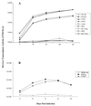
We have previously demonstrated by Gag polyprotein budding assays that the Gag p9 protein of equine infectious anemia virus (EIAV) utilizes a unique YPDL motif as a late assembly domain (L domain) to facilitate release of the budding virus particle from the host cell plasma membrane (B. A. Puffer, L. J. Parent, J. W. Wills, and R. C. Montelaro, J. Virol. 71:6541-6546, 1997). To characterize in more detail the role of the YPDL L domain in the EIAV life cycle, we have examined the replication properties of a series of EIAV proviral mutants in which the parental YPDL L domain was replaced by a human immunodeficiency virus type 1 (HIV-1) PTAP or Rous sarcoma virus (RSV) PPPY L domain in the p9 protein or by proviruses in which the parental YPDL or HIV-1 PTAP L domain was inserted in the viral matrix protein. The replication properties of these L-domain variants were examined with respect to Gag protein expression and processing, virus particle production, and virus infectivity. The data from these experiments indicate that (i) the YPDL L domain of p9 is required for replication competence (assembly and infectivity) in equine cell cultures, including the natural target equine macrophages; (ii) all of the functions of the YPDL L domain in the EIAV life cycle can be replaced by replacement of the parental YPDL sequence in p9 with the PTAP L-domain segment of HIV-1 p6 or the PPPY L domain of RSV p2b; and (iii) the assembly, but not infectivity, functions of the EIAV proviral YPDL substitution mutants can be partially rescued by inclusions of YPDL and PTAP L-domain sequences in the C-terminal region of the EIAV MA protein. Taken together, these data demonstrate that the EIAV YPDL L domain mediates distinct functions in viral budding and infectivity and that the HIV-1 PTAP and RSV PPPY L domains can effectively facilitate these dual replication functions in the context of the p9 protein. In light of the fact that YPDL, PTAP, and PPPY domains evidently have distinct characteristic binding specificities, these observations may indicate different portals into common cellular processes that mediate EIAV budding and infectivity, respectively.
Figures


Similar articles
-
Functional roles of equine infectious anemia virus Gag p9 in viral budding and infection.J Virol. 2001 Oct;75(20):9762-70. doi: 10.1128/JVI.75.20.9762-9770.2001. J Virol. 2001. PMID: 11559809 Free PMC article.
-
Equine infectious anemia virus utilizes a YXXL motif within the late assembly domain of the Gag p9 protein.J Virol. 1997 Sep;71(9):6541-6. doi: 10.1128/JVI.71.9.6541-6546.1997. J Virol. 1997. PMID: 9261374 Free PMC article.
-
Functions of early (AP-2) and late (AIP1/ALIX) endocytic proteins in equine infectious anemia virus budding.J Biol Chem. 2005 Dec 9;280(49):40474-80. doi: 10.1074/jbc.M509317200. Epub 2005 Oct 7. J Biol Chem. 2005. PMID: 16215227
-
Regulation of equine infectious anemia virus expression.J Biomed Sci. 1998;5(1):11-23. doi: 10.1007/BF02253351. J Biomed Sci. 1998. PMID: 9570509 Review.
-
Virulence determinants of equine infectious anemia virus.Curr HIV Res. 2010 Jan;8(1):66-72. doi: 10.2174/157016210790416352. Curr HIV Res. 2010. PMID: 20210781 Review.
Cited by
-
HIV Assembly and Budding: Ca(2+) Signaling and Non-ESCRT Proteins Set the Stage.Mol Biol Int. 2012;2012:851670. doi: 10.1155/2012/851670. Epub 2012 Jun 12. Mol Biol Int. 2012. PMID: 22761998 Free PMC article.
-
PIV5 M protein interaction with host protein angiomotin-like 1.Virology. 2010 Feb 5;397(1):155-66. doi: 10.1016/j.virol.2009.11.002. Epub 2009 Nov 24. Virology. 2010. PMID: 19932912 Free PMC article.
-
Human cytomegalovirus exploits ESCRT machinery in the process of virion maturation.J Virol. 2009 Oct;83(20):10797-807. doi: 10.1128/JVI.01093-09. Epub 2009 Jul 29. J Virol. 2009. PMID: 19640981 Free PMC article.
-
Equine Infectious Anemia Virus Gag Assembly and Export Are Directed by Matrix Protein through trans-Golgi Networks and Cellular Vesicles.J Virol. 2015 Dec 4;90(4):1824-38. doi: 10.1128/JVI.02814-15. Print 2016 Feb 15. J Virol. 2015. PMID: 26637458 Free PMC article.
-
Solution structure of the equine infectious anemia virus p9 protein: a rationalization of its different ALIX binding requirements compared to the analogous HIV-p6 protein.BMC Struct Biol. 2009 Dec 17;9:74. doi: 10.1186/1472-6807-9-74. BMC Struct Biol. 2009. PMID: 20015412 Free PMC article.
References
-
- Bolognesi, D. P., R. C. Montelaro, H. Frank, and W. Schafer. 1978. Assembly of type C oncornaviruses: a model. Science 199:183-186. - PubMed
-
- Freed, E. O. 1998. HIV-1 gag proteins: diverse functions in the virus life cycle. Virology 251:1-15. - PubMed
-
- Garnier, L., J. W. Wills, M. F. Verderame, and M. Sudol. 1996. WW domains and retrovirus budding. Nature 381:744-745. - PubMed
Publication types
MeSH terms
Substances
Grants and funding
LinkOut - more resources
Full Text Sources
Other Literature Sources
Research Materials

