. 2002 Jan 1;16(1):85-100.
doi: 10.1101/gad.925302.

Chromatin assembly factor I and Hir proteins contribute to building functional kinetochores in S. cerevisiae

Affiliations

Chromatin assembly factor I and Hir proteins contribute to building functional kinetochores in S. cerevisiae

Judith A Sharp et al. Genes Dev. .

Abstract

Budding yeast centromeres are comprised of approximately 125-bp DNA sequences that direct formation of the kinetochore, a specialized chromatin structure that mediates spindle attachment to chromosomes. We report here a novel role for the histone deposition complex chromatin assembly factor I (CAF-I) in building centromeric chromatin. The contribution of CAF-I to kinetochore function overlaps that of the Hir proteins, which have also been implicated in nucleosome formation and heterochromatic gene silencing. cacDelta hirDelta double mutant cells lacking both CAF-I and Hir proteins are delayed in anaphase entry in a spindle assembly checkpoint-dependent manner. Further, cacDelta and hirDelta deletions together cause increased rates of chromosome missegregation, genetic synergies with mutations in kinetochore protein genes, and alterations in centromeric chromatin structure. Finally, CAF-I subunits and Hir1 are enriched at centromeres, indicating that these proteins make a direct contribution to centromeric chromatin structures.


Figures
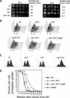
Figure 1
Figure 1
cacΔ hirΔ cells activate the MAD2-dependent spindle assembly checkpoint. (A) Slow growth of cac1Δ hir1Δ cells. Yeast strains PKY346 (wt), PKY1100 (cac1Δ), PKY1154 (hir1Δ), and PKY1168 (cac1Δ hir1Δ) were grown to log phase at 30°C, serially diluted, and grown on YPAD agar at 30°C for 2 d or 16°C for 12 d. Cell doubling time in liquid media for each strain is indicated to the right. (B) cacΔ hirΔ cells experience a MAD2-dependent G2/M phase delay. Yeast cells were grown to log phase at 30°C and arrested in α-factor (10 μg/mL) for 3 h. Cells were washed and released into pheromone-free media at 30°C. Aliquots of cultures were removed at 15-min intervals for a period of 3 h. α-Factor was added back to cultures at 55 min after the initial release to capture cells in the subsequent G1 phase of the cell cycle. DNA content was measured by FACS. Selected time points are marked for clarity. Strains are as in A, with the addition of PKY1340 (cac2Δ hir1Δ) and PKY1257 (cac1Δ hir1Δ mad2Δ). (C) Pronounced G2/M delay in cac1Δ hir1Δ cells at 16°C. Strains are as in A. Yeast cells were grown to log phase at 30°C and synchronized by arrest in 0.2 M hydroxyurea (HU) for 2 h. Cells were washed and released into 16°C media for 24 h. DNA content was measured by FACS. (D) Kinetics of spindle elongation. Yeast cells were grown to log phase at 30°C, arrested in α-factor for 3 h, washed, and released into media containing 0.2 M HU for 2 h at 30°C. Cells were then released from the HU block at 30°C. α-Factor was added back to cultures at 30 min after HU release to capture cells in the subsequent G1 phase of the cell cycle. Aliquots of cultures were taken at 15-min intervals for 3 h after release from HU and processed for tubulin (YOL134) and DNA (DAPI) staining of cells. Approximately 200 cells at each time point were then scored for spindle length and nuclear morphology.Shown at each time point are the percentage of cells in the population with short, bipolar spindles associated with an undivided nucleus at the bud neck. Yeast strains used were PKY346, wt; PKY1168, cac1Δ hir1Δ; PKY1257, cac1Δ hir1Δ mad2Δ; PKY1265, mad2Δ.
Figure 2
Figure 2
Chromosome missegregation in cac1Δ hir1Δ cells. (A) Aberrant DNA segregation in cac1Δ hir1Δ cells at 16°C. Cells treated as in Figure 1C were stained for tubulin (YOL134) and DNA (DAPI). Yeast strains are PKY346 (wild type) and PKY1168 (cac1Δ hir1Δ). Bar, 5 μm. (B) Localization of centromere DNA. After growth at 16°C for 24 h, fixed cells were stained with Hoechst dye to visualize DNA, and CEN4 DNA was visualized using a lacO array/lacI-GFP system. For each genotype, the percentages of mid-to-late anaphase cells with the morphologies shown are indicated below the photographs. Yeast strains used were SBY214, wt; PKY1473, cac1Δ; PKY1475, hir1Δ; PKY1477, cac1Δ hir1Δ.
Figure 3
Figure 3
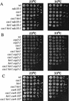
cac1Δ hir1Δ cells display genetic interactions with kinetochore mutations. Yeast strains of the indicated genotypes were grown to log phase at 23°C, a temperature permissive for all kinetochore alleles used. Cells were then serially diluted, plated onto YPAD agar, and incubated at the indicated temperatures (23°C for 3 d; 30°C for 2 d; and 33°C for 2 d). At least two independent segregants for each genotype were tested in this manner. Shown are growth phenotypes of strains containing cac1Δ and hir1Δ gene deletions combined with (A) ndc10-1; (B) cep3-1 and cep3-2; and (C) cse4-107 alleles.
Figure 4
Figure 4
Cac1, Cac2, and Hir1 colocalize with centromere protein Ndc10. All strains were grown to log phase at 30°C and prepared for chromosome spreads as described (Loidl et al. 1998). (A) Spread nuclei of yeast strains PKY028 (CAC1) and PKY1288 (CAC1-FLAG) were stained with anti-FLAG antibodies. Staining of Cac1-FLAG (green) is superimposed on DNA staining (DAPI, blue). (B) Spread nuclei of yeast strains PKY087 (hir1Δ) and PKY028 (HIR1) were stained with anti-Hir1 antibody. Staining of Hir1 (red) is superimposed on DNA staining (DAPI, blue). (C) Spread nuclei of strain PKY1288 (CAC1-FLAG) were processed for double-label immunofluorescence using anti-FLAG and anti-Hir1 antibodies. (D) Spread nuclei of yeast strain PKY2067 (CAC1-FLAG, NDC10-GFP) were stained with anti-FLAG (green) and anti-GFP antibodies (red). (E) Spread nuclei of yeast strain PKY2046 (CAC2-HA, NDC10-GFP) were stained using anti-HA (green) and anti-GFP antibodies (red). (F) Spread nuclei of yeast strain PKY2069 (HIR1, NDC10-GFP) were stained using anti-Hir1 (red) and anti-GFP antibodies (green). Bar, 5μm.
Figure 5
Figure 5
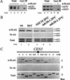
Cac1 is associated with centromeric chromatin. Formaldehyde cross-linked chromatin (1 h fixation) was prepared from cells grown to log phase at 30°C. (A) Chromatin prepared from yeast strain PKY1288 (CAC1-FLAG) was immunoprecipitated in the presence of anti-FLAG antibody or mock treated. PCR was performed on both total input and immunoprecipitated chromatin to visualize recovery of the core centromeric region of CEN3 (249 bp) and CEN16 (310 bp) relative to the ACT1 locus (367 bp). Dilution of total input chromatin: (lanes 1,6) 1:32; (lanes 2,7) 1:64; (lanes 3,8) 1:128. Approximately one-fifth of the eluate from immunoprecipitation was used for PCR analysis: (lanes 4,9) mock immunoprecipitation, (lanes 5,10) the experimental immunoprecipitation. (B) Cac1 localization at CEN3 requires NDC10 but not HIR1. Chromatin was prepared from yeast strains (lanes 1,3) PKY2199 (CAC1-FLAG); (lane 2) PKY2385 (CAC1-FLAG hir1Δ); and (lanes 4,5) PKY2200 (CAC1-FLAG ndc10-1); and analyzed as in A. Total input chromatin was diluted 1:64 prior to PCR analysis. (C) Distribution of CAC1 across the CEN3 region. Chromatin was prepared from yeast strain PKY1288 (CAC1-FLAG), and PCR was performed as in A. As in all experiments, CEN DNA in these FLAG antibody immunoprecipitations was enriched relative to the ACT1 negative control locus (data not shown). (Row 1) Anti-FLAG precipitated chromatin, (row 2) mock-precipitated chromatin, and (row 3) total input chromatin (1:64 dilution) was analyzed for recovery of fragments at or flanking the core centromeric region of CEN3 as indicated on the diagram (not to scale).
Figure 6
Figure 6
Centromeric chromatin phenotypes in cac1Δ hir1Δ cells. Nuclei were prepared from yeast strains PKY346 (wt), PKY1100 (cac1Δ), PKY1154 (hir1Δ), and PKY1168 (cac1Δ hir1Δ) as indicated and digested with nucleases as follows. (AC) Indirect end-label analysis of CEN3 chromatin. Nuclei were incubated with 0.6 U MNase (Sigma) at 32°C for 0 min (lanes 1,5,9,13), 5 min (lanes 2,6,10,14), 10 min (lanes 3,7,11,15), or 15 min (lanes 4,8,12,16) prior to isolation of genomic DNA, restriction enzyme digestion, and DNA blot hybridization with probes as described in the Materials and Methods. (A) CDEIII-proximal side of CEN3 in cells grown at 30°C. Samples were digested with ClaI. (B) CDEIII-proximal side of CEN3 in cells shifted to 16°C for 36 h prior to isolation of nuclei. Samples were digested with ClaI. (C) CDEI-proximal side of CEN3 in nuclei prepared from cells grown at 16°C. Samples were digested with BamHI. (D) DraI accessibility to CDEII within CEN3. Nuclei were incubated with DraI (0, 50, 100, or 150 U/mL) at 37°C for 30 min. DNA was purified, digested with EcoRI, and subjected to Southern blot hybridization. The fold differences in digestion relative to wild-type cells represent the average of three independent experiments with standard deviations indicated by the error bars. (Lower panel) A region of the same gel used for the Southern blot visualized by ethidium bromide staining, indicating similar extents of DraI digestion of total chromatin in all four strains.
Figure 7
Figure 7
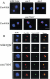
Extracentromeric localization of Cse4 in cac1Δ hir1Δ cells. (A) Dispersion of Cse4. Yeast strains PKY2084 (CSE4-HA), PKY2296 (cac1Δ CSE4-HA), PKY2298 (hir1Δ CSE4-HA), and PKY2179 (cac1Δ hir1Δ CSE4-HA) were grown to log phase at 30°C and prepared for chromosome spreads. Nuclei were stained with anti-HA antibody. Staining of Cse4-HA (red) is superimposed on DNA staining (DAPI, blue). Bar, 5 μm. (B) Extracentromeric localization of Cse4. Yeast strains PKY2444 (CSE4-HA NDC10-GFP) and PKY2453 (cac1Δ hir1Δ CSE4-HA NDC10-GFP) were treated as in A and were stained to detect Cse4-HA (red) and Ndc10-GFP (green).

Similar articles

Cited by

References

    1. Allshire RC, Nimmo ER, Ekwall K, Javerzat JP, Cranston G. Mutations derepressing silent centromeric domains in fission yeast disrupt chromosome segregation. Genes & Dev. 1995;9:218–233. - PubMed
    1. Amon A. The spindle checkpoint. Curr Opin Genet Dev. 1999;9:69–75. - PubMed
    1. Biggins S, Severin FF, Bhalla N, Sassoon I, Hyman AA, Murray AW. The conserved protein kinase Ipl1 regulates microtubule binding to kinetochores in budding yeast. Genes & Dev. 1999;13:532–544. - PMC - PubMed
    1. Blat Y, Kleckner N. Cohesins bind to preferential sites along yeast chromosome III, with differential regulation along arms versus the centric region. Cell. 1999;98:249–259. - PubMed
    1. Bloom KS, Carbon J. Yeast centromere DNA is in a unique and highly ordered structure in chromosomes and small circular minichromosomes. Cell. 1982;29:305–317. - PubMed

Publication types

MeSH terms

Substances

LinkOut - more resources