. 2001 Dec 17;20(24):7271-83.
doi: 10.1093/emboj/20.24.7271.

A novel transferable nuclear export signal mediates CRM1-independent nucleocytoplasmic shuttling of the human cytomegalovirus transactivator protein pUL69

Affiliations

A novel transferable nuclear export signal mediates CRM1-independent nucleocytoplasmic shuttling of the human cytomegalovirus transactivator protein pUL69

P Lischka et al. EMBO J. .

Abstract

The best studied nuclear export processes are mediated by classical leucine-rich nuclear export signals that specify recognition by the CRM1 export receptor. However, details concerning alternative nuclear export signals and pathways are beginning to emerge. Within the family of Herpesviridae, a set of homologous regulatory proteins that are exemplified by the ICP27 of herpes simplex virus were described recently as nucleocytoplasmic shuttling proteins. Here we report that pUL69 of the beta-herpesvirus human cytomegalovirus is a nuclear protein that is able to shuttle between the nucleus and the cytoplasm independently of virus-encoded cofactors. In contrast to proteins containing a leucine-rich export signal, the shuttling activity of pUL69 was not affected by leptomycin B, indicating that pUL69 trafficking is not mediated by the export receptor CRM1. Importantly, we identified and characterized a novel type of transferable, leptomycin B-insensitive export signal that is distinct from other export signals described previously and is required for pUL69-mediated activation of gene expression. These data suggest that pUL69 is exported via a novel nuclear export pathway, based on a so far unique nuclear export signal of 28 amino acids.


Figures

None
Fig. 1. Nucleocytoplasmic shuttling of pUL69 in HCMV-infected primary human fibroblasts. Heterokaryons were generated by fusion of HCMV-infected human fibroblasts and NIH 3T3 mouse cells. Protein synthesis was blocked with cycloheximide (50 µg/ml) 30 min prior to cell fusion and throughout the experiment. Two hours after fusion, cells were fixed and a double immunofluorescence analysis was performed with a polyclonal antiserum directed against pUL69 (B) and a monoclonal antibody against the IE1 protein (C). Staining with Hoechst 33258 (A) was used to differentiate between human and murine nuclei within the heterokaryon. Murine nuclei display a characteristic punctate pattern, whereas human nuclei are stained diffusely with this reagent; murine nuclei are indicated by arrows. (D) The phase contrast image of the heterokaryons.
None
Fig. 2. Nucleocytoplasmic shuttling of pUL69 in transfected HeLa cells. (A) Schematic representation of the wild-type (wt) UL69 protein and β-Gal fusion proteins that were used as internal controls in this heterokaryon analysis. The region of highest homology to the HSV-1 ICP27 and the location of basic, acidic and proline/glutamine-rich amino acid clusters are indicated for pUL69. Plasmid CFNrev-βgal (expressing β-Gal fused to the NLS from SV40 T-Ag and the NES from HIV 1-Rev) served as a positive control for nucleocytoplasmic shuttling in heterokaryon analysis. Plasmid CFN-βGal, which lacks the Rev NES, served as a negative control (Roth and Dobbelstein, 1997). (B) HeLa cells were co-transfected with the UL69 expression plasmid pHM160 and one of the internal control plasmids as indicated (a–d, UL69 + CFNrev-βGal; e–f, UL69 + CFN-βGal). The transfected cells were subjected to the interspecies heterokaryon assay as described in Materials and methods. Double immunofluorescence analysis with polyclonal anti-pUL69 serum and a monoclonal antibody against β-Gal was performed in order to detect the expressed proteins. Hoechst, staining with Hoechst 33258 (a and e); α UL69, staining with anti-pUL69 antiserum (b and f); α β-Gal, staining with anti-β-galactosidase antibody; phase, phase contrast image of the heterokaryon; murine nuclei are indicated by arrows.
None
Fig. 3. Effect of LMB on the shuttling of pUL69 in interspecies heterokaryon assays. HeLa cells were co-transfected with expression constructs for wild-type pUL69 together with plasmid CFNrev-βGal. Three hours prior to fusion and throughout the experiment, cells were incubated in the absence (AD) or presence of 2.5 ng/ml LMB (EH). Two hours after fusion, the proteins were detected by double-label immunofluorescence analysis as described in the legend of Figure 2.
None
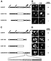
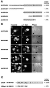
Fig. 4. Delineation of a putative NES within the C-terminus of pUL69 by deletion mutagenesis. (A) Schematic diagram illustrating N-terminal deletion mutants of pUL69. The N-terminal cluster of basic amino acids shows homology to a bipartite NLS. (B) HeLa cells transfected with plasmids expressing N-terminal pUL69 deletion mutants were analyzed for the subcellular localization of the indicated mutants via indirect immunofluorescence analysis. (C) Schematic diagram illustrating C-terminal or internal deletion mutants of pUL69; in the expanded section at the bottom of (C), the putative NES (amino acids 574–624) of pUL69 is shown. (D) HeLa cells transfected with plasmids expressing the pUL69 mutants as shown in (C) were subjected to heterokaryon analyses. Hoechst, DNA staining of transfected cells (B) or heterokaryons (D) with Hoechst 33258 (a, c, e, g, i, k and m); α UL69, transfected cells (B) or heterokaryons (D) stained for pUL69 are shown in b, d, f, h, j, l and n.
None
Fig. 5. Nuclear export of a heterologous protein by the putative pUL69 NES. (A) Schematic representation of the wild-type UL69 coding region showing the putative NES which was inserted into plasmid CFNrev-βGal, thus replacing the NES of HIV-1 Rev. The resulting expression vector encoding β-Gal in fusion with the NLS of SV40 T-Ag and the putative NES of pUL69 was termed CFNUL69-βGal. (B) Heterokaryon experiments as described in the legend of Figure 2 were performed to visualize the nuclear export activity of the expressed β-Gal fusion protein.
None
Fig. 6. Confirmation of the autonomous function of the pUL69 nuclear export sequence by microinjection experiments. (A) Schematic representation of the UL69 coding region showing the putative NES fused to the C-terminus of GST to produce GST–UL69NES. (B) Prokaryotic expression and purification of GST–UL69NES. Shown is a Coomassie Blue-stained gel: lane 1, extracts from E.coli cells grown without isopropyl-β-d-thiogalactopyranoside (IPTG); lane 2, extracts of E.coli cells grown in the presence of IPTG; lane 3, purified GST fusion protein. (C) HeLa cells were co-microinjected into the nucleus (a and b) or the cytoplasm (c and d) with GST–UL69NES and rabbit IgG (IgG, injection control). Cells were incubated for 1 h, fixed and analyzed for GST- and rabbit IgG-specific localization by double-label immunofluorescence microscopy. The injected GST fusion protein was visualized by using a GST-specific mouse monoclonal antibody followed by a Cy3-conjugated goat anti-mouse antibody (b and d). The co-microinjected rabbit IgG was detected with a Cy2-conjugated goat anti-rabbit polyclonal antibody (a and c).
None
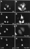
Fig. 7. Effect of LMB on nuclear export of GST–UL69NES in microinjection experiments. HeLa cells were co-microinjected into the nucleus with the indicated GST fusion protein and rabbit IgG and analyzed as described in the legend of Figure 6. Cell cultures in (C), (D), (G) and (H) were supplemented 2 h before microinjection and throughout the experiment with LMB at a concentration of 2.5 ng/ml. IgG, rabbit IgG, used as a control for nuclear injection; pUL69 NES, GST fusion protein with the putative NES of pUL69; Rex NES, GST fusion protein with the NES of Rex.
None
Fig. 8. Delineation of a minimal pUL69 export sequence. (A) Schematic representation of N- and C-terminal deletions of the pUL69 NES generated to identify a minimal domain that is able to mediate nuclear export. The shuttling activity of each pUL69 NES mutant is indicated by + or . Numbers in parentheses indicate the number of heterokaryons showing positive staining/the number of heterokaryons examined. (B) pUL69 NES deletion mutants shown in (A) were inserted in plasmid CFNrev-βGal between the SV40 T-Ag NLS and β-Gal, thus replacing the HIV-1 Rev NES. HeLa cells were transfected and analyzed by interspecies heterokaryon analysis as described in the legend of Figure 2. (C) Comparison of nuclear shuttling mediated by the pUL69 NES and the HDAg-L NES by heterokaryon analysis. The HDAg-L NES was inserted in plasmid CFNrev-βGal and analyzed as described in (A) for the pUL69 NES.
None
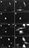
Fig. 9. The HDAg-L NES is not able to compete for nuclear export mediated by the pUL69 NES. HeLa cells were co-microinjected into the nucleus with the respective GST fusion proteins, rabbit IgG and either with or without an excess amount of the HDAg-L NES peptide cross-linked to BSA (as indicated in the figure). Cells were incubated at 37°C for 2 h and analyzed by indirect immunofluorescence. The injected GST fusion proteins were visualized by using a GST-specific mouse monoclonal antibody followed by a Cy3-conjugated goat anti-mouse antibody (B, D, F and H). The co-microinjected rabbit IgG was detected with a Cy2-conjugated goat anti-rabbit polyclonal antibody (A, C, E and G). UL69-NES, GST fusion protein with the putative NES of pUL69; HDAg-NES, GST fusion protein with the NES of HDAg-L; HDAg-Pep, HDAg-L NES peptides covalently coupled to BSA.
None
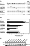
Fig. 10. Identification of amino acids important for pUL69 export activity and transactivation. (A) A series of single or double alanine replacement mutations were generated in the context of wild-type pUL69 to identify the amino acids that are critical for pUL69 nuclear export activity. The resulting mutants were then tested in heterokaryon analyses for nuclear shuttling. The shuttling activity of each pUL69 NES mutant is indicated by + or –. Numbers in parentheses indicate the number of heterokaryons showing positive staining/the number of heterokaryons examined. (B) Luciferase analysis after co-transfection of a pUL69-responsive luciferase reporter construct carrying the IE1/2 enhancer-promoter together with expression plasmids for the pUL69 alanine replacement mutants. Lane 1, co-transfection was performed with the empty expression vector pCB6; lane 2, co-transfection was performed with an expression vector for wild-type pUL69; lanes 3–10, co-transfections were performed with expression vectors representing the mutants indicated in (A). Fold activation was calculated relative to the basal activity of the reporter construct after co-transfection with the empty vector pCB6. Each experiment was performed in triplicate and was repeated at least three times. (C) Western blot analysis of U373MG cell extracts after transfection of the pUL69 mutants indicated in (A) using the pUL69-specific monoclonal antibody 69-66.
None
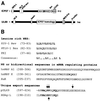
Fig. 11. (A) Schematic representation of ICP27 and pUL69 showing the various protein domains as described by Bryant et al. (2001), Winkler et al. (2000) and this study. NES-L, leucine-rich nuclear export signal; NES-P/A, proline-rich/acidic nuclear export signal; NLS, nuclear localization signal; RGG box, arginine-rich RNA-binding region; KH1-3, hnRNP K homology domains; Sm, Sm protein homology domain; CCHC, cysteine–histidine zinc finger-like domain; basic, cluster of basic amino acids; Pro/Glu, cluster of prolines and glutamines; ICP27 homology, domain of pUL69 with high homology to ICP27. (B) Sequence comparison of various types of NES. Amino acid sequences of NES of the classical leucine-rich type are compared with bidirectional shuttling signals and unique NES. The NES of HIV-1 Rev (Fischer et al., 1995), HTLV-1 Rex (Palmeri and Malim, 1996), protein kinase inhibitor (PKI) (Wen et al., 1995) and a derived consensus NES (Henderson and Eleftheriou, 2000) are listed for representation of leucine-rich NES; hnRNP A1 (Michael et al., 1995), hnRNP K (Michael et al., 1997) and HuR (Fan and Steitz, 1998) represent proteins containing bidirectional signals; the hepatitis D antigen HDAg-L NES (Lee et al., 2001) recently has been described as a unique export sequence. Identical residues in the unique export sequences identified in HDAg-L and pUL69 are shown in bold. The residues that are essential for nuclear export in HDAg-L and pUL69 are indicated by arrows. Numbers refer to the positions of the amino acid sequences within each protein.

Similar articles

Cited by

References

    1. Andreoni M., Faircloth,M., Vugler,L. and Britt,W.J. (1989) A rapid microneutralization assay for the measurement of neutralizing antibody reactive with human cytomegalovirus. J. Virol. Methods, 23, 157–167. - PubMed
    1. Bello L.J., Davison,A.J., Glenn,M.A., Whitehouse,A., Rethmeier,N., Schulz,T.F. and Barklie,C.J. (1999) The human herpesvirus-8 ORF 57 gene and its properties. J. Gen. Virol., 80, 3207–3215. - PubMed
    1. Brewer C.B. (1994) Cytomegalovirus plasmid vectors for permanent lines of polarized epithelial cells. Methods Cell Biol., 43, 233–245. - PubMed
    1. Bryant H.E., Wadd,S.E., Lamond,A.I., Silverstein,S.J. and Clements,J.B. (2001) Herpes simplex virus IE63 (ICP27) protein interacts with spliceosome-associated protein 145 and inhibits splicing prior to the first catalytic step. J. Virol., 75, 4376–4385. - PMC - PubMed
    1. Caceres J.F., Screaton,G.R. and Krainer,A.R. (1998) A specific subset of SR proteins shuttles continuously between the nucleus and the cytoplasm. Genes Dev., 12, 55–66. - PMC - PubMed

Publication types

MeSH terms