. 2002 Jan;22(1):332-42.
doi: 10.1128/MCB.22.1.332-342.2002.

Role for the related poly(ADP-Ribose) polymerases tankyrase 1 and 2 at human telomeres

Affiliations

Role for the related poly(ADP-Ribose) polymerases tankyrase 1 and 2 at human telomeres

Brandoch D Cook et al. Mol Cell Biol. 2002 Jan.

Abstract

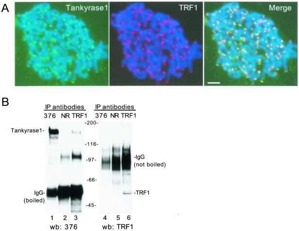
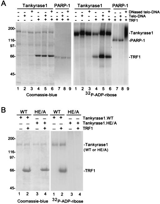
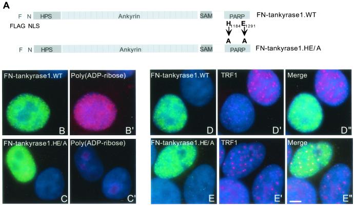
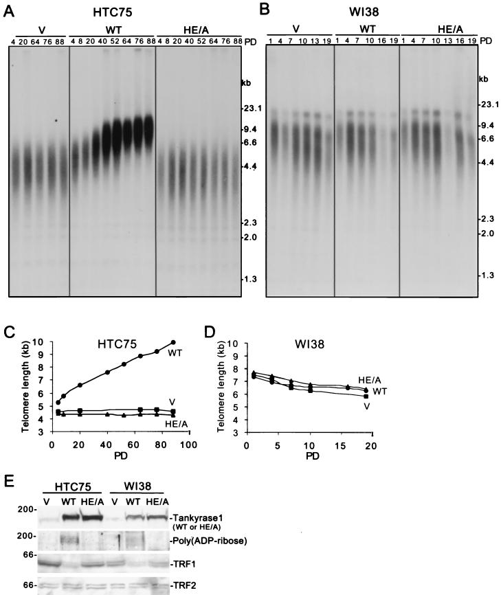
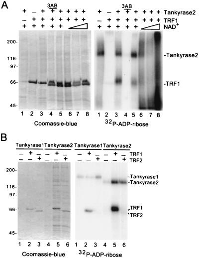
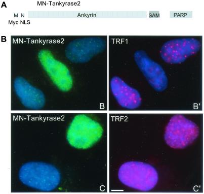
Telomere maintenance is essential for the continuous growth of tumor cells. In most human tumors telomeres are maintained by telomerase, a specialized reverse transcriptase. Tankyrase 1, a human telomeric poly(ADP-ribose) polymerase (PARP), positively regulates telomere length through its interaction with TRF1, a telomeric DNA-binding protein. Tankyrase 1 ADP-ribosylates TRF1, inhibiting its binding to telomeric DNA. Overexpression of tankyrase 1 in the nucleus promotes telomere elongation, suggesting that tankyrase 1 regulates access of telomerase to the telomeric complex. The recent identification of a closely related homolog of tankyrase 1, tankyrase 2, opens the possibility for a second PARP at telomeres. We therefore sought to establish the role of tankyrase 1 at telomeres and to determine if tankyrase 2 might have a telomeric function. We show that endogenous tankyrase 1 is a component of the human telomeric complex. We demonstrate that telomere elongation by tankyrase 1 requires the catalytic activity of the PARP domain and does not occur in telomerase-negative primary human cells. To investigate a potential role for tankyrase 2 at telomeres, recombinant tankyrase 2 was subjected to an in vitro PARP assay. Tankyrase 2 poly(ADP-ribosyl)ated itself and TRF1. Overexpression of tankyrase 2 in the nucleus released endogenous TRF1 from telomeres. These findings establish tankyrase 2 as a bona fide PARP, with itself and TRF1 as acceptors of ADP-ribosylation, and suggest the possibility of a role for tankyrase 2 at telomeres.


Figures

FIG. 1.
FIG. 1.
Generation of tankyrase 1-specific antibodies. (A) Schematic representation of tankyrase 1 and 2. Percent identities to tankyrase 2 are indicated. Lines show the domains against which the indicated antibodies (Ab) were raised. HPS, homopolymeric tracts of histidine, proline, and serine. SAM, sterile alpha module. (B) Immunoprecipitation analysis of in vitro-translated proteins. Plasmids encoding tankyrase 1 (T1), tankyrase 2 (T2) or vector control (C) were subjected to in vitro transcription-translation. Reaction products were applied directly to the gel (Input; 40% of total) or immunoprecipitated (IP) with normal rabbit serum (NRS) or anti-tankyrase 1 antibodies 465, 609, or 376. Products were analyzed by SDS-PAGE and fluorography.
FIG. 2.
FIG. 2.
Tankyrase 1 is localized to telomeres and complexed to TRF1 in vivo. (A) Colocalization of endogenous tankyrase 1 and TRF1 at telomeres. Indirect immunofluorescence analysis of swollen-formaldehyde fixed metaphase spreads from HeLaI.2.11 cells stained with anti-tankyrase 1 antibody 609 (green) and anti-TRF1 (red). Merge represents superimposition of the red and green images. DAPI staining of DNA is shown in blue. Scale bar, 5 μm. (B) Coimmunoprecipitation of an endogenous tankyrase 1-TRF1 complex from HelaI.2.11 cells. Cell extracts were immunoprecipitated (IP) with anti-tankyrase 1 antibody 376 (lanes 1 and 4), normal rabbit IgG (NR) (lanes 2 and 5), or anti-TRF1 antibody 415 (lanes 3 and 6). Samples were processed as described in Materials and Methods, suspended in Laemmli buffer, and boiled (left panel) or not boiled (right panel). Reaction products were fractionated by SDS-PAGE and subjected to Western blot (wb) analysis with anti-tankyrase 1 antibody 376 (left panel) or anti-TRF1 antibody 415 (right panel).
FIG. 3.
FIG. 3.
Characterization of wild-type and mutant tankyrase 1 activity in an in vitro PARP assay. (A) Tankyrase 1 is not activated by telomeric DNA. Reaction mixtures containing 4 μg of recombinant tankyrase 1 (lanes 1 to 6) or 0.2 μg of recombinant PARP-1 (lanes 7 to 9) were subjected to an in vitro PARP assay containing 1.3 μM [32P]NAD+ substrate without (-) (lanes 1 to 3) or with (+) (lanes 4 to 9) 4 μg of TRF1 and without (-) (lanes 1, 4, and 7) or with (+) 0.8 μg of telomeric DNA (Telo-DNA) (lanes 2, 5, and 8) or 0.8 μg of DNase-treated telomeric (DNased telo-DNA) (lanes 3, 6, and 9). (B) The tankyrase 1.HE/A protein is catalytically inactive in vitro. Reaction mixtures containing 4 μg of recombinant tankyrase 1.WT (lanes 1 and 2) or 4 μg of tankyrase 1.HE/A (lanes 3 and 4) were subjected to an in vitro PARP assay containing 1.3 μM [32P]NAD+ substrate with (+) (lanes 2 and 4) or without (-) 4 μg of TRF1 (lanes 1 and 3). Reaction products in panels A and B were fractionated on SDS-PAGE gels and visualized by Coomassie blue staining (left panel) or autoradiography (right panel).
FIG. 4.
FIG. 4.
Tankyrase 1.HE/A is inactive in vivo. (A) Schematic representation of tankyrase 1 expression constructs. F, FLAG epitope tag; N, NLS, nuclear localization signal from SV40; WT, wild type; HE/A, double point mutation converting the histidine (H) at position 1184 and the glutamic acid (E) at position 1291 to alanine (A) residues. (B to E) FN-tankyrase 1.HE/A does not synthesize ADP-ribose polymers in vivo or release TRF1 from telomeres, as shown by indirect immunofluorescence analysis of HeLaI.2.11 cells transiently transfected with FN-tankyrase 1.WT (B and D) or FN-tankyrase 1.HE/A (C and E), formaldehyde fixed, and stained with anti-FLAG antibody (green) (B, C, D, and E) and anti-poly(ADP-ribose) antibody (red) (B′ and C′), or anti-TRF1 antibody (red) (D′ and E′). Merge represents superimposition of the red and green images. DAPI staining of DNA is shown in blue. Scale bar, 5 μm.
FIG. 5.
FIG. 5.
Analysis of stable cell lines expressing FN-tankyrase 1.WT or HE/A. (A and B) Southern blot analysis of HinfI/RsaI-digested genomic DNA from telomerase-positive HTC75 cell lines (A) or telomerase-negative WI38 cell lines (B) expressing vector control (V), FN-tankyrase 1.WT (WT), or FN-tankyrase 1.HE/A (HE/A). Cell lines were grown for 88 (HTC75) or 19 (WI38) population doublings (PD), and DNA samples were analyzed at the indicated PDs. Blots were probed with TTAGGG-repeat probe to detect telomeric restriction fragments. (C and D) Graphical representations of telomere length changes in HTC75 (C) or WI38 (D) cell lines expressing FN-tankyrase 1.WT, HE/A, or vector. Plots represent the mean telomere length values derived from the Southern blots analyzed in panels A and B. (E) Western blot analysis of whole-cell extracts from HTC75 (PD 52) or WI38 (PD 4) cells expressing vector control (V), FN-tankyrase 1.WT (WT), or FN-tankyrase 1.HE/A (HE/A). Blots were probed with the following antibodies: anti-tankyrase 1 376, anti-poly(ADP-ribose), anti-TRF1 415, and anti-TRF2.
FIG. 6.
FIG. 6.
Two-hybrid analysis of tankyrase 1 and 2-TRF1 interactions. (A) β-Galactosidase concentrations (Miller units; average of three independent transformations) were measured for strains expressing the indicated fusion proteins. GAD, GAL4 activation domain; AR, ank repeats. (B) Northern blots of RNAs from a variety of human adult and fetal tissues and human primary and cancer cells, probed with tankyrase 1, tankyrase 2, or β-actin DNA as a control. f., fetal; PD, population doubling.
FIG. 7.
FIG. 7.
Tankyrase 2 is a PARP that modifies itself and TRF1, but not TRF2, in vitro. (A) Tankyrase 2 ADP-ribosylates itelf and TRF1 in vitro. Reaction mixtures containing 2 μg of recombinant tankyrase 2 (lanes 1 and 3 to 8) were subjected to an in vitro PARP assay containing 1.3 μM [32P]NAD+ substrate without (-) (lane 1) or with (+) 4 μg of TRF1 (lanes 2 to 8). Three reaction mixtures were supplemented with unlabeled NAD+ (0.04, 0.2, and 1 mM, triangle) (lanes 6 to 8) and one reaction mixture contained 1 mM 3-amino benzamide (3AB) (lane 4). (B) Tankyrase 2 does not modify TRF2 in vitro. Reactions containing 0.5 μg of recombinant tankyrase 1 (lanes 1 to 3) or 2 μg of recombinant tankyrase 2 (lanes 4 to 6) were subjected to an in vitro PARP assay containing 1.3 μM [32P]NAD+ substrate with (+) 1 μg TRF1 (lanes 2 and 5) or (+) 1 μg TRF2 (lanes 3 and 6). Reaction products in panels A and B were fractionated by SDS-PAGE and visualized by Coomassie blue staining (left panel) or autoradiography (right panel).
FIG. 8.
FIG. 8.
Overexpression of tankyrase 2 in the nucleus releases TRF1, but not TRF2, from telomeres. (A) Schematic diagram of the tankyrase 2 expression construct. M, Myc epitope tag; N, SV40 NLS. (B) Indirect immunofluorescence of HelaI.2.11 cells transiently transfected with MN-tankyrase 2, formaldehyde fixed, and stained with anti-myc antibody (green) (B and C) and anti-TRF1 antibody 415 (red) (B′) or anti-TRF2 antibody (red) (C′). DAPI staining of DNA is shown in blue. Scale bar, 5 μm.

Similar articles

Cited by

References

    1. Ame, J., E. Jacobson, and M. Jacobson. 2001. ADP-ribose polymer metabolism, p. 1–34. In G. D. Murcia, and S. Shall (ed.), From DNA damage and stress signalling to cell death: poly(ADP-ribosyl)ation reactions. Oxford University Press, Oxford, England.
    1. Bianchi, A., S. Smith, L. Chong, P. Elias, and T. de Lange. 1997. TRF1 is a dimer and bends telomeric DNA. EMBO J. 16:1785–1794. - PMC - PubMed
    1. Bianchi, A., R. M. Stansel, L. Fairall, J. D. Griffith, D. Rhodes, and T. de Lange. 1999. TRF1 binds a bipartite telomeric site with extreme spatial flexibility. EMBO J 18:5735–5744. - PMC - PubMed
    1. Bilaud, T., C. Brun, K. Ancelin, C. E. Koering, T. Laroche, and E. Gilson. 1997. Telomeric localization of TRF2, a novel human telobox protein. Nat. Genet. 17:236–239. - PubMed
    1. Broccoli, D., A. Smogorzewska, L. Chong, and T. de Lange. 1997. Human telomeres contain two distinct Myb-related proteins, TRF1 and TRF2. Nat. Genet. 17:231–235. - PubMed

Publication types

MeSH terms