. 2001 Dec 3;20(23):6627-36.
doi: 10.1093/emboj/20.23.6627.

Apoptosis-associated release of Smac/DIABLO from mitochondria requires active caspases and is blocked by Bcl-2

Affiliations

Apoptosis-associated release of Smac/DIABLO from mitochondria requires active caspases and is blocked by Bcl-2

C Adrain et al. EMBO J. .

Abstract

Smac/DIABLO is a mitochondrial protein that potentiates some forms of apoptosis, possibly by neutralizing one or more members of the IAP family of apoptosis inhibitory proteins. Smac has been shown to exit mitochondria and enter the cytosol during apoptosis triggered by UV- or gamma-irradiation. Here, we report that Smac/DIABLO export from mitochondria into the cytosol is provoked by cytotoxic drugs and DNA damage, as well as by ligation of the CD95 death receptor. Mitochondrial efflux of Smac/DIABLO, in response to a variety of pro-apoptotic agents, was profoundly inhibited in Bcl-2-overexpressing cells. Thus, in addition to modulating apoptosis-associated mitochondrial cytochrome c release, Bcl-2 also regulates Smac release, suggesting that both molecules may escape via the same route. However, whereas cell stress-associated mitochondrial cytochrome c release was largely caspase independent, release of Smac/DIABLO in response to the same stimuli was blocked by a broad-spectrum caspase inhibitor. This suggests that apoptosis-associated cytochrome c and Smac/DIABLO release from mitochondria do not occur via the same mechanism. Rather, Smac/DIABLO efflux from mitochondria is a caspase-catalysed event that occurs downstream of cytochrome c release.


Figures

None
Fig. 1. Tissue distribution of Smac/DIABLO. (A) Characterization of Smac/DIABLO antibody. Whole-cell extracts from 293T cells transfected with vector plasmid, or an expression plasmid encoding full-length human Smac/DIABLO were immunoblotted with purified anti-Smac/DIABLO antibodies. (B) One hundred micrograms of the indicated human tissue lysates were subjected to SDS–PAGE, followed by immunoblotting with Smac/DIABLO antibodies. The left-hand lane contains 25 ng of bacterially expressed Smac/DIABLO as a control. (C) Fifty micrograms of the indicated tumour cell lysates were subjected to SDS–PAGE and immunoblotted with Smac/DIABLO antibodies. The left-hand lane contains 25 ng of bacterially expressed Smac/DIABLO as a control.
None
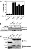
Fig. 2. Smac/DIABLO release from mitochondria is a general feature of apoptosis. Jurkat cells, either untreated or treated with 250 nM staurosporine (STS), 10 µM actinomycin D (Act. D) or 10 µM daunorubicin (Dauno), were harvested at the indicated timepoints. (A) The percentage of cells exhibiting apoptotic features was determined as described in Materials and methods. (B) Cells (107) were subjected to a digitonin-based subcellular fractionation procedure as described in Materials and methods. Cytosolic fractions (∼50 µg per lane) were subjected to SDS–PAGE, followed by immunoblotting with the indicated antibodies. (C) Cytosolic and pellet fractions prepared from the same cells were subjected to SDS–PAGE, followed by probing with cytochrome c and Smac/DIABLO-specific antibodies, as indicated.
None
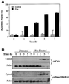
Fig. 3. Timecourse analysis of Fas-induced cytochrome c and Smac/DIABLO release. Jurkat cells, treated with 100 ng/ml of the agonistic anti-Fas antibody CH-11, were harvested at the indicated timepoints. (A) The percentage of cells exhibiting apoptotic features was determined as described in Materials and methods. (B) Subcellular fractions were subjected to SDS–PAGE, followed by immunoblotting with anti-cytochrome c and anti-Smac/DIABLO antibodies, as indicated.
None
Fig. 4. Bcl-2 blocks Smac/DIABLO translocation. CEM cells stably transfected with either vector (left panels) or a Bcl-2 expression plasmid (right panels) were treated with 250 nM staurosporine (STS), 10 µM actinomycin D (Act. D) or UVB-irradiated for 90 s on a transilluminator (UVB). Cells were harvested after 7 h and subjected to subcellular fractionation as described in Materials and methods. Cytosolic and pellet fractions were subjected to SDS–PAGE, followed by immunoblotting with the indicated antibodies. Pellet fractions were probed with a Bcl-2-specific antibody to indicate the relative levels of endogenous versus overexpressed Bcl-2.
None
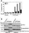
Fig. 5. Timecourse analysis of Bcl-2-regulated Smac/DIABLO release. CEM.vector and CEM.Bcl-2 cells were UVB-irradiated for 90 s (A and B) or treated with 10 µM actinomycin D (C and D) and harvested at the indicated timepoints. (A) The percentage of cells exhibiting apoptotic features was determined as described in Materials and methods. (B) Cytosolic and pellet subcellular fractions were subjected to SDS–PAGE and immunoblotted with the indicated antibodies. Cytosolic fractions were also probed with an antibody that recognizes only the active form of caspase-9. (C) The percentage of cells exhibiting apoptotic features was determined as described in Materials and methods. (D) Cytosolic fractions from the same cells were subjected to SDS–PAGE, followed by immunoblotting with anti-cytochrome c and Smac/DIABLO-specific antibodies, as indicated. Cytosols were also probed for active caspase-9 as described in (B).
None
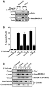
Fig. 6. Smac/DIABLO release requires active caspases. (A) Jurkat cells, pre-incubated in medium containing z-VAD-fmk (50 µM) where indicated, were induced to undergo apoptosis by treatment with 100 ng/ml anti-Fas (CH-11). Cytosolic and pellet fractions were prepared and subjected to SDS–PAGE, followed by immunoblotting with the indicated antibodies. (B) Jurkat cells were induced to undergo apoptosis (in the presence or absence of 50 µM z-VAD-fmk) by treatment with 10 µM daunorubicin (Dauno), 10 µM actinomycin D (Act. D) or UVB-irradiation (90 s) (UVB) for 7 h. The percentage of cells exhibiting apoptotic features was determined as described in Materials and methods. (C) Cytosolic fractions from the same cells were subjected to SDS–PAGE, followed by immunoblotting with the indicated antibodies. (D) Jurkat cells, pre-treated with 50 µM z-VAD-fmk where indicated, were induced to undergo apoptosis by the addition of 20 µM actinomycin D to culture media and harvested at the indicated timepoints. The percentage of cells exhibiting apoptotic features was determined as described in Materials and methods. (E) Cytosolic and pellet fractions from the same cells were subjected to SDS–PAGE, followed by immunoblotting with the indicated antibodies.
None
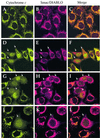
Fig. 7. Immunolocalization of Smac/DIABLO and cytochrome c in HeLa cells. (AC) Viable (non-apoptotic) HeLa cells were stained with antibodies to cytochrome c (green) and Smac/DIABLO (red) as indicated. Note that both proteins clearly co-localize within punctate, mitochondrial structures. (DF) HeLa cells, pre-incubated with z-VAD-fmk (100 µM), were UV-irradiated followed by further incubation for 8 h. Note the cells where cytochrome c has exited mitochondria and can be seen in a diffuse pattern throughout the cell, including the nucleus (white arrows). In the latter case, note the retention of Smac/DIABLO in punctate mitochondrial structures within the cytoplasm. (GI) HeLa cells, pre-incubated with z-VAD-fmk (100 µM), were then treated with 10 µM actinomycin D for 8 h. Once again note the retention of Smac/DIABLO in mitochondrial structures. (JL) HeLa cells were treated as described in (G–I). A close-up of a single cell that has undergone mitochondrial cytochrome c release, but where Smac/DIABLO is retained within mitochondria, is shown.
None
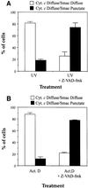
Fig. 8. Quantitative analysis of cytochrome c versus Smac/DIABLO release in apoptotic cells. HeLa cells, pre-incubated where indicated in the presence of 100 µM z-VAD-fmk, were either UV-irradiated (A), or treated with 10 µM actinomycin D (B). Upon appearance of apoptotic cells in the cultures (8 h after UV-irradiation or actinomycin D treatment) cells were immunostained with anti-cytochrome c and anti-Smac/DIABLO antibodies as described in Materials and methods. Cells exhibiting a diffuse (extra-mitochondrial) cytochrome c staining pattern were evaluated for their pattern of Smac/DIABLO staining (diffuse or punctate). Open bars represent the proportion of cells that exhibited doubly diffuse cytochrome c and Smac/DIABLO staining patterns. Filled bars indicate the percentage of cells that exhibited diffuse cytochrome c staining but retained punctate (mitochondrial) Smac/DIABLO staining patterns. Error bars denote the standard error of the mean of three fields of at least 100 cells within a representative experiment.
None
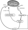
Fig. 9. Hypothetical model of Bcl-2- and caspase-regulated Smac/DIABLO release from mitochondria. The capacity of Bcl-2 to regulate Smac/DIABLO release from mitochondria may derive from its ability to block cytochrome c efflux. Once released from mitochondria, cytochrome c promotes the assembly of the apoptosome, which results in caspase-9 activation and the propagation of a caspase cascade. Under these conditions, XIAP may attenuate caspase activity by integration into the apoptosome complex, or repress the activity of active executioner caspases-3 and -7. As a counter-measure, caspase-mediated attack of a (unidentified) mitochondrial component may result in the opening of a Smac/DIABLO-conducting pore. Once released into the cytosol, Smac/DIABLO may enhance caspase activity by binding to IAPs, including XIAP, thereby neutralizing their caspase-inhibitory properties.

Similar articles

Cited by

References

    1. Adrain C. and Martin,S.J. (2001) The mitochondrial apoptosome: a killer unleashed by the cytochrome seas. Trends Biochem. Sci., 26, 390–397. - PubMed
    1. Adrain C., Slee,E.A., Harte,M.T. and Martin,S.J. (1999) Regulation of apoptotic protease activating factor-1 oligomerization and apoptosis by the WD-40 repeat region. J. Biol. Chem., 274, 20855–20860. - PubMed
    1. Antonsson B., Montessuit,S., Lauper,S., Eskes,R. and Martinou,J.C. (2000) Bax oligomerization is required for channel-forming activity in liposomes and to trigger cytochrome c release from mitochondria. Biochem. J., 345, 271–278. - PMC - PubMed
    1. Antonsson B., Montessuit,S., Sanchez,B. and Martinou,J.C. (2001) Bax is present as a high molecular weight oligomer/complex in the mitochondrial membrane of apoptotic cells. J. Biol. Chem., 276, 11615–11623. - PubMed
    1. Bossy-Wetzel E. and Green,D.R. (1999) Caspases induce cytochrome c release from mitochondria by activating cytosolic factors. J. Biol. Chem., 274, 17484–17490. - PubMed

Publication types

MeSH terms