. 2001 Dec;75(24):12209-19.
doi: 10.1128/JVI.75.24.12209-12219.2001.

Intracellular trafficking of the UL11 tegument protein of herpes simplex virus type 1

Affiliations

Intracellular trafficking of the UL11 tegument protein of herpes simplex virus type 1

J S Loomis et al. J Virol. 2001 Dec.

Abstract

Growing evidence indicates that herpes simplex virus type 1 (HSV-1) acquires its final envelope in the trans-Golgi network (TGN). During the envelopment process, the viral nucleocapsid as well as the envelope and tegument proteins must arrive at this site in order to be incorporated into assembling virions. To gain a better understanding of how these proteins associate with cellular membranes and target to the correct compartment, we have been studying the intracellular trafficking properties of the small tegument protein encoded by the U(L)11 gene of HSV-1. This 96-amino-acid, myristylated protein accumulates on the cytoplasmic face of internal membranes, where it is thought to play a role in nucleocapsid envelopment and egress. When expressed in the absence of other HSV-1 proteins, the UL11 protein localizes to the Golgi apparatus, and previous deletion analyses have revealed that the membrane-trafficking information is contained within the first 49 amino acids. The goal of this study was to map the functional domains required for proper Golgi membrane localization. In addition to N-terminal myristylation, which allows for weak membrane binding, UL11 appears to be palmitylated on one or more of three consecutive N-terminal cysteines. Using membrane-pelleting experiments and confocal microscopy, we show that palmitylation of UL11 is required for both Golgi targeting specificity and strong membrane binding. Furthermore, we found that a conserved acidic cluster within the first half of UL11 is required for the recycling of this tegument protein from the plasma membrane to the Golgi apparatus. Taken together, our results demonstrate that UL11 has highly dynamic membrane-trafficking properties, which suggests that it may play multiple roles on the plasma membrane as well as on the nuclear and TGN membranes.


Figures
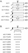
FIG. 1
FIG. 1
Mutational analysis of the acidic cluster region of UL11. (A) Inactivation of the wild-type sequence. The 96-amino-acid UL11 sequence (open rectangles) is shown with the position of its acidic cluster (AC) (solid black box). N-terminal myristylation (wavy line) is a natural modification of UL11 and was also provided by the first 10 amino acids of the Src oncoprotein (solid rectangle with wavy line). All constructs have the GFP protein (open oval) fused to their C termini. (B) Replacement with foreign acidic cluster sequences. The wild-type sequence of the UL11 acidic cluster is shown (residues 37 to 43). Wild-type and mutant acidic cluster sequences from furin and Nef were inserted in place of the UL11 sequence as shown. All constructs were expressed as UL11-GFP chimeras.
FIG. 2
FIG. 2
Expression of UL11 mutants with inactivated acidic clusters. (A) Biochemical analysis. A7 melanoma cells transfected with the indicated constructs were labeled for 2.5 h with l-[35S]methionine, and UL11-GFP chimeras were immunoprecipitated from cell lysates with a polyclonal antibody specific for GFP. Proteins were separated by SDS-PAGE and visualized by autoradiography. The position of the 45-kDa molecular mass marker is indicated. (B) Subcellular localization. Plasmids were transfected into A7 cells as in panel A, and UL11-GFP chimeras were visualized by live-cell confocal microscopy at approximately 18 h following transfection.
FIG. 2
FIG. 2
Expression of UL11 mutants with inactivated acidic clusters. (A) Biochemical analysis. A7 melanoma cells transfected with the indicated constructs were labeled for 2.5 h with l-[35S]methionine, and UL11-GFP chimeras were immunoprecipitated from cell lysates with a polyclonal antibody specific for GFP. Proteins were separated by SDS-PAGE and visualized by autoradiography. The position of the 45-kDa molecular mass marker is indicated. (B) Subcellular localization. Plasmids were transfected into A7 cells as in panel A, and UL11-GFP chimeras were visualized by live-cell confocal microscopy at approximately 18 h following transfection.
FIG. 3
FIG. 3
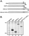
Recovery of CD4-UL11 chimeras from the plasma membrane. (A) CD4-UL11 constructs. The 458-amino-acid human CD4 protein (shaded rectangle) contains a large extracellular domain, a hydrophobic transmembrane domain (tm), and a short cytoplasmic domain. The wild-type UL11 sequence or the acidic cluster (AC) deletion mutant (open rectangles) was attached to the CD4 protein in place of the last 29 residues of the cytoplasmic tail, but in this case, GFP was not included. (B) Biochemical analysis. A7 cells were transfected and metabolically labeled with [35S]methionine for 2.5 h. The CD4 derivatives were immunoprecipitated with a monoclonal antibody specific for CD4, separated by SDS-PAGE, and visualized by autoradiography. The positions of molecular mass markers (in kilodaltons) are indicated. (C) Subcellular localization. A7 cells transfected with the indicated constructs were incubated with a monoclonal antibody specific for CD4 for 40 min on ice. After excess antibody was washed away, the cells were shifted to 37°C for 60 min to allow for endocytosis. To detect internalized antibody, the cells were fixed, permeabilized, stained with fluorescein isothiocyanate-labeled secondary antibody, and visualized by confocal microscopy.
FIG. 3
FIG. 3
Recovery of CD4-UL11 chimeras from the plasma membrane. (A) CD4-UL11 constructs. The 458-amino-acid human CD4 protein (shaded rectangle) contains a large extracellular domain, a hydrophobic transmembrane domain (tm), and a short cytoplasmic domain. The wild-type UL11 sequence or the acidic cluster (AC) deletion mutant (open rectangles) was attached to the CD4 protein in place of the last 29 residues of the cytoplasmic tail, but in this case, GFP was not included. (B) Biochemical analysis. A7 cells were transfected and metabolically labeled with [35S]methionine for 2.5 h. The CD4 derivatives were immunoprecipitated with a monoclonal antibody specific for CD4, separated by SDS-PAGE, and visualized by autoradiography. The positions of molecular mass markers (in kilodaltons) are indicated. (C) Subcellular localization. A7 cells transfected with the indicated constructs were incubated with a monoclonal antibody specific for CD4 for 40 min on ice. After excess antibody was washed away, the cells were shifted to 37°C for 60 min to allow for endocytosis. To detect internalized antibody, the cells were fixed, permeabilized, stained with fluorescein isothiocyanate-labeled secondary antibody, and visualized by confocal microscopy.
FIG. 4
FIG. 4
Phosphorylation of UL11. (A) Biochemical analysis. Duplicate plates of COS-1 cells were transfected with the indicated constructs. Approximately 18 h later, cells were labeled with either [35S]methionine for 2.5 h or [32P]orthophosphate for 16 h. The labeled proteins were immunoprecipitated with anti-GFP serum, subjected to SDS-PAGE analysis, and visualized by autoradiography. (B) Subcellular localization. The indicated constructs were transfected into A7 cells, and 18 h later, the sUL11-GFP chimeras were visualized by confocal microscopy.
FIG. 4
FIG. 4
Phosphorylation of UL11. (A) Biochemical analysis. Duplicate plates of COS-1 cells were transfected with the indicated constructs. Approximately 18 h later, cells were labeled with either [35S]methionine for 2.5 h or [32P]orthophosphate for 16 h. The labeled proteins were immunoprecipitated with anti-GFP serum, subjected to SDS-PAGE analysis, and visualized by autoradiography. (B) Subcellular localization. The indicated constructs were transfected into A7 cells, and 18 h later, the sUL11-GFP chimeras were visualized by confocal microscopy.
FIG. 5
FIG. 5
Expression of UL11 chimeras having foreign acidic cluster sequences. (A) Biochemical analysis. A7 cells were transfected with the indicated constructs and labeled with [35S]methionine for 2.5 h. The labeled proteins were immunoprecipitated with anti-GFP serum, subjected to SDS-PAGE analysis, and visualized by autoradiography. (B) Subcellular localization. Constructs were transfected into A7 cells, and 18 h later, the UL11-GFP derivatives were visualized by confocal microscopy.
FIG. 5
FIG. 5
Expression of UL11 chimeras having foreign acidic cluster sequences. (A) Biochemical analysis. A7 cells were transfected with the indicated constructs and labeled with [35S]methionine for 2.5 h. The labeled proteins were immunoprecipitated with anti-GFP serum, subjected to SDS-PAGE analysis, and visualized by autoradiography. (B) Subcellular localization. Constructs were transfected into A7 cells, and 18 h later, the UL11-GFP derivatives were visualized by confocal microscopy.
FIG. 6
FIG. 6
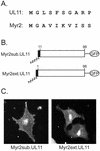
Analysis of the first 10 residues of UL11. (A) Sequence comparison. The first 10 amino acids of the UL11 protein and a myristylated form of the Rous sarcoma virus Gag protein are shown. The glycine residue at the second position is the site of myristylation for both proteins, but otherwise the sequences are highly divergent. (B) Myr2-UL11 chimeras. The first 10 residues of the Myr2.Gag protein (black rectangle) and its associated myristate (wavy line) were either substituted in place of the first 10 residues of UL11 (Myr2sub.UL11) or attached to the N terminus as an extension (Myr2ext.UL11). Both constructs have the GFP protein (open oval) fused to their C termini. (C) Subcellular localization. The constructs were transfected into A7 cells, and 18 h later, the UL11-GFP chimeras were visualized by live-cell confocal microscopy.
FIG. 7
FIG. 7
Palmitylation of UL11. (A) Mutational analysis of the CCC motif. The wild-type UL11 protein (top) is myristylated (wavy line) and contains the acidic cluster (AC) (residues 37 to 43, indicated by a black box) along with a cluster of three cysteines (residues 11 to 13). An N-terminal 10-amino-acid sequence from the Fyn protein (hatched box) is known to be sufficient for both myristylation and palmitylation (double wavy line). All constructs have the GFP protein (open oval) fused to their C termini. (B) Subcellular localization. The indicated constructs were transfected into COS-1 cells, and 18 h later, the UL11-GFP chimeras were visualized by confocal microscopy. (C) Biochemical analysis. Transfected COS-1 cells were labeled with either [35S]methionine for 2.5 h, [3H]myristic acid for 10 min, or [3H]palmitic acid for 60 min. Cell lysates were prepared, and UL11-GFP proteins were immunoprecipitated, mixed with sample buffer (without β-mercaptoethanol), resolved by SDS-PAGE, and visualized by autoradiography.
FIG. 7
FIG. 7
Palmitylation of UL11. (A) Mutational analysis of the CCC motif. The wild-type UL11 protein (top) is myristylated (wavy line) and contains the acidic cluster (AC) (residues 37 to 43, indicated by a black box) along with a cluster of three cysteines (residues 11 to 13). An N-terminal 10-amino-acid sequence from the Fyn protein (hatched box) is known to be sufficient for both myristylation and palmitylation (double wavy line). All constructs have the GFP protein (open oval) fused to their C termini. (B) Subcellular localization. The indicated constructs were transfected into COS-1 cells, and 18 h later, the UL11-GFP chimeras were visualized by confocal microscopy. (C) Biochemical analysis. Transfected COS-1 cells were labeled with either [35S]methionine for 2.5 h, [3H]myristic acid for 10 min, or [3H]palmitic acid for 60 min. Cell lysates were prepared, and UL11-GFP proteins were immunoprecipitated, mixed with sample buffer (without β-mercaptoethanol), resolved by SDS-PAGE, and visualized by autoradiography.
FIG. 8
FIG. 8
Membrane binding of UL11 derivatives. Transfected A7 cells were harvested and washed in NTE buffer and then allowed to swell for 1 h in hypotonic buffer. Cells were disrupted by Dounce homogenization, and nuclei were removed by a low-speed spin. To separate membrane-bound molecules from soluble forms, the supernatants were subjected to centrifugation at 100,000 × g. The soluble (S) and pellet (P) fractions were collected and analyzed for the presence of UL11 protein by fluorometry (A) or Western blot analysis (B). The fraction pelleted was calculated by dividing the amount pelleted by the total amount of protein (soluble plus pellet). Error bars indicate standard deviations.
FIG. 8
FIG. 8
Membrane binding of UL11 derivatives. Transfected A7 cells were harvested and washed in NTE buffer and then allowed to swell for 1 h in hypotonic buffer. Cells were disrupted by Dounce homogenization, and nuclei were removed by a low-speed spin. To separate membrane-bound molecules from soluble forms, the supernatants were subjected to centrifugation at 100,000 × g. The soluble (S) and pellet (P) fractions were collected and analyzed for the presence of UL11 protein by fluorometry (A) or Western blot analysis (B). The fraction pelleted was calculated by dividing the amount pelleted by the total amount of protein (soluble plus pellet). Error bars indicate standard deviations.

Similar articles

Cited by

References

    1. Alconada A, Bauer U, Hoflack B. A tyrosine-based motif and a casein kinase II phosphorylation site regulate the intracellular trafficking of the varicella-zoster virus glycoprotein I, a protein localized in the trans-Golgi network. EMBO J. 1996;15:6096–6110. - PMC - PubMed
    1. Baines J D, Jacob R J, Simmerman L, Roizman B. The herpes simplex virus 1 UL11 proteins are associated with cytoplasmic and nuclear membranes and with nuclear bodies of infected cells. J Virol. 1995;69:825–833. - PMC - PubMed
    1. Baines J D, Roizman B. The UL11 gene of herpes simplex virus 1 encodes a function that facilitates nucleocapsid envelopment and egress from cells. J Virol. 1992;66:5168–5174. - PMC - PubMed
    1. Berthiaume L, Resh M D. Biochemical characterization of a palmitoyl acyltransferase activity that palmitoylates myristoylated proteins. J Biol Chem. 1995;270:22399–22405. - PubMed
    1. Bowzard J B, Visalli R J, Wilson C B, Loomis J S, Callahan E M, Courtney R J, Wills J W. Membrane targeting properties of a herpesvirus tegument protein-retrovirus Gag chimera. J Virol. 2000;74:8692–8699. - PMC - PubMed

Publication types

LinkOut - more resources