. 2001 Nov;13(11):2525-37.
doi: 10.1105/tpc.010231.

Repression of the defense gene PR-10a by the single-stranded DNA binding protein SEBF

Affiliations

Repression of the defense gene PR-10a by the single-stranded DNA binding protein SEBF

B Boyle et al. Plant Cell. 2001 Nov.

Abstract

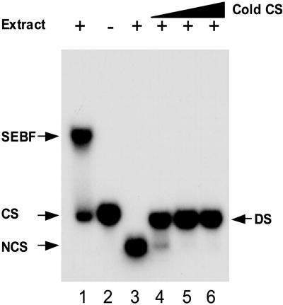
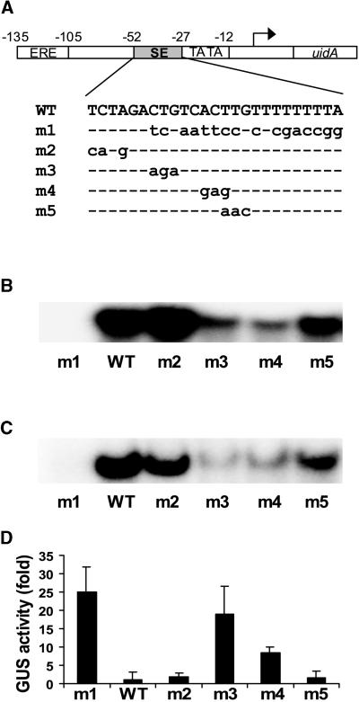
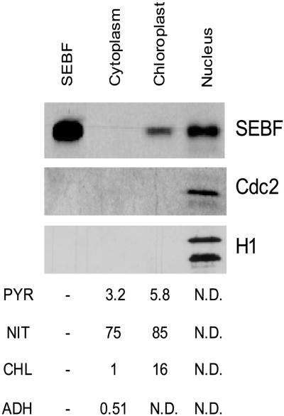
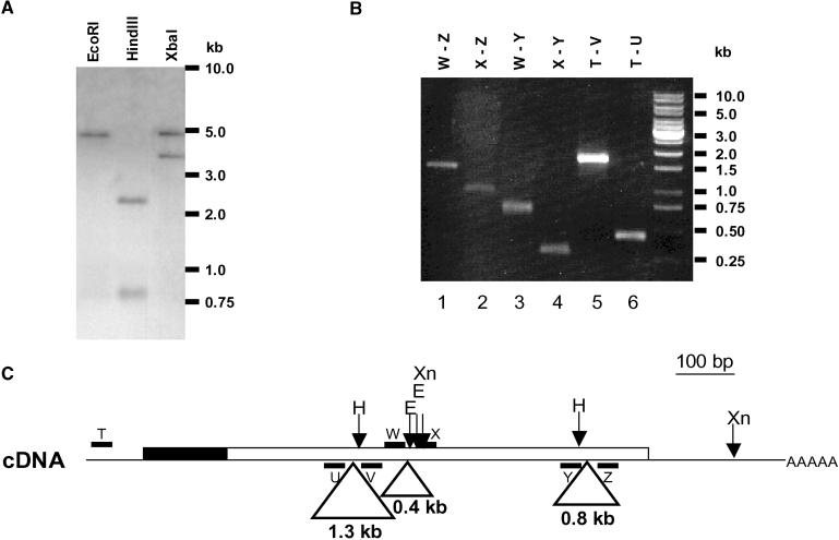
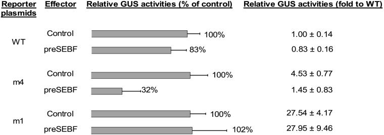
The potato pathogenesis-related gene PR-10a is transcriptionally activated in response to pathogen infection or elicitor treatment. Characterization of the cis-acting elements of the PR-10a promoter revealed the presence of a silencing element between residues -52 and -27 that contributes to transcriptional regulation. In this study, we have isolated a silencing element binding factor (SEBF) from potato tuber nuclei that binds to the coding strand of the silencing element in a sequence-specific manner. The consensus binding site of SEBF, PyTGTCNC, is present in a number of PR genes and shows striking similarity to the auxin response element. Mutational analysis of the PR-10a promoter revealed an inverse correlation between the in vitro binding of SEBF and the expression of PR-10a. SEBF was purified to homogeneity from potato tubers, and sequencing of the N terminus of the protein led to the isolation of a cDNA clone. Sequence analysis revealed that SEBF is homologous with chloroplast RNA binding proteins that possess consensus sequence-type RNA binding domains characteristic of heterogeneous nuclear ribonucleoproteins (hnRNPs). Overexpression of SEBF in protoplasts repressed the activity of a PR-10a reporter construct in a silencing element-dependent manner, confirming the role of SEBF as a transcriptional repressor.


Figures

Figure 1.
Figure 1.
SEBF Binds Single-Stranded SE. EMSA was performed with 10 μg of crude nuclear preparation and 20,000 cpm of 32P-labeled SE coding strand (CS; lane 1) or noncoding strand (NCS; lane 3). The double-stranded probe (DS) was made by annealing radiolabeled noncoding and nonlabeled (cold) coding strands. The ratio of CS to NCS is 0.75:1, 1.5:1, and 3:1 for lanes 4, 5, and 6, respectively. No extract was added in lane 2. Arrows indicate the positions of the CS, NCS, and DS probes and of the SEBF shift in the gel.
Figure 2.
Figure 2.
Mutational Analysis of the SE. (A) Scheme of the reporter constructs and mutant oligonucleotides used in this study. Reporter constructs contain the PR-10a promoter region from −135 to +136 fused to the bacterial gene uidA encoding β-glucuronidase. The positions of the ERE, the SE, and the TATA box are shown. The transcriptional start site is indicated with an arrow. The common sequence between the wild-type (WT) and mutant oligonucleotides is indicated by dashed lines, and mutated nucleotides are represented by lowercase letters. (B) EMSA studies using 10 μg of crude potato tuber nuclear preparation and 20,000 cpm of the 32P-labeled single-stranded CS oligonucleotides presented in (A). (C) EMSA studies using 10 ng of purified recombinant mature SEBF and 20,000 cpm of the 32P-labeled single-stranded CS oligonucleotides presented in (A). (D) The sequence from −52 to −27 of the PR-10a promoter fused to the uidA gene was replaced with the sequences presented in (A). The resulting plasmids were electroporated in potato leaf protoplasts, and the β-glucuronidase (GUS) activity was measured. The histogram represents fold activity to wild type (WT = 1). Transfection efficiencies were corrected by coelectroporating a luciferase reporter gene. Results represent the mean from a minimum of six individual electroporations. Error bars indicate ±sem.
Figure 3.
Figure 3.
Determination of the Consensus SEBF Binding Site. (A) Two-by-two mutational analysis of the SEBF binding site. (B) Single-nucleotide analysis of the SEBF binding site. Oligonucleotides used for fine mapping of the SEBF binding site are listed in each panel. The common sequence between wild-type (WT) and mutant oligonucleotides is indicated by dashed lines, and mutated nucleotides are represented by lowercase letters. EMSA results show the binding of SEBF to oligonucleotides containing the mutated nucleotides. Studies were performed using 10 μg of crude nuclear preparation and 20,000 cpm of the 32P-labeled single- stranded oligonucleotides listed in each panel.
Figure 4.
Figure 4.
Purification of SEBF. Coomassie blue staining of proteins from each step of the purification of SEBF (lanes 1 to 4). The crude nuclear extract (Crude; lane 1) was loaded onto a Q-Sepharose column. SEBF was eluted at 400 mM NaCl (Q-Seph; lane 2) before two rounds of affinity purification (Aff.1 and Aff.2; lanes 3 and 4). Lane 5 shows the results of a DNA probed protein gel blot (Dp) experiment performed with affinity 2–purified SEBF and the wild-type coding strand as a radiolabeled probe. Arrows indicate the two purified bands. Molecular mass markers are indicated at left.
Figure 5.
Figure 5.
Sequence of SEBF and Alignment with a Family of Nucleus-Encoded Chloroplast RNA Binding Proteins. The amino acid sequence derived from the SEBF cDNA is presented. A member of each group of nucleus-encoded chloroplast RNA binding proteins was chosen for sequence comparison (cp29A from group I, GenBank accession number Q08935; cp31 from group II, GenBank accession number P19683; cp33 from group III, GenBank accession number P19684). Amino acids obtained through N-terminal sequencing of the purified protein are indicated by stars. The beginning of the mature form of SEBF is indicated by the arrowhead above the sequence. The putative transit peptide (T), the acidic region (A), and the cs-RBDs (RI and RII) are underlined. The conserved amino acids that define the cs-RBD are indicated by triangles below the sequence. Black boxes indicate sequence identity, and gray shading indicates conservative substitutions.
Figure 6.
Figure 6.
Cellular Localization of SEBF. Potato leaves were fractionated into cytoplasm, nuclei, and chloroplasts. Ten micrograms of each fraction were separated by 12% SDS-PAGE. Proteins were transferred to nitrocellulose, and the presence of SEBF and the nuclear proteins histone H1 and Cdc2 was revealed with specific antibodies. The first lane (10 ng of recombinant mature SEBF) shows the protein that served to immunize the rabbits. Chlorophyll (CHL), nitrite reductase (NIT), and alkaline pyrophosphatase (PYR) were used as chloroplastic markers (data are presented as μg chlorophyll·mg−1 protein, nmol nitrite·mg−1 protein·min−1, and μmol PO4·mg−1 protein·min−1, respectively), and alcohol dehydrogenase (ADH) was used as a cytoplasmic marker (data are presented as increased OD·mg−1 protein·min−1). N.D., not detectable; −, not determined.
Figure 7.
Figure 7.
Genomic Organization of SEBF. (A) DNA gel blot analysis of SEBF. Five micrograms of digested genomic DNA was loaded per lane and probed with a randomly labeled XmnI fragment from the SEBF cDNA shown in (C). Molecular mass markers are indicated at right. (B) PCR analysis of the genomic DNA. The positions of the oligonucleotides (T to Z; lanes 1 to 6) on the cDNA are presented in (C). The differences in size between the amplification products of lanes 5 and 6, lanes 3 and 4, and lanes 2 and 4 define the sizes of introns 1, 2, and 3, respectively. Molecular mass markers are indicated at right. (C) Deduced genomic organization of SEBF. The cDNA is represented as a line, and the coding region is represented as a box. The N-terminal transit sequence is presented in black. Introns are indicated as triangles emerging from the cDNA. The oligonucleotides used in the PCR reactions are designated by letters (T, U, V, W, X, Y, Z). The positions and sizes of the introns are deduced from the PCR analysis (B). Restriction sites are indicated by arrows: H, HindIII; E, EcoRI; Xn, XmnI.
Figure 8.
Figure 8.
Overexpression of SEBF Represses PR-10a Expression. The coding sequence of pre-SEBF and the coding sequence of a control protein were each inserted into plasmid pBI223D (effector plasmids). These plasmids were coelectroporated in potato leaf protoplasts with the reporter plasmids described in Figure 2D (reporter plasmids). The histogram represents the effect of pre-SEBF overexpression on reporter activity compared with the overexpression of the control protein (control = 100). Fold activity to wild-type SE (WT = 1) is presented for easier reference to Figure 2D. Transfection efficiencies were corrected by coelectroporating a luciferase reporter gene. Results represent means from a minimum of three individual electroporations. GUS, β-glucuronidase. Error bars indicate ±sem.

Similar articles

Cited by

References

    1. Arnon, D.I. (1949). Copper enzymes in isolated chloroplasts: Polyphenoloxidase in Beta vulgaris. Plant Physiol. 24 1–15. - PMC - PubMed
    1. Ausubel, F.M., Brent, R., Kingston, R.E., Moore, D.D., Seidman, J.G., Smith, J.A., and Struhl, K. (2001). Current Protocols in Molecular Biology. (New York: Wiley).
    1. Barnes, W.M. (1990). Variable patterns of expression of luciferase in transgenic tobacco leaves. Proc. Natl. Acad. Sci. USA 87 9183–9187. - PMC - PubMed
    1. Blumwald, E., Gilad, S.A., and Lam, B.C.H. (1998). Early signal transduction pathways in plant–pathogen interactions. Trends Plant Sci. 3 342–346.
    1. Brisson, N., Giroux, H., Zollinger, M., Camirand, A., and Simard, C. (1989). Maturation and subcellular compartmentation of potato starch phosphorylase. Plant Cell 1 559–566. - PMC - PubMed

Publication types

MeSH terms

Associated data