. 2001 Nov;12(11):3375-85.
doi: 10.1091/mbc.12.11.3375.

The N-terminal domain of the t-SNARE Vam3p coordinates priming and docking in yeast vacuole fusion

Affiliations
Free PMC article

The N-terminal domain of the t-SNARE Vam3p coordinates priming and docking in yeast vacuole fusion

R Laage et al. Mol Biol Cell. 2001 Nov.
Free PMC article

Abstract

Homotypic fusion of yeast vacuoles requires a regulated sequence of events. During priming, Sec18p disassembles cis-SNARE complexes. The HOPS complex, which is initially associated with the cis-SNARE complex, then mediates tethering. Finally, SNAREs assemble into trans-complexes before the membranes fuse. The t-SNARE of the vacuole, Vam3p, plays a central role in the coordination of these processes. We deleted the N-terminal region of Vam3p to analyze the role of this domain in membrane fusion. The truncated protein (Vam3 Delta N) is sorted normally to the vacuole and is functional, because the vacuolar morphology is unaltered in this strain. However, in vitro vacuole fusion is strongly reduced due to the following reasons: Assembly, as well as disassembly of the cis-SNARE complex is more efficient on Vam3 Delta N vacuoles; however, the HOPS complex is not associated well with the Vam3 Delta N cis-complex. Thus, primed SNAREs from Vam3 Delta N vacuoles cannot participate efficiently in the reaction because trans-SNARE pairing is substantially reduced. We conclude that the N-terminus of Vam3p is required for coordination of priming and docking during homotypic vacuole fusion.


Figures
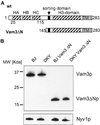
Figure 1
Figure 1
Deletion of the N-terminal domain of Vam3p. (A) Domain structure of wild-type Vam3p according to coiled-coil prediction (SwissProt Expasy “coils”) and the NMR structure (Dulubova et al., 2001). The approximate sites of coiled-coil domains are indicated by striped boxes, transmembrane domains (TM) by gray boxes, and the sorting domain (amino acids 154–160) by black boxes. Vam3ΔN is deleted from amino acids 1–145. (B) Expression and sorting of the truncated Vam3p. Vacuoles were prepared as described and a fraction (10 μg) of each wild-type and mutant tester strain was analyzed by SDS-PAGE followed by Western blotting. Immunoblots were decorated with antibodies to Vam3p and Nyv1p.
Figure 2
Figure 2
Vacuolar morphology is unaltered in the Vam3ΔN mutant. Wild-type or mutant BJ3505 strains were incubated with 10 μM of the dye FM4–64 (Molecular Probes, Eugene, OR) for 20 min at 30°C in YPD. Cells were centrifuged briefly (1 min at 5000 × g), washed twice with YPD medium, and chased for 15 min at 30°C (Vida and Emr, 1995). Stained vacuoles were analyzed in a standard fluorescence microscope. (A and B) BJ3505 wild-type cells; (C and D) BJ3505 VAM3ΔN cells; (E and F) BJ3505 vam3Δ cells as a negative control.
Figure 3
Figure 3
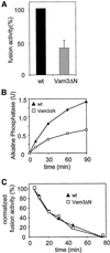
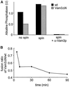
Decreased fusion of Vam3ΔN vacuoles. (A) Fusion activity of Vam3ΔN- compared with wild-type-vacuoles. Standard fusion reactions with cytosol and ATP were incubated for 90 min at 26°C, and alkaline phosphatase activity was determined as described in MATERIALS AND METHODS. To allow comparison of independent experiments, wild-type vacuole fusion was set to 100%. Fusion of Vam3ΔN vacuoles was reduced to 40 ± 11% (mean ± SEM, n = 14). (B) Time course of the fusion reaction. Standard fusion reactions (150 μl) containing wild-type or Vam3ΔN tester vacuoles were started with cytosol and ATP. At the indicated time points a 30-μl aliquot was removed and placed on ice to stop the reaction. Fusion activity was measured after 90 min. A representative experiment is shown. (C) Loss of fusion competence over time. Wild-type or mutant BJ3505 and DKY6281 vacuoles were separately incubated with cytosol and ATP at 26°C. At the indicated time points 20 μl of BJ3505 and DKY6281 vacuoles were mixed and incubated for an additional 70 min. Then fusion activity was assayed.
Figure 4
Figure 4
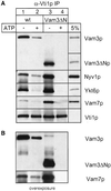
Vam3ΔN assembles into cis-SNARE complexes. Wild-type or mutant vacuoles (60 μg each) were incubated in reaction buffer with cytosol, with or without ATP at 26°C. After 10 min the vacuoles were collected by centrifugation. Solubilization and immunoprecipitation was performed as described in MATERIALS AND METHODS. Immunoprecipitation was done with a polyclonal anti-Vti1p antiserum covalently coupled to Protein A–Sepharose beads (Ungermann et al., 1999). Proteins were eluted with 0.1 M glycine, pH 2.6, precipitated with TCA, separated on 15% SDS-polyacrylamide gels, and detected with the indicated antibodies (A). A fraction of the wild-type detergent extract (5%; for Vam3p, Vam7p, Vti1p, and Ykt6p) as well as a fraction of the mutant extract (Vam3ΔN) was precipitated and loaded as a controls. The amount of coprecipitated SNAREs was quantified densitometrically (NIH image 1.6): wild-type − ATP, 100 ± 16; wild-type + ATP, 46 ± 10; Vam3ΔN − ATP, 225 ± 14; Vam3ΔN + ATP, 24 ± 4 (mean density after background subtraction in arbitrary units ± SEM, n = 4). (B) An overexposure of the immunoblots decorated with anti-Vam3p and anti-Vam7p.
Figure 5
Figure 5
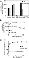
Effect of fusion inhibitors and activators. (A) Standard fusion reactions (30 μl) containing cytosol and ATP were incubated at 26°C for 90 min with or without the indicated inhibitors (64 ng/μl Gdi1p [GDI]; 10 mM BAPTA) or activators (10 μM CoA; 5 ng/μl Sec18p). To compare the results obtained for wild-type (black bars) and Vam3ΔN (gray bars) vacuoles, the fusion measured for control conditions of both strains was set to 100%. (B and C) Effect of Sec18p on the fusion reaction. Purified recombinant His6-Sec18p (Haas and Wickner, 1996) was added at the indicated concentrations to 30 μl fusion reactions to stimulate priming. The reaction mixture was incubated at 26°C for 90 min and then fusion was measured. (A and C) Representative experiments; (B) average values of three independent experiments ± SEM expressed as percentage.
Figure 6
Figure 6
Vam3ΔN vacuoles show a delay in docking. Standard fusion reactions (200 μl), containing wild-type or Vam3ΔN vacuoles, were incubated in the presence of cytosol and ATP; at the indicated time points a 30-μl aliquot was mixed with an inhibitor and incubation at 26°C was continued for a total of 90 min. Concentration of antibodies to Sec18p and Vti1p IgGs was 0.1 μg/μl, Gdi1p was used at 64 μg/ml. (A) Sensitivity to α-Sec18p of wild-type and Vam3ΔN vacuoles. Priming kinetics obtained were normalized by setting fusion at 90 min to 100%. An inlet shows the alkaline phosphatase units before normalization. Average values of nine independent experiments are shown. (B) Sensitivity to α-Vti1p or Gdi1p of wild-type and Vam3ΔN vacuoles. Average values of 20 independent experiments are shown. (C) Time point, where fusion inhibition by α-Sec18 is 50%. Wild-type 6.5 ± 1.7 min (SEM), n = 9; Vam3ΔN 6.2 ± 2.2 min (SEM), n = 9. Difference not significant (Student's t test = 0.4). (D) Time point, where fusion inhibition by α-Vti1 or Gdi1p is 50%. Wild-type 11.5 ± 2.9 min (SEM), n = 20; Vam3ΔN 15.3 ± 2.4 min (SEM), n = 20. Difference is highly significant (Student's t test < 0.00001).
Figure 7
Figure 7
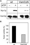
Vam3ΔN vacuoles form less trans-SNARE complexes. (A) Coimmunoprecipitation of Nyv1p with anti-Vam3p. Vacuoles from BJ3505 nyv1Δ or BJ3505 nyv1Δ VAM3ΔN were fused with DKY6281 vam3Δ as described in MATERIALS AND METHODS. After the incubation vacuoles were collected by centrifugation, solubilized and immunoprecipitated with α-Vam3p. Eluted proteins were separated by SDS-PAGE and detected by Western blotting with antibodies to Nyv1p and Vam3p. The amount of coprecipitated SNAREs was quantified densitometrically (NIH image 1.6): wild-type, 100 ± 10; Vam3ΔN, 34 ± 16 (mean density after background subtraction in arbitrary units ± SEM, n = 3). (B) A 30 μl aliquot was removed from the identical fusion reaction described in (A) and incubated for 90 min at 26°C to measure fusion activity. Fusion of BJ3505 nyv1Δ with DKY6281 vam3Δ (black bar) was set to 100%.
Figure 8
Figure 8
Efficient recruitment of HOPS/Vps33p to Vam3p depends on the N-terminal domain. Vacuoles (300 μg) prepared from BJ VPS33:: ProtA or BJ VAM3ΔN VPS33:: ProtA strains, were solubilized, and Protein A–tagged Vps33p was purified as described in MATERIALS AND METHODS. Fractions of the Protein-A-purification were precipitated with TCA, separated on a 15% SDS gel and blotted onto nitrocellulose. The Western blot probed with antibodies against Vam3p (A and B) or Vps11 (C). L = 1% of total protein loaded on the column; FT = 1% of the flow-through; E = 30% of the eluate. The left panel shows the isolation from vacuoles with full length Vam3p and the right panel the isolation from Vam3ΔN vacuoles (The high-molecular-weight band is Vps33p, which was detected by the secondary antibody because of the Protein A–tag).
Figure 9
Figure 9
Preclustering rescues the reduced fusion of Vam3ΔN vacuoles. (A) Vacuoles prepared from wild-type or Vam3ΔN tester strains were incubated in a standard fusion reaction with cytosol and ATP at 26°C. Alternatively, tester vacuoles were mixed and centrifuged for 4 min at 9000 × g at 4°C, the supernatant was removed and the vacuoles were briefly resuspended in reaction buffer containing cytosol and ATP and incubated for 90 min at 26°C. Where indicated, IgGs to Vam3p (0.1 μg/μl) were added to the reaction. Fusion activity of wild-type vacuoles was set to 100%. (B) Time course of preclustering by centrifugation. Standard fusion reactions with wild-type or Vam3ΔN vacuoles where started by incubation at 26°C, at the indicated time points an aliquot was withdrawn, centrifuged for 4 min, 9000 × g, 4°C and resuspended, incubation at 26°C was continued to a total of 90 min. To illustrate the time-dependent effect of centrifugation, the ratio of fusion signals obtained for Vam3ΔN versus wild-type vacuoles is shown.

Similar articles

Cited by

References

    1. Banta LM, Vida TA, Herman PK, Emr SD. Characterization of yeast Vps33p, a protein required for vacuolar protein sorting and vacuole biogenesis. Mol Cell Biol. 1990;10:4638–4649. - PMC - PubMed
    1. Bryant NJ, James DE. Vps45p stabilizes the syntaxin homologue Tlg2p and positively regulates SNARE complex formation. EMBO J. 2001;20:3380–3388. - PMC - PubMed
    1. Burd CG, Peterson M, Cowles CR, Emr SD. A novel Sec18p/NSF-dependent complex required for Golgi-to-endosome transport in yeast. Mol Biol Cell. 1997;8:1089–1104. - PMC - PubMed
    1. Carr CM, Grote E, Munson M, Hughson FM, Novick PJ. Sec1p binds to SNARE complexes and concentrates at sites of secretion. J Cell Biol. 1999;146:333–344. - PMC - PubMed
    1. Chen YA, Scales SJ, Patel SM, Doung YC, Scheller RH. SNARE complex formation is triggered by Ca2+ and drives membrane fusion. Cell. 1999;97:165–174. - PubMed

Publication types

MeSH terms

LinkOut - more resources