. 2001 Nov 1;20(21):5982-90.
doi: 10.1093/emboj/20.21.5982.

Recruitment of protein kinase D to the trans-Golgi network via the first cysteine-rich domain

Affiliations

Recruitment of protein kinase D to the trans-Golgi network via the first cysteine-rich domain

Y Maeda et al. EMBO J. .

Abstract

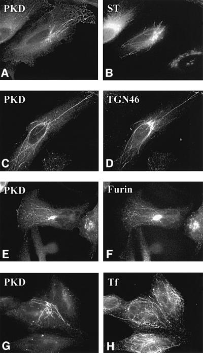
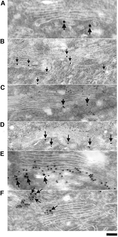
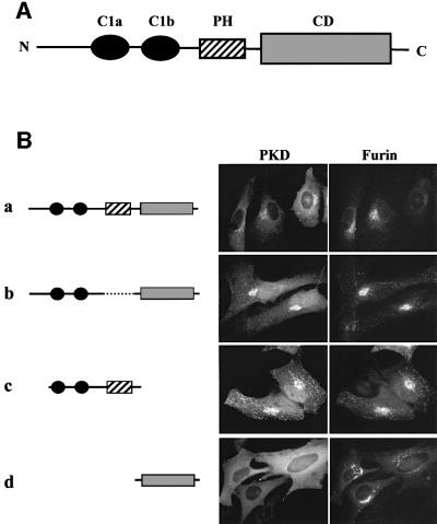
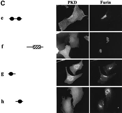
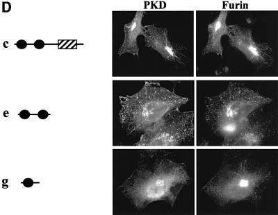
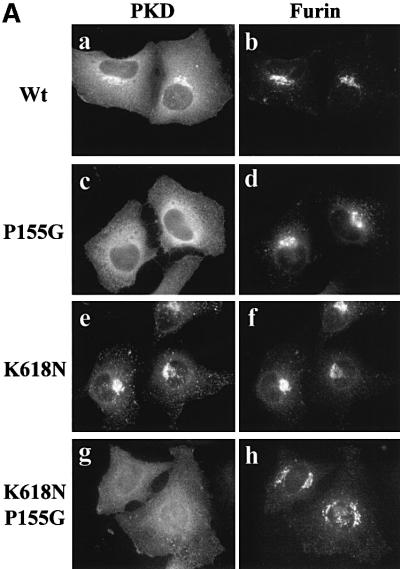
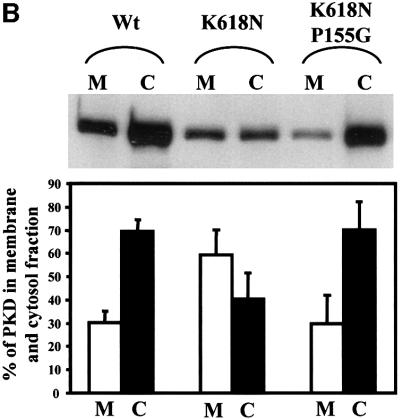
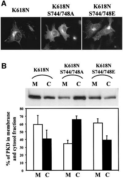
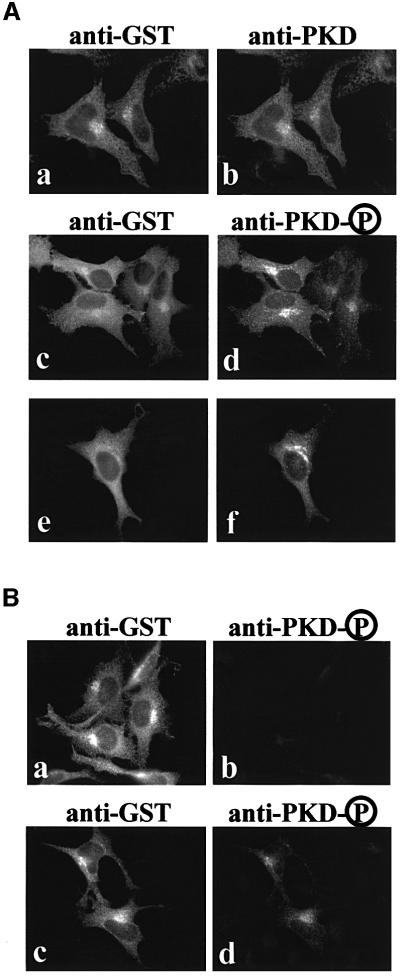
Protein kinase D (PKD) is a cytosolic protein, which upon binding to the trans-Golgi network (TGN) regulates the fission of transport carriers specifically destined to the cell surface. We have found that the first cysteine-rich domain (C1a), but not the second cysteine-rich domain (C1b), is sufficient for the binding of PKD to the TGN. Proline 155 in C1a is necessary for the recruitment of intact PKD to the TGN. Whereas C1a is sufficient to target a reporter protein to the TGN, mutation of serines 744/748 to alanines in the activation loop of intact PKD inhibits its localization to the TGN. Moreover, anti-phospho-PKD antibody, which recognizes only the activated form of PKD, recognizes the TGN-bound PKD. Thus, activation of intact PKD is important for binding to the TGN.


Figures

None
Fig. 1. Wild-type PKD localizes to TGN. HeLa cells were transfected with GST–PKD (C, D, G and H) alone or co-transfected with GFP–sialyltransferase (A and B) or EGFP–furin (E and F). The cells were treated with BFA for 6 min (A and B) or 10 min (C–H), fixed and stained with anti-phospho-PKD (A, C, E and G), anti-TGN46 (D) and anti-transferrin (H) antibodies. For early endosome staining, as described in Materials and methods, the cells were incubated with pre-incubation medium for 1 h, followed by incubation with iron-saturated transferrin for 15 min prior to BFA treatment (G and H). BFA induced tubulation of the early Golgi cisternae, TGN and the endosomes. PKD co-localizes with TGN46 (C and D) and furin (E and F), but not sialyltransferase (A and B) and transferrin (G and H). Thus, PKD is contained in the portion of the TGN containing furin and TGN46. Interestingly, under these conditions, the peripheral Golgi proteins COPI coats are detached; PKD, however, remains attached to theTGN-derived tubes.
None
Fig. 2. PKD-K618N is localized to specific domains of the TGN. HeLa cells stably transfected with GST–FLAG-tagged PKD-K618N (GF17 cells) were fixed at steady state and prepared for immunogold labeling with anti-GST antibody to visualize PKD-K618N (10 nm particles, arrows), and the Golgi markers GM130 (for cis/medial compartment), galactosyl transferase (for trans-Golgi compartment and TGN) and TGN46 (for the TGN) (5 nm particles). (A and B) GST–FLAG-tagged PKD-K618N is located to the tubular-vesicular profiles attached to the trans side of the Golgi stack and rarely to its lateral side (B). (C and D) GST–FLAG-tagged PKD-K618N does not co-localize withGM-130. Gold labeling for GST–FLAG-tagged PKD-K618N (arrows) is located on the opposite side of the Golgi stack compared with GM130 (small 5 nm gold particles). (E) GST–FLAG-tagged PKD-K618N (arrows) is partially co-localized with the galactosyl transferase (small 5 nm gold particles). (F) GST–FLAG-tagged PKD-K618N (arrows) co-localizes with TGN46 (5 nm gold particles) on the trans side of the Golgi stack. Bar: (A, E, F), 70 nm; (B), 250 nm; (C), 150 nm; (D), 300 nm.
None
Fig. 3. The first cysteine-rich domain (C1a) is sufficient for TGN localization of a reporter molecule GST. (A) Schematic presentation of the domains of PKD. PKD is composed of two cysteine-rich domains (C1a and C1b), a PH domain and a CD at the C-terminus. (B) The indicated GST-tagged PKD domains and pEGFP-furin were transiently co-expressed in HeLa cells. Two days after transfection, the cells were stained with anti-GST antibody followed by Texas Red-conjugated secondary antibody to visualize GST–PKD domains. The results show that the PH domain and the CD of PKD are not required for TGN localization. (C) The indicated GST–PKD domains and pEGFP-furin were transiently co-expressed in HeLa cells. After 2 days, the cells were stained with anti-GST antibody followed by Texas Red-conjugated secondary antibody as shown in (B). The C1a domain of PKD is sufficient for TGN localization. (D) The indicated GST–PKD domains and pEGFP-furin were co-expressed transiently in HeLa cells. In these transient transfection experiments, the levels of the proteins being expressed vary. Whereas the co-localization of the GST–PKD and pEGFP-furin is seen in all cells expressing these two proteins, cells expressing higher levels of GST–PKD domains show tubulation and the tubules contain furin.
None
Fig. 4. Proline 155 in the first cysteine-rich domain (C1a) is necessary for TGN localization. (A) HeLa cells were co-transfected with pEGFP-furin and GST–wild-type PKD (a and b), GST–PKD-P155G (c and d), GST–PKD-K618N (e and f) or GST–PKD-K618N/P155G (g and h). The cells were stained with anti-GST antibody to localize GST–PKD and the results show that replacement of proline 155 in the first CRD in wild-type and the kinase-inactive PKD prevents the recruitment of the corresponding PKD to the TGN. (B) HeLa cells transfected with GST–wild-type PKD, GST–PKD-K618N or GST–K618N/P155G were separated into the membrane (M) and the cytosolic (C) fractions. GST-tagged PKD-derivatives were affinity purified from each fraction using glutathione–beads and western blotted with anti-GST antibodies. The corresponding bands were quantitated and the results show that replacing proline with glycine causes a quantitative loss in the recruitment of PKD to the membrane fraction. These data represent the mean ± SD of three independent experiments.
None
Fig. 5. Phosphorylation of serines 744/748 in the CD is required for the recruitment of intact PKD to the TGN. (A) HeLa cells were transfected with GST–PKD-K618N, GST–PKD-K618N/S744/748A or GST–PKD-K618N/S744/748E. The cells were stained with anti-GST antibody. Replacing serines 744/748 with alanines prevents the recruitment of PKD-K618N to the TGN. (B) HeLa cells transfected with the same expression vectors shown in (A) were fractionated into cytosolic (C) and membrane (M) fractions, and the quantity of PKD contained in these fractions was determined as described in the legend to Figure 4B. The results reveal that replacement of serines 744/748 with alanines quantitatively inhibits recruitment of PKD-K618N to the membrane fractions. These data represent the mean ± SD of three independent experiments.
None
Fig. 6. The TGN-associated pool of PKD is in an active form. (A) HeLa cells were transfected with GST–wild-type PKD and double stained with anti-GST (a) and anti-PKD (b) or anti-GST (c and e) and anti-phospho-PKD (d and f) antibodies. Two different fields (c and d, e and f) are shown here for double staining with anti-GST and anti-phospho-PKD antibodies. The TGN-associated pool of PKD is highly reactive with the phospho-PKD antibody, which recognizes the activated form of PKD (d and f). (B) HeLa cells were transfected with GST–PKD-S744/748E/S916A (a and b) or GST–PKD-S744/748E (c and d) and double stained with anti-GST (a and c) and anti-phospho-PKD (b and d) antibodies. GST–PKD-S744/748E/S916A does not undergo autophosphorylation at serine 916 and is, therefore, not recognized by the phospho-PKD antibody (b), but GST antibody recognizes the TGN-attached pool of PKD (a), whereas expression of the constitutively activated form of PKD in which serines 744 and 748 are replaced with glutamic acid is recognized by both the GST and the phospho-PKD antibodies. These four images were taken under exactly the same conditions. The results confirm the specificity of the anti-phospho-PKD antibody.

Similar articles

Cited by

References

    1. Beck K.A., Buchanan,J.A., Malhotra,V. and Nelson,W.J. (1994) Golgi spectrin: identification of an erythroid β-spectrin homolog associated with the Golgi complex. J. Cell Biol., 127, 707–723. - PMC - PubMed
    1. Dieterich S., Herget,T., Link,G., Bottinger,H., Pfizenmaier,K. and Johannes,F.J. (1996) In vitro activation and substrates of recombinant, baculovirus expressed human protein kinase C µ. FEBS Lett., 381, 183–187. - PubMed
    1. Hausser A., Storz,P., Link,G., Stoll,H., Liu,Y.C., Altman,A., Pfizenmaier,K. and Johannes,F.J. (1999) Protein kinase C µ is negatively regulated by 14-3-3 signal transduction proteins. J. Biol. Chem., 274, 9258–9264. - PubMed
    1. Huijbregts R.P., Topalof,L. and Bankaitis,V.A. (2000) Lipid metabolism and regulation of membrane trafficking. Traffic, 1, 195–202. - PubMed
    1. Iglesias T. and Rozengurt,E. (1998) Protein kinase D activation by mutations within its pleckstrin homology domain. J. Biol. Chem., 273, 410–416. - PubMed

Publication types

MeSH terms