. 2001 Nov 1;20(21):5898-907.
doi: 10.1093/emboj/20.21.5898.

Immunoproteasome assembly and antigen presentation in mice lacking both PA28alpha and PA28beta

Affiliations

Immunoproteasome assembly and antigen presentation in mice lacking both PA28alpha and PA28beta

S Murata et al. EMBO J. .

Abstract

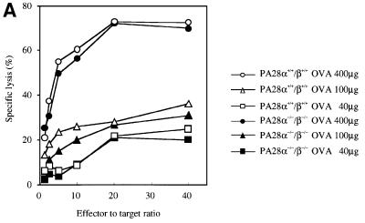
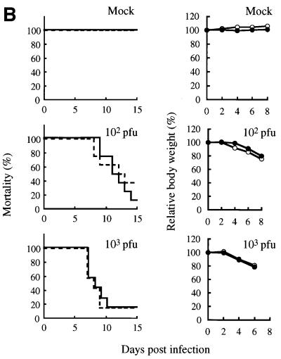
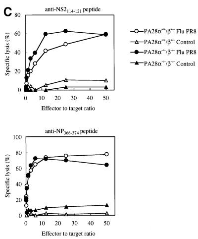
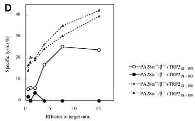
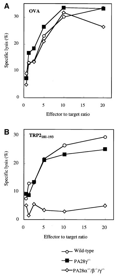
Two members of the proteasome activator, PA28alpha and PA28beta, form a heteropolymer that binds to both ends of the 20S proteasome. Evidence in vitro indicates that this interferon-gamma (IFN-gamma)-inducible heteropolymer is involved in the processing of intracellular antigens, but its functions in vivo remain elusive. To investigate the role of PA28alpha/beta in vivo, we generated mice deficient in both PA28alpha and PA28beta genes. The ATP-dependent proteolytic activities were decreased in PA28alpha(-/-)/beta(-/-) cells, suggesting that 'hybrid proteasomes' are involved in protein degradation. Treatment of PA28alpha(-/-)/beta(-/-) cells with IFN-gamma resulted in sufficient induction of the 'immunoproteasome'. Moreover, splenocytes from PA28alpha(-/-)/beta(-/-) mice displayed no apparent defects in processing of ovalbumin. These results are in marked contrast to the previous finding that immunoproteasome assembly and immune responses were impaired in PA28beta(-/-) mice. PA28alpha(-/-)/beta(-/-) mice also showed apparently normal immune responses against infection with influenza A virus. However, they almost completely lost the ability to process a melanoma antigen TRP2-derived peptide. Hence, PA28alpha/beta is not a prerequisite for antigen presentation in general, but plays an essential role for the processing of certain antigens.


Figures

None
Fig. 1. Disruption of the PA28α and PA28β genes by homologous recombination. (A) Structure of the targeting vector, the wild-type PA28α/β genes and the mutated PA28α/β genes following homologous recombination. Relevant restriction enzyme sites are indicated. Exons are depicted as closed boxes. The first and the last exons of the PA28α/β genes are numbered 1 and 11, respectively. The probes used for Southern blot analysis are shown as α- and β-probes. (B) Southern blot analysis. Genomic DNA extracted from mouse tails was digested with BamHI or EcoRV, blotted, and hybridized with the α- or the β-probe shown in (A), respectively. The wild-type allele (WT) gave a 2.5 kb fragment for the α-probe and a 9.0 kb fragment for the β-probe, while the mutant allele (KO) gave a 4.8 kb fragment for the α-probe and a 7.5 kb fragment for the β-probe. (C) Northern blot analysis. Total RNAs isolated from MEFs cultured with or without IFN-γ for 36 h were hybridized with the full-length mouse PA28α or PA28β cDNA probe. 28S and 18S rRNAs were stained with ethidium bromide to monitor the integrity of RNA. (D) Western blot analysis. Protein lysates from MEFs cultured with or without IFN-γ for 72 h were used to examine the expression of PA28α, β and γ.
None
Fig. 2. Expression of proteasomal components in the cytosolic and membrane fractions of various tissues from wild-type and PA28α–/––/– mice. Cytosolic (C) and membrane (M) proteins were isolated from the liver, spleen and brain of wild-type and knockout mice as described in Materials and methods. The ratios of protein content in cytosolic and membrane fractions were 2.4:1 in the liver, 5.6:1 in the spleen and 3.0:1 in the brain. Samples (10 µg of proteins) were analyzed by SDS–PAGE and western blotting using antibodies against PA28α, PA28β, PA28γ, X, LMP2 and Mss1. Asterisks indicate artifact bands produced by the PA28α and PA28β antibodies.
None
Fig. 3. ATP-dependent protein degradation activity, peptide hydrolysis activities and immunoproteasome formation in wild-type and PA28α–/––/– cells. (A) ATP- and AZ-dependent degradation of [35S]ODC was assayed using crude extracts from wild-type and knockout MEFs cultured with (filled bars) or without (open bars) IFN-γ for 72 h. The results are the mean of determinations performed in triplicate. Error bars represent one standard deviation on each side of the mean. There are statistically significant differences between wild-type and PA28α–/––/– cells (P <0.01, shown as asterisks). The experiment was repeated three times and consistently yielded statistically significant differences between wild-type and knockout MEFs (data not shown). (B) Sedimentation velocity analysis. Samples (2 mg of proteins) from wild-type (open circles) and knockout (filled circles) MEFs were fractionated by glycerol density gradient centrifugation (10–40% glycerol from fraction 1 to fraction 30). Aliquots (20 µl) of individual fractions were used for assay of [35S]ODC degradation activities. Western blot analysis of each fraction was performed using antibodies against X, LMP2 and PA28α. Asterisks indicate artifact bands. Numbers correspond to fraction numbers in the upper and lower panels. (C) Peptide hydrolysis activities. Aliquots of individual fractions prepared in (B) were subjected to peptide hydrolysis assays using three kinds of substrates. Suc-LLVY-AMC and Cbz-LLE-AMC were hydrolyzed in the presence of 0.05% SDS, whereas Boc-LRR-AMC was hydrolyzed without SDS. Open circles, wild-type; filled circles, knockout. (D) Initial assembly of immunoproteasomes. MEFs were cultured in the presence of IFN-γ. After the indicated times (hours), MEFs were harvested for western blot analysis with anti-LMP2 and anti-X antibodies. (E) Two-dimensional gel electrophoresis. MEFs were cultured in the presence or absence of IFN-γ for 48 h, and then metabolically labeled for 4 h followed by a 16 h chase. Cell lysates were immunoprecipitated with anti-20S proteasome antibodies and subjected to isoelectric focusing followed by SDS–PAGE.
None
Fig. 4. Role of PA28α/β in antigen processing and immune responses against viral infection. (A) Presentation of the OVA257–264 epitope. LPS blasts from wild-type (open symbols) and PA28α–/––/– (filled symbols) mice were osmotically loaded with the indicated doses of OVA proteins and labeled with 51Cr. The labeled cells were used as target cells for the lysis by OVA257–264-specific CTLs. (B) Mortality rates and relative body weight loss after infection with the influenza A virus strain PR8. Mice (n = 8 per group) were infected intranasally with the indicated doses of Flu PR8. Mortality rates in wild-type (dashed line) and PA28α–/––/– (solid line) mice are shown on the left. Relative body weight loss in wild-type (open circles) and PA28α–/––/– (filled circles) mice is shown on the right. Data represent the mean percentage body weight relative to the weight before infection. (C) CTL induction after Flu PR8 infection. Two weeks after infection with Flu PR8 (circles), splenocytes from wild-type (open symbols) and PA28α–/––/– (filled symbols) mice were isolated, stimulated with synthetic peptide NS2114–121 or NP366–374 for 6 days, and used as effector cells. 51Cr-labeled EL-4 cells pulsed with NS2114–121 or NP366–374 were used as target cells. Splenocytes from non-infected mice (triangles) were used as a control. (D) Presentation of the TRP2181–188 epitope. LPS blasts from wild-type (open symbols) and PA28α–/––/– (filled symbols) mice were osmotically loaded with the TRP2181–193 peptide (circles) or the TRP2181–188 peptide (triangles) and labeled with 51Cr. The labeled cells were used as target cells for the lysis by TRP2181–188-specific CTLs.
None
Fig. 5. Role of PA28γ in antigen processing. (A) Presentation of the OVA257–264 epitope. (B) Presentation of the TRP2181–188 epitope. Analyses were performed as in Figure 4A and D, respectively. Open circles, wild type; filled squares, PA28γ–/–; open diamonds, PA28α–/––/––/–.

Similar articles

Cited by

References

    1. Ben-Shahar S., Komlosh,A., Nadav,E., Shaked,I., Ziv,T., Admon,A., DeMartino,G.N. and Reiss,Y. (1999) 26S proteasome-mediated production of an authentic major histocompatibility class I-restricted epitope from an intact protein substrate. J. Biol. Chem., 274, 21963–21972. - PubMed
    1. Bloom M.B., Perry-Lalley,D., Robbins,P.F., Li,Y., El-Gamil,M., Rosenberg,S.A. and Yang,J.C. (1997) Identification of tyrosinase-related protein 2 as a tumor rejection antigen for the B16 melanoma. J. Exp. Med., 185, 453–459. - PMC - PubMed
    1. Bochtler M., Ditzel,L., Groll,M., Hartmann,C. and Huber,R. (1999) The proteasome. Annu. Rev. Biophys. Biomol. Struct., 28, 295–317. - PubMed
    1. Brooks P. et al. (2000) Subcellular localization of proteasomes and their regulatory complexes in mammalian cells. Biochem. J., 346, 155–161. - PMC - PubMed
    1. Coux O., Tanaka,K. and Goldberg,A.L. (1996) Structure and functions of the 20S and 26S proteasomes. Annu. Rev. Biochem., 65, 801–847. - PubMed

Publication types

MeSH terms