. 2001 Nov;21(22):7537-44.
doi: 10.1128/MCB.21.22.7537-7544.2001.

The notch intracellular domain can function as a coactivator for LEF-1

Affiliations

The notch intracellular domain can function as a coactivator for LEF-1

D A Ross et al. Mol Cell Biol. 2001 Nov.

Abstract

Notch signaling commences with two ligand-mediated proteolysis events that release the Notch intracellular domain, NICD, from the plasma membrane. NICD then translocates into the nucleus and interacts with the DNA binding protein CSL to activate transcription. We found that NICD expression also potentiates activity of the transcription factor LEF-1. NICD stimulation of LEF-1 activity was context dependent and occurred on a subset of promoters distinct from those activated by beta-catenin. Importantly, the effect of NICD does not appear to be mediated through canonical components of the Wnt signaling pathway or downstream components of the Notch pathway. In vitro assays show a weak association between the C-terminal transactivation domain of NICD and the high-mobility group domain of LEF-1, suggesting that the two proteins interact in vivo. Our data therefore describe a new nuclear target of Notch signaling and a new coactivator for LEF-1.


Figures
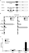
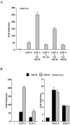
FIG. 1
FIG. 1
Notch potentiates LEF-1 activity. (A) Schematic representations of reporters used to assay LEF-1 activity. Binding sites for LEF-1 are shown as gray (consensus) or black (mutant) ovals. (B) Effects of LEF-1, β-catenin, and NICD on 7xLEF-luc and LEF-OT in Neuro-2A cells. Relative luciferase values for reporters containing seven LEF-1 binding sites (7xLEF-luc; gray bars) or no LEF-1 binding sites (fos-luc; black bars) are shown in the left panel. Values for reporters having three consensus LEF-1 binding sites (LEF-OT; gray bars) or three mutant LEF-1 binding sites (LEF-OF; black bars) are shown in the right panel. Values for fold induction were determined relative to those obtained for each reporter in the absence of any expression plasmids. (C) Effects of LEF-1 and NICD on naturally occurring promoters. Transfections of Neuro-2A cells were carried out with reporters containing promoters from the WISP-1 (white bars), Cyclin D1 (striped bars), or Xtwn (black bars) genes. Expression plasmids that were cotransfected with each set of reporters are indicated. Values are given as fold induction relative to the reporter alone.
FIG. 2
FIG. 2
Specific Notch proteins activate a subset of HMG domain transcription factors. (A) Ability of Notch 1, 2, and 3 to potentiate LEF-1. LEF-1 activity was assayed in NIH 3T3 cells using the LEF-OT reporter in the presence of Notch1 (N1) NICD, full-length Notch1 (FL N1), Notch2 (N2) NICD, or Notch3 (N3) NICD. Values are given as fold induction relative to LEF-OT alone. (B) Effects of NICD on other HMG box transcription factors. Neuro-2A cells were transfected with LEF-OT and expression vectors for LEF-1, TCF-1, HAF-2, or HAF-1 in the presence (striped bars) or absence (black bars) of NICD. Values are give as fold induction as for panel A.
FIG. 3
FIG. 3
NICD activation of LEF-1 is independent of β-catenin. NICD augments activity of Δ56LEF. Neuro-2A cells were transfected with 7xLEF-luc and expression plasmids for Δ56LEF (LEF-1 lacking the β-catenin interaction domain) and β-catenin as indicated (left panel, black bars). Cells were also transfected with LEF-OT and expression plasmids for Δ56LEF and NICD as indicated (right panel, gray bars). Results are given as fold induction relative to the reporters alone.
FIG. 4
FIG. 4
NICD activates LEF-1 independently of CSL. (A) Schematic diagrams of the NICD deletion fragments are shown. R, RAM domain; A, Ankyrin repeats; TAD, C-terminal transcription activation domain; VP16 A.D., VP16 transcription activation domain. (B) NICDΔTAD-VP16 does not augment activity of LEF-1. Neuro-2A cells were transfected with a CSL-dependent reporter, CSL-luc (black bars), or LEF-OT (gray bars) and the expression vectors as indicated. NICDΔTAD-VP16 carries the VP16 TAD in place of the Notch TAD. Results are given as fold induction relative to the reporters alone. (C) The Notch TAD is sufficient for LEF-1 activation. NIH 3T3 cells were transfected with LEF-OT (gray bars) or LEF-OF (black bars) and the NICD fragments indicated. Results are presented as fold induction relative to the reporter alone.
FIG. 5
FIG. 5
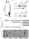
The Notch TAD physically interacts with the LEF-1 HMG domain. (A) LEF-1 activates Gal4-NICDΔRA. Neuro-2A cells were transfected with a Gal4 responsive reporter and either the DNA binding domain of Gal4 (Gal4) or a Gal4 fused to the TAD of Notch (Gal4-NICDΔRA) in the presence (gray bars) or absence (black bars) of LEF-1 as indicated. Results are shown as fold induction relative to the reporter plus the Gal4 DNA binding domain. (B) LEF-1 interacts with the Notch TAD in vitro. 293T cells were transfected with plasmids expressing HA-LEF-1 and FLAG-CSL, extracts were incubated with GST, GST-NICDΔTAD, or GST-NICDΔRA, and bound proteins were analyzed by Western analysis using anti-HA antibodies (top panel) or anti-FLAG antibodies (lower panel). Samples of untreated cell extracts were loaded in each of the far left lanes, corresponding to 1/350 of the input analyzed for HA-LEF-1 (top) and 1/5 of the input analyzed for FLAG-CSL (bottom). (C) LEF interacts with the Notch TAD and ALY with comparable affinities. 293T cells were transfected with HA-LEF, extracts were incubated with GST, GST-NICDΔRA or GST-ALY as indicated, and bound proteins were analyzed by Western analysis with an anti-HA antibody. Lane 1 contains untreated extract corresponding to 1/300 of the input. (D) Schematic diagram of the LEF-1 fragments used to map the interaction with NICD. Amino acid positions and several functionally defined domains are indicated. βCID, β-catenin interaction domain; CAD, context-dependent activation domain; HMG, HMG domain. (E) The LEF-1 HMG box mediates interactions with the Notch TAD. The indicated radiolabeled LEF-1 fragments were generated by in vitro transcription and translation (lanes 1, 4, 7, 10, and 13) and analyzed for binding to GST (lanes 2, 5, 8, 11, and 14) and GST-NICDΔRA (lanes 3, 6. 9, 12, and 15). Untreated samples represent 1/100 of the input used for each binding analysis. Positions of molecular mass standards (in kilodaltons) are shown at the left.
FIG. 6
FIG. 6
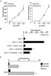
High-level expression of NICD is required for LEF-1 activation. (A) Low levels of NICD stimulate a CSL reporter but do not potentiate LEF-1. Fold induction of CSL-luc, with increasing amounts of NICD expression plasmid, is shown relative to the reporter alone (left graph). Stimulation of LEF-1 in the presence of increasing amounts of NICD compared to the LEF-OT reporter and LEF-1 alone is shown (right graph). (B) Excess CSL inhibits NICD's ability to stimulate LEF-1 activity. Neuro-2A cells were transfected with the LEF-OT reporter and the indicated expression vectors. Results are given as fold induction relative to the reporter alone. (C) Activity of NICD in the presence of endogenous LEF-1. Jurkat cells were transfected with reporters containing wild-type (LEF-OT) or mutant (LEF-OF) LEF-1 binding sites, plus or minus an expression vector for NICD as indicated. Data are represented as fold induction relative to LEF-OF alone.
FIG. 7
FIG. 7
Retrovirally expressed NICD stimulates LEF-1 activity. (A) NIH 3T3 cells transduced with either the MIGR (black bars) or MIGR-NICD (gray bars) retroviruses and then transfected with LEF-OF or LEF-OT reporters and a LEF-1 expression vector. Results for each transduced cell population are shown as fold induction relative to the LEF-OF plus LEF-1. (B) Tamoxifen-responsive NICD stimulates LEF-1 activity. NIH 3T3 cells were transduced with retroviruses expressing either the parental ER element (ER cells) or NICD fused to the estrogen receptor element (NICD-ER cells). LEF-1 activity was assayed in the absence (black bars) or presence (gray bars) of 25 nM tamoxifen, following the transfection of ER and NICD-ER cells with the LEF-OF or LEF-OT reporters and a LEF-1 expression vector. Results are shown as fold induction relative to LEF-OF and LEF-1 with no tamoxifen.

Similar articles

Cited by

References

    1. Aster J C, Robertson E S, Hasserjian R P, Turner J R, Kieff E, Sklar J. Oncogenic forms of NOTCH1 lacking either the primary binding site for RBP-Jκ or nuclear localization sequences retain the ability to associate with RBP-Jκ and activate transcription. J Biol Chem. 1997;272:11336–11343. - PubMed
    1. Axelrod J D, Matsuno K, Artavanis-Tsakonas S, Perrimon N. Interaction between Wingless and Notch signaling pathways mediated by dishevelled. Science. 1996;271:1826–1832. - PubMed
    1. Beatus P, Lundkvist J, Oberg C, Lendahl U. The notch 3 intracellular domain represses notch 1-mediated activation through Hairy/Enhancer of split (HES) promoters. Development. 1999;126:3925–3935. - PubMed
    1. Beckman H, Kadesch T. The leucine zipper of TFE3 dictates helix-loop-helix dimerization specificity. Genes Dev. 1991;5:1057–1066. - PubMed
    1. Behrens J, von Kries J P, Kuhl M, Bruhn L, Wedlich D, Grosschedl R, Birchmeier W. Functional interaction of beta-catenin with the transcription factor LEF-1. Nature. 1996;382:638–642. - PubMed

Publication types

MeSH terms