. 2001 Oct 15;194(8):1081-96.
doi: 10.1084/jem.194.8.1081.

Legionella pneumophila is internalized by a macropinocytotic uptake pathway controlled by the Dot/Icm system and the mouse Lgn1 locus

Affiliations

Legionella pneumophila is internalized by a macropinocytotic uptake pathway controlled by the Dot/Icm system and the mouse Lgn1 locus

M Watarai et al. J Exp Med. .

Abstract

The products of the Legionella pneumophila dot/icm genes enable the bacterium to replicate within a macrophage vacuole. This study demonstrates that the Dot/Icm machinery promotes macropinocytotic uptake of L. pneumophila into mouse macrophages. In mouse strains harboring a permissive Lgn1 allele, L. pneumophila promoted formation of vacuoles that were morphologically similar to macropinosomes and dependent on the presence of an intact Dot/Icm system. Macropinosome formation appeared to occur during, rather than after, the closure of the plasma membrane about the bacterium, since a fluid-phase marker preloaded into the macrophage endocytic path failed to label the bacterium-laden macropinosome. The resulting macropinosomes were rich in GM1 gangliosides and glycosylphosphatidylinositol-linked proteins. The Lgn1 allele restrictive for L. pneumophila intracellular replication prevented dot/icm-dependent macropinocytosis, with the result that phagosomes bearing the microorganism were targeted into the endocytic network. Analysis of macrophages from recombinant inbred mouse strains support the model that macropinocytotic uptake is controlled by the Lgn1 locus. These results indicate that the products of the dot/icm genes and Lgn1 are involved in controlling an internalization route initiated at the time of bacterial contact with the plasma membrane.


Figures
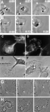
Figure 1
Figure 1
(A and B) Phase transparent puffs are associated with Dot/Icm-promoted uptake by macrophages. Displayed are images captured at various times after contact of L. pneumophila with macrophages. Times refer to seconds after leftmost image was captured. Thin arrows point to bacteria, whereas thick arrowheads point to expanding ruffles. (A) LP02 (intact Dot/Icm) observed after contact with macrophage. (B) LP03 (dotA) observed after contact with macrophage (online supplemental video titled “DOTA3”). (C–F) Diffuse ruffles and actin aggregation are dependent on an intact Dot/Icm system. Displayed are incubations of macrophages with LP02-GFP (C) and LP03-GFP (D) stained by phalloidin. Also shown are phase contrast micrographs of the identical microscope fields for LP02-GFP (E) and LP03-GFP (F). (G and H) Videomicroscopy of LP02-GFP introduced onto bone marrow macrophages. Supplemental online videos (G) “Ruffle (LP02)” and (H) “Macropin (LP02).”
Figure 2
Figure 2
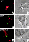
Macropinosome formation by L. pneumophila. Bone marrow–derived macrophages were incubated for 10 min at 37°C in the presence of GFP-expressing bacteria and Rh-Dx155, fixed, and processed for phase and immunofluorescence microscopy. Displayed are macrophages from A/J mice incubated with LP02 (Dot/Icm intact; A and B) or LP03 (dotA; C and D) as well as macrophages from C57Bl/6J mice incubated with LP02 (Dot/Icm intact; E and F). Shown are GFP and rhodamine channels merged (A, C, and E) or phase contrast images (B, D, and F). LP, L. pneumophila; MP, macropinosome.
Figure 3
Figure 3
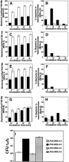
Delayed uptake and macropinosome formation promoted by the L. pneumophila Dot/Icm system. Black bar, LP02 (dot/icm +). White bar, LP03 (dotA). Uptake (A, C, E, and G) or macropinosome formation (B, D, F, and H) were quantitated as described in Materials and Methods. Incubation periods were either after short centrifugation (A–F) or in the absence of centrifugation (G and H). (A and B) No opsonization. (C and D) Opsonization with anti-L. pneumophila serum (Materials and Methods). (A–D) Bacterial strains harbor GFP. (E–H) Bacterial strains harboring pAY68 (pH 6.0 Ag). 100 macrophages were examined per coverslip. Data are the mean of triplicate samples ± SE. MOI is indicated on the y axis. (I) Intracellular growth of L. pneumophila after infection at MOI = 10. L. pneumophila strains LP02 (intact Dot/Icm) or LP03 (dotA) were introduced at MOI = 0.1 or 10 by centrifugation onto monolayers of A/J mouse bone marrow macrophages and assayed for increase in viable counts over a 24-h period (Materials and Methods). t24/t0 = bacterial counts at 24 h of incubation/bacterial counts determined after allowing for initial uptake of the bacteria (Materials and Methods).
Figure 4
Figure 4
Compartments preloaded with a fluid-phase marker do not fuse with macropinosomes harboring L. pneumophila with intact Dot/Icm system. Bone marrow–derived macrophages were incubated at an MOI = 10 along with 1 mg/ml Rh-Dx155 for 30 min at 37°C and then incubated with bacteria either in the continued presence (A) or absence (B) of the fluid-phase marker (Materials and Methods). Number of bacteria in compartments staining with TRITC was determined as described in Material and Methods. Gray bar, LP02-GFP (intact Icm/Dot system); black bar, Lp03-GFP (dotA).
Figure 5
Figure 5
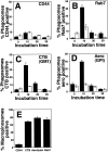
Association of membrane-localized markers with L. pneumophila macropinosomes. Bone marrow–derived macrophages were incubated with L. pneumophila, and membrane associated markers were localized by immunofluorescence, as described in Materials and Methods. Shown are merged images of the GFP and TRITC channels side-by-side with phase-contrast images of the identical cells. CD44, fixed cells probed with anti-CD44; GPI, fixed cells probed with Alexa594-aerolysin; GM1, cells coincubated with biotin-CTB subunit and bacteria (Materials and Methods); Rab7, fixed cells probed with anti-Rab7. Arrows, edge of phagosomal membranes.
Figure 6
Figure 6
Kinetics of colocalization of membrane-associated markers with L. pneumophila-bearing phagosomes. (A–D) LP02-GFP (Dot/Icm intact) or LP03-GFP (dotA) at an MOI = 10 were centrifuged onto macrophage monolayers, then incubated for the noted periods of time at 37°C before fixation and probing with indicated reagents (Materials and Methods). “% phagosomes positive” refers to percentage of internalized bacteria that show costaining with the noted markers, based on observation of 100 bacteria per coverslip (Materials and Methods). The data represent the mean ± SE of three coverslips. (E) Macrophages incubated with LP02-GFP for 10 min at 37°C were probed for noted markers, and macropinosomes harboring GFP-labeled bacteria were identified by fluorescence and phase microscopy. The macropinosomes were then observed for the presence of the noted markers, as in A–D. “% positive” refers only to those phagosomes with a macropinocytotic morphology, and represents percentage of macropinosomes that show costaining with the noted markers. Data for macropinosomes are from triplicate coverslips representing 50 macropinosomes per coverslip. CD44, anti-CD44; Aero, Alexa594-aerolysin; Rab7, anti-Rab7. Black bar, LP02 (Dot/Icm intact); white bar, LP03 (dotA).
Figure 7
Figure 7
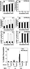
Defective targeting and macropinosome formation in the Lgn-restrictive strain C57Bl/6J. Bone marrow–derived macrophages were isolated from either C57Bl/6J or A/J mice and challenged with either LP02-GFP (black bar, intact Dot/Icm) or LP03-GFP (white bar, dotA; Materials and Methods). In A and B, the MOI = 10 bacteria/macrophage. In C–F, the MOI = 1.0 bacteria/macrophage. (A) Uptake efficiency by C57Bl/6J macrophages was determined by antibody probing, as in Fig. 3. (B) Macropinosome formation by C57Bl/6J macrophages was determined by colocalization of bacteria with Rh-Dx155, as in Fig. 3. (C and D) Uptake efficiency by A/J (C) and C57Bl/6J (D) macrophages was determined by protection of internalized bacteria from gentamicin killing (reference 34). (E and F) Ability of L. pneumophila to bypass entry into the endocytic pathway was determined by indirect immunofluorescence probing of fixed samples with anti–LAMP-1 (reference 36). Colocalization of LAMP-1 with bacteria was determined for bone marrow macrophages derived from A/J (E) or C57BL/6J mice (F). ND, no colocalization detectable. Data are mean of triplicate coverslips ± SE, 100 bacteria per coverslip. (G) Macropinosome formation in permissive macrophages is not the result of enhanced cytotoxicity. Coverslips of macrophages from either A/J or C57Bl/6J mice were incubated at the indicated MOI with either LP02 (dot+) or LP03 (dotA) and assayed for cytotoxicity by enumerating EtdBr-permeable cells microscopically (Materials and Methods; reference 34). Cytotoxicity or percentage of EtdBr permeable equals the percentage of cells found to be EtdBr permeable in rhodamine fluorescence channel relative to total number of cells on coverslip (Materials and Methods).
Figure 8
Figure 8
RI strains having STSs of C57Bl/6J do not support L. pneumophila macropinosome formation. Displayed is polyacrylamide gel of PCR analysis of three STSs located within the Lgn1 interval (D13Die6, D13Die36-A,B, and D13Die3), using DNA prepared from A/J, C57Bl/6J, and A×B RI strains (Materials and Methods). Arrows next to gel note bands corresponding to A/J or C57Bl/6J (also called B6) genotypes. Below the gel is the genotype of each strain, as determined by this analysis (A, Lgn1 locus of A/J; B, Lgn1 locus of C57Bl/6J). Asterisk refers to RI strain that has both high levels of macropinosome formation and high levels of uptake. Macropinosome formation is from Table . Below the gel is the chromosomal location of each STS relative to the Lgn1 locus.

Similar articles

Cited by

References

    1. Nash T.W., Libbey D.M., Horowitz M.A. Interaction between the Legionnaires' disease bacterium (Legionella pneumophila) and human alveolar macrophages. J. Clin. Invest. 1984;74:771–782. - PMC - PubMed
    1. Fields B.S., Sanden G.N., Barbaree J.M., Morrill W.E., Wadowsky R.M., White E.H., Feeley J.C. Intracellular multiplication of Legionella pneumophilia in an amoebae isolated from hospital hot water tanks. Curr. Microbiol. 1998;18:131–137.
    1. Wadowsky R.M., Butler L.J., Cook M.K., Verma S.M., Paul M.A., Fields B.S., Keleti G., Sykora J.L., Yee R.B. Growth-supporting activity for Legionella pneumophilia in tap water cultures and implication of hartmannellid amoebae as growth factors. Appl. Environ. Microbiol. 1988;54:2677–2682. - PMC - PubMed
    1. Gao L.Y., Abu Kwaik Y. Apoptosis in macrophages and alveolar epithelial cells during early stages of infection by Legionella pneumophila and its role in cytopathogenicity. Infect. Immun. 1999;67:862–870. - PMC - PubMed
    1. Cianciotto N., Eisenstein B.I., Engelberg N.C. Genetics and molecular pathogenesis of Legionella pneumophilia, an intracellular parasite of macrophages. Mol. Biol. Med. 1989;6:409–424. - PubMed

Publication types