. 2001 Oct 15;20(20):5692-702.
doi: 10.1093/emboj/20.20.5692.

PKCbeta modulates antigen receptor signaling via regulation of Btk membrane localization

Affiliations

PKCbeta modulates antigen receptor signaling via regulation of Btk membrane localization

S W Kang et al. EMBO J. .

Abstract

Mutations in Bruton's tyrosine kinase (Btk) result in X-linked agammaglobulinemia (XLA) in humans and X-linked immunodeficiency (xid) in mice. While targeted disruption of the protein kinase C-beta (PKCbeta) gene in mice results in an immunodeficiency similar to xid, the overall tyrosine phosphorylation of Btk is significantly enhanced in PKCbeta-deficient B cells. We provide direct evidence that PKCbeta acts as a feedback loop inhibitor of Btk activation. Inhibition of PKCbeta results in a dramatic increase in B-cell receptor (BCR)-mediated Ca2+ signaling. We identified a highly conserved PKCbeta serine phosphorylation site in a short linker within the Tec homology domain of Btk. Mutation of this phosphorylation site led to enhanced tyrosine phosphorylation and membrane association of Btk, and augmented BCR and FcepsilonRI-mediated signaling in B and mast cells, respectively. These findings provide a novel mechanism whereby reversible translocation of Btk/Tec kinases regulates the threshold for immunoreceptor signaling and thereby modulates lymphocyte activation.


Figures

None
Fig. 1. Pharmacological inhibition of PKC isoforms results in enhanced BCR-induced Ca2+ signaling. A20 and Ramos B cells were pretreated with either general (Ro318425) or PKCβ-specific inhibitor (LY379196) at doses indicated for 5 min, then activated with anti-Ig cross-linking. (A) In A20 and Ramos B cells, Ca2+ mobilization was monitored by spectrofluorimetry after a 30 s baseline measurement. (B) Ramos cells are stimulated with anti-IgM for 2 min or unstimulated after pretreatment with 5 µM Ro318425 or DMSO. Cells were fractionated by hypotonic lysis, and membrane and cytosolic fractions were analyzed by western blot analysis. Membrane enrichment of Btk was detected by immunoblotting with anti-Btk antibody (left). Fractionation efficiency was evaluated by immunoblotting with anti-transferrin receptor (TrfR). Total cell lysates (TCL) were immunoblotted with anti-phosphotyrosine antibody (middle). Enhancement of pp130 signal was observed in both membrane and cytosolic fractions by anti-phosphotyrosine (PY) blotting (right) and the same membrane was stripped and reblotted with anti-PLCγ2 antibody. (C) Ramos cells were treated as in (B), and lysed in BBS following BCR stimulation. Syk (left) or Lyn (right) was immunoprecipitated and immunoblotted with anti-PY antibody and the same membrane was stripped and reblotted with anti-Syk or anti-Lyn antibodies.
None
Fig. 2. PKCβ specifically down-modulates both Btk transphosphorylation and autophosphorylation. (A) Btk, Lyn and PKCβ proteins were coordinately expressed in NIH 3T3 cells using recombinant vaccinia virus. Btk was immunoprecipitated and the total tyrosine phosphorylation content was measured by immunoblotting. Btk loading was measure by anti-Btk antibody. Relative tyrosine phosphorylation levels were quantified by densitometric analysis. (B) Left panel: Btk, Lyn and Akt proteins were co-expressed and Btk tyrosine phosphorylation was analyzed as in (A). Right panel: Lyn was immunoprecipitated with anti-Lyn antibody and in vitro kinase assay (IVK) was performed. Bottom: Btk and Lyn were co-expressed with increasing dosage of PKCµ. Btk phosphorylation was analyzed as in (A). (C) Btk-Wt and Btk-E41K were co-expressed with Lyn and increasing dosage of PKCβ as in (A). Btk protein was immunoprecipitated and sequentially immunoblotted with anti-PY, anti-PY551, anti-PY223 and anti-Btk specific antibodies. (D) Btk and Lyn were co-expressed with high dosage PKCβ and cells were treated with increasing doses of Ro318425 for 30 min. Btk was immunoprecipitated and analyzed as in (A).
None
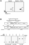
Fig. 3. Phosphopeptide mapping of the PKCβ-induced phosphorylation site on Btk. (A) Kinase inactive Btk (KI; Btk-K430R) was expressed in NIH 3T3 cells using recombinant vaccinia virus in the presence or absence of co-expressed PKCβ. Cells (1 × 10⁷) were labeled with [32P]orthophosphate and Btk was immunoprecipitated, digested with trypsin, and tryptic peptides were separated by thin-layer electro phoresis at pH 1.9 followed by chromatography (see Materials and methods). (B) Identification of the domain phosphorylated by PKCβ. Upper panel: schematic representation of IgA cleavage sites within Btk. Bottom panel: partially purified Btk from (A) was incubated with IgA protease. The digested fragments were resolved by SDS–PAGE, and visualized by autoradiography (first panel) and western blot analysis using antibodies against N-terminal (middle) and C-terminal (third panel) regions of Btk.
None
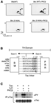
Fig. 4. PKCβ phosphorylates S180 in the Tec-linker of Btk. (A) Phosphopeptide mapping analysis was performed on Btk-Wt and Btk-S180A, with or without the co-expression of PKCβ. As shown in Figure 3A, Btk-Wt displays two predominant phospho-tryptic fragments (P1, P2), and P1 is increased with PKCβ co-expression. The putative PKCβ phosphorylation site mutant Btk-S180A fails to induce P1, while P2 is still intact (panel 4). (B) Sequence alignment of murine Tec family kinases and Raja eglanteria Btk ortholog, skate PTK. Alignment of the conserved PKC consensus sequence is shown. (C) PKCβ-mediated negative regulation of Tec was examined in NIH 3T3 cells coordinately expressing Tec, Lyn and PKCβ. Tec was immunoprecipitated and blotted with anti-PY and reblotted with anti-Tec antibodies.
None
Fig. 5. Btk-S180A is hyperphosphorylated and exhibits enhanced BCR-induced Ca2+ signaling and FcεRI-induced JNK activation. (A) Btk-Wt or Btk-S180A was coordinately expressed as indicated. Btk is partially purified by immunoprecipitation, and its phosphotyrosine content was evaluated by anti-PY blotting. Duplicate membranes were immunoblotted with anti-Btk antibody to demonstrate equal loading. (B) BCR-induced Ca2+ flux was analyzed from A20 cells expressing Btk-Wt, S180A, or E41K. A20 cells were infected with vaccinia viruses for 8 h, followed by loading with Indo-1. BCR was cross-linked with anti-IgG [10 µg/ml F(ab)2] at the time indicated (arrow), and Ca2+ mobilization was monitored by spectrofluorimetry. Btk expression was evaluated by western blot analysis. The data shown are a representative of five independent experiments. (C) Btk-deficient DT40 B cells were reconstituted with Btk-Wt, S180A, E41K or GFP vector control. FACS sorted cell populations with equivalent GFP expression were obtained and Ca2+ mobilization was monitored by spectrofluorimetry as in (A). GFP expression by FACS and Btk expression by western blot analysis are shown (upper panels). (D) MAPK activation was examined in BMMC derived from Btk–/– mice reconstituted with retroviruses expressing vector alone, Btk-Wt or Btk-S180A. BMMCs were sensitized overnight with 1 µg/ml anti-DNP monoclonal IgE antibody, then stimulated with DNP-HAS (100 ng/ml) for the indicated times. For JNK assay, in vitro kinase assay was performed with GST–c-Jun as substrate. p38 and ERK activity were evaluated using phosphospecific antibodies against phosphorylated p38 (anti-pp38) or ERK (anti-pERK). Protein expression was analyzed by immunoblotting with anti-JNK, anti-p38 or anti-ERK. Equal Btk protein expression was examined by immunoblotting with anti-Btk antibody (right).
None
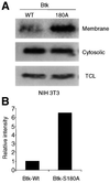
Fig. 6. Increased membrane targeting of Btk-S180A. (A) NIH 3T3 cells expressing Btk-Wt or Btk-S180A were fractionated by hypotonic lysis and Dounce homogenization (Li et al., 1995). Membrane and cytosolic fractions, and total cell lysates were analyzed by immunoblotting with anti-Btk antibody. (B) Graphical representation of Btk membrane localization. Relative intensity of Btk band was analyzed by densitometry. The data shown are representative of two independent experiments.
None
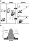
Fig. 7. Modulation of antigen receptor signaling by PKCβ. (A) Model for the PKCβ-mediated inhibition of Btk membrane localization. (B) Schematic representation of the Btk activity requirement for B-cell survival and function. Btk-XLAi (inactivated) represents loss-of-function Btk mutations. Btk-XLAa (activated) represents gain-of-function Btk mutations.

Similar articles

Cited by

References

    1. Andreotti A.H., Bunnell,S.C., Feng,S., Berg,L.J. and Schreiber,S.L. (1997) Regulatory intramolecular association in a tyrosine kinase of the Tec family. Nature, 385, 93–97. - PubMed
    1. Bagowski C.P., Stein-Gerlach,M., Choidas,A. and Ullrich,A. (1999) Cell-type specific phosphorylation of threonines T654 and T669 by PKD defines the signal capacity of the EGF receptor. EMBO J., 18, 5567–5576. - PMC - PubMed
    1. Baraldi E., Carugo,K.D., Hyvonen,M., Surdo,P.L., Riley,A.M., Potter,B.V., O’Brien,R., Ladbury,J.E. and Saraste,M. (1999) Structure of the PH domain from Bruton’s tyrosine kinase in complex with inositol 1,3,4,5-tetrakisphosphate. Structure, 7, 449–460. - PubMed
    1. Bolland S., Pearse,R.N., Kurosaki,T. and Ravetch,J.V. (1998) SHIP modulates immune receptor responses by regulating membrane association of Btk. Immunity, 8, 509–516. - PubMed
    1. Buhl A.M. and Cambier,J.C. (1997) Co-receptor and accessory regulation of B-cell antigen receptor signal transduction. Immunol. Rev., 160, 127–138. - PubMed

Publication types

MeSH terms