. 2001 Oct 15;20(20):5678-91.
doi: 10.1093/emboj/20.20.5678.

TRAF6 is a critical mediator of signal transduction by the viral oncogene latent membrane protein 1

Affiliations

TRAF6 is a critical mediator of signal transduction by the viral oncogene latent membrane protein 1

U Schultheiss et al. EMBO J. .

Abstract

The oncogenic latent membrane protein 1 (LMP1) of the Epstein-Barr virus recruits tumor necrosis factor-receptor (TNFR)-associated factors (TRAFs), the TNFR-associated death domain protein (TRADD) and JAK3 to induce intracellular signaling pathways. LMP1 serves as the prototype of a TRADD-binding receptor that transforms cells but does not induce apoptosis. Here we show that TRAF6 critically mediates LMP1 signaling to p38 mitogen-activated protein kinase (MAPK) via a MAPK kinase 6-dependent pathway. In addition, NF-kappaB but not c-Jun N-terminal kinase 1 (JNK1) induction by LMP1 involves TRAF6. The PxQxT motif of the LMP1 C-terminal activator region 1 (CTAR1) and tyrosine 384 of CTAR2 together are essential for full p38 MAPK activation and for TRAF6 recruitment to the LMP1 signaling complex. Dominant-negative TRADD blocks p38 MAPK activation by LMP1. The data suggest that entry of TRAF6 into the LMP1 complex is mediated by TRADD and TRAF2. In TRAF6-knockout fibroblasts, significant induction of p38 MAPK by LMP1 is dependent on the ectopic expression of TRAF6. We describe a novel role of TRAF6 as an essential signaling mediator of a transforming oncogene, downstream of TRADD and TRAF2.


Figures

None
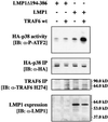
Fig. 1. Dominant-negative TRAF6 efficiently blocks LMP1 signaling to p38. (A) Transient p38 MAPK assays in HEK 293 cells. Cells were transfected with 1.0 µg of HA-p38 MAPK together with 1.0 µg of wild-type (wt) LMP1 or LMP1(Δ194–386) lacking the C-terminal signaling domain and 1.0 µg of wt TRAF6 or dominant-negative TRAF6(300–524) expression vectors, as indicated. Total amounts of transfected DNAs were adjusted to 3.0 µg using pcDNA3.1 empty vector. At 24 h post-transfection, non-radioactive HA-p38 MAPK immunocomplex kinase assays were performed. Top panels, HA-p38 MAPK activity. Immunoblot analysis (IB) of in vitro GST–ATF2 phosphorylation by the precipitated HA-p38 MAPK using a phospho (P)-ATF2-specific antibody. Second panels from top, immunoblot of the immunoprecipitated (IP) HA-p38 MAPK. Third panels from top, immunoblot analysis of TRAF6 expression using the rabbit H-274 antibody. One asterisk, wt TRAF6; two asterisks, TRAF6(300–524). Bottom panels, LMP1 expression. Apparent molecular weights are given in kDa. The left and right colums are derived from two separate and independent experiments. (B) Transient p38 MAPK assays in HEK 293 cells. Transfections and kinase assays were performed essentially as described in (A) with the following alterations. As indicated, 1.0 µg of pSV-LMP1:CD40 was co-transfected. In vitro HA-p38 MAPK assays were performed in the presence of [γ-32P]ATP. Top panel, autoradiograph of GST–ATF2 phosphorylation. Middle panel, phosphoimager quantitation of GST–ATF2 phosphorylation normalized to the immunoprecipitated HA-p38 MAPK. Bottom panel, precipitated HA-p38 MAPK.
None
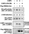
Fig. 2. MKK6 mediates p38 MAPK induction by LMP1. Transient p38 MAPK assays in HEK 293 cells. As indicated, cells were transfected with 1.0 µg of HA-p38 MAPK together with 0.5 µg of wild-type LMP1 or LMP1(Δ194–386) expression vectors and 1.0 µg of MKK6(Ala), a dominant-negative Flag-tagged MKK6 mutant. Total amounts of transfected DNAs were adjusted to 2.5 µg using carrier DNA. At 24 h post-transfection, non-radioactive HA-p38 MAPK immunocomplex kinase assays were performed. Top panel, HA-p38 MAPK activity. Second panel from top, quantitation of HA-p38 MAPK activity normalized to HA-p38 MAPK IP. Third panel from top, immunoprecipitated HA-p38 MAPK. Second panel from bottom, LMP1 expression. Bottom panel, Flag-MKK6(Ala) expression. One out of three independent experiments with identical results is shown.
None
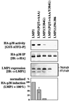
Fig. 3. The TRAF-interaction motif of CTAR1 and tyrosine 384 of CTAR2 cooperate to induce p38 MAPK. Transient HA-p38 MAPK kinase assays in HEK 293 cells. As indicated, 1.0 µg of HA-p38 MAPK was transfected together with 1.0 µg of LMP1, LMP1(PQT→AAA) mutated in CTAR1, LMP1(Y384G) mutated in CTAR2, LMP1(PQT→AAA/Y384G) double mutant expression vectors, or LMP1Δ194–386 lacking the complete C-terminal signaling domain as a negative control. At 24 h post-transfection, non-radioactive HA-p38 MAPK immunocomplex kinase assays were performed. Blots of one representative experiment out of four is shown. Top panel, immunoblot of GST–ATF2 in vitro phosphorylated by the precipitated HA-p38 MAPK. Second panel from top, immunoprecipitated HA-p38 MAPK. Third panel from top, LMP1 expression. Bar graph, p38 MAPK activation by LMP1 and LMP1 mutants given as a percentage relative to wild-type LMP1. HA-p38 MAPK induction by wild-type LMP1 was set to 100%. Data are mean p38 MAPK activities of four independent experiments normalized to the immunoprecipitated HA-p38 MAPK and to the expression of LMP1. In all assays, LMP1(PQT→AAA/Y384G) was completely inactive with respect to p38 MAPK activation.
None
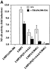
Fig. 4. Dominant-negative TRAF6 interferes with NF-κB but not with JNK1 induction by LMP1. (A) TRAF6(300–524) expression partially inhibits NF-κB signaling triggered by both CTAR1 and CTAR2 of LMP1. Transient NF-κB luciferase reporter assays in HEK 293 cells. Transfected expression vectors: 0.5 µg of LMP1Δ194–386, wild-type LMP1, LMP1(PQT→AAA), LMP1(Y384G), or LMP1(PQT→AAA/Y384G), 1.5 µg of TRAF6(300–524) or pcDNA3.1 empty vector, as indicated. (B) Control experiment: TRAF6(300–524) blocks LMP1:CD40 but not NIK-induced NF-κB activity. Tranfections were performed as described in (A). pSV-LMP1:CD40 (0.5 µg) or pcDNA3-NIK (0.5 µg) were co-transfected. For (A) and (B), luciferase activities were corrected for transfection efficiencies. NF-κB activities (y-axis) are given as x-fold induction calculated versus the signaling-defect LMP1Δ194–386 mutant. LMP1Δ194–386 values were set to 1. Data are mean values of three independent experiments. (C) JNK1 activation by LMP1 is independent of TRAF6. Transient HA-JNK1 assays in HEK 293 cells. Transfections were performed as described in the legend to Figure 1 with 1.0 µg pSRα-HA-JNK1 transfected instead of pCMV-HA-p38. Top panel, HA-JNK1 activity. Autoradiograph of GST–c-Jun phosphorylation. Middle panel, phosphoimager quantitation of GST–c-Jun phosphorylation normalized to the immunoprecipitated HA-JNK1. Bottom panel, immunoprecipitated HA-JNK1.
None
Fig. 5. TRAF6 interaction with the LMP1 signaling complex. (A) LMP1 and TRAF6 co-localize in clusters at the plasma membrane. HEK 293 cells were transfected with pcDNA3.1-TRAF6 and pSV-HA-LMP1–EGFP expressing an HA-LMP1 variant that is C-terminally fused to EGFP. The day after transfection, cells were processed for immunostaining, fluorescence microscopy and confocal imaging. Confocal images were generated from a single cell with HA-LMP1–EGFP autofluorescence in green (a), TRAF6 immunofluorescence in red (b) and the overlay of both images (c). Arrows indicate clusters of HA-LMP1–EGFP molecules within the plasma membrane (a). TRAF6 co-localizes with HA-LMP1–EGFP clusters (b and c). n, nucleus. Clustering of TRAF6 at the plasma membrane could not be detected in control transfections lacking HA-LMP1–EGFP expression (data not shown). (B) HA-LMP1–EGFP is fully functional with respect to p38 MAPK activation. Transient HA-p38 MAPK assays were performed in HEK 293 cells. Equal amounts of LMP1 and HA-LMP1–EGFP expression vectors were transfected. (C) TRAF6 rapidly translocates to induced clusters of a conditional NGFR:LMP1 chimera at the plasma membrane. HeLa cells were transiently transfected with 1 µg of pSV-NGFR:LMP1 expressing a conditional chimera of the low affinity p75 NGFR extracellular and transmembrane domains and the intracellular signaling domain of LMP1. Oligomerization and signaling activity of NGFR:LMP1 can be induced by crosslinking with specific antibodies. Twenty-four hours post-transfection, all cells were incubated with a mouse anti-NGFR antibody specific for the extracellular NGFR portion of the NGFR:LMP1 chimera. Subsequently, cells were either treated with an anti-mouse IgG secondary antibody for 15 min to crosslink NGFR:LMP1 (three lower panels), or left untreated as a negative control (two upper panels). The cells were fixed and immunofluorescence analysis was performed. Left column, NGFR:LMP1 immunofluorescence (red). Middle column, TRAF6 immunofluorescence (green). Right column, overlay of NGFR:LMP1 and TRAF6. The yellow/orange color of the induced NGFR:LMP1 clusters in the overlay images (three lower panels, right column) clearly demonstrate the co-localization of TRAF6 with activated NGFR:LMP1 at the plasma membrane. In contrast, unstimulated cells display a homogenous NGFR:LMP1 plasma membrane stain without detectable TRAF6 co-localization (upper two panels). (D) TRAF6 and TRAF6(300–524) are recruited to HA-LMP1 depending on the PxQxT motif and tyrosine 384 of LMP1. HEK 293 cells were transfected with pcDNA3.1-TRAF6 (upper graph) or pcDNA3.1-TRAF6(300–524) (lower graph) together with wild-type pSV-HA-LMP1, pSV-HA-LMP1(PQT→AAA/Y384G) or pcDNA3.1. Binding of TRAF6 to wild-type or mutated HA-LMP1 was assayed by a novel sandwich ELISA assay as described in the text. Wild-type TRAF6 was detected by the anti-TRAF6 H274 antibody, the specificity of which was shown by immunoblotting (inlay). m, mock-transfected HEK 293 cells; tr., TRAF6-transfected HEK 293 cells. Xpress-tagged TRAF6(300–524) was detected by an anti-Xpress-tag antibody. The cartoons to the right illustrate the set-up of the ELISA assays, the ‘X’ representing factors that mediate TRAF6 binding to HA-LMP1. POX, peroxidase.
None
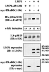
Fig. 6. Dominant-negative TRADD(1–194) blocks p38 MAPK signaling by LMP1. Transient HA-p38 MAPK assays in HEK 293 cells. Cells were transfected with 1.0 µg of HA-p38 MAPK together with 1.0 µg of wild-type LMP1, LMP1(Δ194–386) or myc-TRADD(1–194) expression vectors, as indicated. pcDNA3-p35 (0.5 µg) was co-transfected to prevent apoptosis. Total amounts of transfected DNA were adjusted to 3.0 µg using pRK5 empty vector. Top panel, HA-p38 MAPK activity. Second panel from top, quantitation of HA-p38 MAPK activity normalized to the immunoprecipitated HA-p38 MAPK. Third panel from top, immunoprecipitated HA-p38 MAPK. Second panel from bottom, LMP1 expression. Bottom panel, immunoblot analysis of myc-TRADD(1–194) expression using the anti-myc tag antibody 9E10. One out of three independent experiments with identical results is shown.
None
Fig. 7. Ectopic expression of TRAF6 enables LMP1 to strongly induce p38 MAPK in TRAF6-deficient mouse fibroblasts. TRAF6–/– mouse fibroblasts were transfected in 10 cm tissue culture plates with 3.3 µg of HA-p38 MAPK together with 9.9 µg of wild-type LMP1 or LMP1(Δ194–386) and 6.6 µg of TRAF6 expression vectors, as indicated. Transfected DNA levels were adjusted using 6.6 µg of pcDNA3.1 empty vector. At 24 h post-transfection, non-radioactive HA-p38 MAPK immunocomplex kinase assays were performed. Top panel, HA-p38 MAPK activity. Second panel from top, immunoprecipitated HA-p38 MAPK. Third panel from top, TRAF6 expression. TRAF6 was immunoprecipitated from cleared cell lysates using the anti-TRAF6 D10 antibody. Bottom panel, LMP1 expression. Immunoblot from total cell lysates.
None
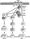
Fig. 8. Model of TRAF6 function in LMP1 signal transduction. For a detailed explanation refer to the text.

Similar articles

Cited by

References

    1. Aicher A., Shu,G.L., Magaletti,D., Mulvania,T., Pezzutto,A., Craxton,A. and Clark,E.A. (1999) Differential role for p38 mitogen-activated protein kinase in regulating CD40-induced gene expression in dendritic cells and B cells. J. Immunol., 163, 5786–5795. - PubMed
    1. Arch R.H., Gedrich,R.W. and Thompson,C.B. (1998) Tumor necrosis factor receptor-associated factors (TRAFs) — a family of adapter proteins that regulates life and death. Genes Dev., 12, 2821–2830. - PubMed
    1. Aviel S., Winberg,G., Massucci,M. and Ciechanover,A. (2000) Degradation of the Epstein–Barr virus latent membrane protein 1 (LMP1) by the ubiquitin-proteasome pathway. Targeting via ubiquitination of the N-terminal residue. J. Biol. Chem., 275, 23491–23499. - PubMed
    1. Baichwal V.R. and Sugden,B. (1988) Transformation of Balb 3T3 cells by the BNLF-1 gene of Epstein–Barr virus. Oncogene, 2, 461–467. - PubMed
    1. Baud V., Liu,Z.G., Bennett,B., Suzuki,N., Xia,Y. and Karin,M. (1999) Signaling by proinflammatory cytokines: oligomerization of TRAF2 and TRAF6 is sufficient for JNK and IKK activation and target gene induction via an amino-terminal effector domain. Genes Dev., 13, 1297–1308. - PMC - PubMed

Publication types

MeSH terms

Substances