. 2001 Oct;69(10):6348-63.
doi: 10.1128/IAI.69.10.6348-6363.2001.

High extracellular levels of Mycobacterium tuberculosis glutamine synthetase and superoxide dismutase in actively growing cultures are due to high expression and extracellular stability rather than to a protein-specific export mechanism

Affiliations

High extracellular levels of Mycobacterium tuberculosis glutamine synthetase and superoxide dismutase in actively growing cultures are due to high expression and extracellular stability rather than to a protein-specific export mechanism

M V Tullius et al. Infect Immun. 2001 Oct.

Abstract

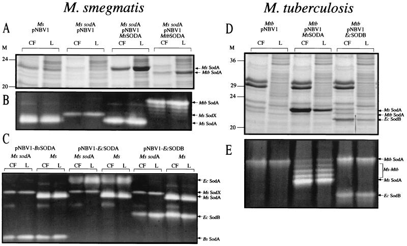
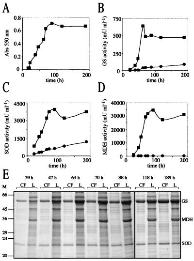
Glutamine synthetase (GS) and superoxide dismutase (SOD), large multimeric enzymes that are thought to play important roles in the pathogenicity of Mycobacterium tuberculosis, are among the bacterium's major culture filtrate proteins in actively growing cultures. Although these proteins lack a leader peptide, their presence in the extracellular medium during early stages of growth suggested that they might be actively secreted. To understand their mechanism of export, we cloned the homologous genes (glnA1 and sodA) from the rapid-growing, nonpathogenic Mycobacterium smegmatis, generated glnA1 and sodA mutants of M. smegmatis by allelic exchange, and quantitated expression and export of both mycobacterial and nonmycobacterial GSs and SODs in these mutants. We also quantitated expression and export of homologous and heterologous SODs from M. tuberculosis. When each of the genes was expressed from a multicopy plasmid, M. smegmatis exported comparable proportions of both the M. tuberculosis and M. smegmatis GSs (in the glnA1 strain) or SODs (in the sodA strain), in contrast to previous observations in wild-type strains. Surprisingly, recombinant M. smegmatis and M. tuberculosis strains even exported nonmycobacterial SODs. To determine the extent to which export of these large, leaderless proteins is expression dependent, we constructed a recombinant M. tuberculosis strain expressing green fluorescent protein (GFP) at high levels and a recombinant M. smegmatis strain coexpressing the M. smegmatis GS, M. smegmatis SOD, and M. tuberculosis BfrB (bacterioferritin) at high levels. The recombinant M. tuberculosis strain exported GFP even in early stages of growth and at proportions very similar to those of the endogenous M. tuberculosis GS and SOD. Similarly, the recombinant M. smegmatis strain exported bacterioferritin, a large (approximately 500-kDa), leaderless, multimeric protein, in proportions comparable to GS and SOD. In contrast, high-level expression of the large, leaderless, multimeric protein malate dehydrogenase did not lead to extracellular accumulation because the protein was highly unstable extracellularly. These findings indicate that, contrary to expectations, export of M. tuberculosis GS and SOD in actively growing cultures is not due to a protein-specific export mechanism, but rather to bacterial leakage or autolysis, and that the extracellular abundance of these enzymes is simply due to their high level of expression and extracellular stability. The same determinants likely explain the presence of other leaderless proteins in the extracellular medium of actively growing M. tuberculosis cultures.


Figures

FIG. 1
FIG. 1
Nonpolar kanamycin resistance cassette cloned into the SmaI site of pUC19. The cassette contains the kanamycin resistance gene (aphA-2) coding region but lacks both a promoter and a transcriptional terminator. The stop codons in all three frames immediately upstream of aphA-2 are underlined, as is the stop codon of the gene. The start codon of aphA-2 and the start codon provided at the 3′ end of the cassette are shown in boldface type. The 15 nucleotides immediately downstream of the BclI site are identical to the sequence directly upstream of the M. tuberculosis dnaK gene (the ribosomal binding site [rbs] is indicated). NdeI and BclI sites were included to facilitate cloning of other resistance genes into the cassette. Abbreviations for restriction sites: H, HindIII; Sh, SphI; P, PstI; Sl, SalI; X, XbaI; B, BamHI; K, KpnI; Sc, SacI; E, EcoRI.
FIG. 2
FIG. 2
Maps of the M. smegmatis 1-2c glnA1 and sodA genomic loci. Both the glnA1 (A) and the sodA (C) loci were isolated as BamHI→EcoRI (5′-to-3′) genomic fragments. Both genes have putative transcriptional terminators (T) directly downstream of their coding regions. The glnA1 locus contains a truncated open reading frame (orf) with a high degree of similarity to the M. tuberculosis Rv1897c gene. The sodA locus contains a truncated open reading frame upstream of sodA with a predicted protein sequence that is highly similar (75% identity) to a truncated open reading frame in the same location in the M. fortuitum sodA locus (44). The glnA1 and the sodA loci were cloned into the multiple cloning site of pNBV1 for expression of GS and SOD in M. smegmatis and M. tuberculosis. The Kmr cassette (Fig. 1) was inserted into the unique XhoI site of glnA1 (B) and the BstEII site of sodA (D) to generate the disrupted loci used for allelic exchange.
FIG. 3
FIG. 3
ECHXB adapter. EcoRI adapter with several restriction sites useful for cloning into the multiple cloning site of pNBV1 and other vectors.
FIG. 4
FIG. 4
Southern analysis of M. smegmatis glnA1 and sodA mutants. Genomic DNA from M. smegmatis 1-2c (wild type [wt]), M. smegmatis glnA1, and M. smegmatis sodA strains was digested with BamHI and EcoRI and probed with the entire glnA1 locus (Fig. 2A) (A) or the entire sodA locus (Fig. 2C) (B). M, molecular mass markers in kilobases.
FIG. 5
FIG. 5
Maps of the mycobacterial expression constructs. All of the constructs were cloned into the HindIII and BamHI or XbaI sites of the multiple cloning site of pNBV1. (A) The M. tuberculosis mdh gene was cloned downstream of the M. tuberculosis glnA1 promoter (pGS). The following genes were cloned in the same way as mdh: E. coli sodA, E. coli sodB, B. subtilis sodA, and S. enterica serovar Typhimurium glnA. (B) The M. tuberculosis bfrB gene was cloned downstream of an M. tuberculosis glnA1 promoter (pGS′) modified to have an NdeI site at the start codon. (C) The UV-optimized GFP gene (gfpuv) was cloned downstream of an M. bovis BCG hsp60 promoter (pHSP60′) modified to contain an NdeI site at the start codon. (D and E) A 3.3-kb HindIII fragment containing the M. smegmatis glnA1 and sodA loci was cloned into the HindIII site of pNBV1-MDH (A) or pNBV1-BFRB (B). The M. smegmatis glnA1 and sodA genes were transcribed from their own promoters.
FIG. 6
FIG. 6
GS expression and export in M. smegmatis wild-type and M. smegmatis glnA1 strains. SDS-PAGE analysis of culture filtrates (CF) and lysates (L). Tenfold more CF than L was loaded on the gel (the equivalent of 2 ml versus 0.2 ml of the original culture volume). The results are representative of three independent cultures. Arrows indicate the positions of the M. smegmatis (Ms) and M. tuberculosis (Mtb) GSs. M, molecular mass markers in kilodaltons.
FIG. 7
FIG. 7
SOD expression and export in M. smegmatis wild type, M. smegmatis sodA, and M. tuberculosis wild-type strains. M. smegmatis strains were analyzed for SOD expression by SDS-PAGE (A) and SOD activity gel (B and C). M. tuberculosis strains were also analyzed for SOD expression by SDS-PAGE (D) and SOD activity gel (E). Tenfold more culture filtrate (CF) than lysate (L), based on the original culture volume, was loaded on the gels for each analysis (4 ml of CF and 0.4 ml of L [A and D], 6 ml of CF and 0.6 ml of L [B and C], and 2 ml of CF and 0.2 ml of L [E]). The results are representative of two or three independent cultures. Arrows indicate the positions of the M. smegmatis (Ms) SodA, M. tuberculosis (Mtb) SodA, M. smegmatis-M. tuberculosis hybrid SODs (Ms-Mtb), E. coli (Ec) SodA, E. coli SodB, B. subtilis (Bs) SodA, and the M. smegmatis SOD activity in the sodA mutant (Ms SodX). M, molecular mass markers in kilodaltons.
FIG. 8
FIG. 8
Expression and export of GS, SOD, and MDH by M. smegmatis glnA1 pNBV1-MsGS-MsSODA-MDH. (A) Growth of M. smegmatis glnA1 pNBV1-MsGS-MsSODA-MDH. (B, C, and D) Enzyme activity of GS, SOD, and MDH in the culture filtrate (●) and lysate (▪) over time. (E) SDS-PAGE analysis of culture filtrates (CF) and lysates (L). Tenfold more CF than L, based on the original culture volume, was loaded on the gels (at 39 and 47 h, 4 ml of CF and 0.4 ml of L; at 63 to 189 h, 2 ml of CF and 0.2 ml of L). The positions of the M. smegmatis GS, M. smegmatis SOD, and the M. tuberculosis MDH are indicated. M, molecular mass markers in kilodaltons.
FIG. 9
FIG. 9
Stability of MDH in 7H9 culture filtrate. (A) SDS-PAGE analysis of purified MDH (lane 1) and its stability after addition to culture filtrate. An amount equivalent to 2 ml of the original culture volume was loaded for each culture filtrate time point. The positions of the M. smegmatis GS, M. smegmatis SOD, and the M. tuberculosis MDH are indicated. The purified MDH was judged to be 85% pure by analysis with NIH Image software (version 1.62). (B) The percentage of initial activity of MDH and GS is listed beneath each lane, with the activity at day zero defined to be 100%. Abbreviations: ND, not determined; M, molecular mass markers in kilodaltons.
FIG. 10
FIG. 10
Expression and export of GS, SOD, and BfrB by M. smegmatis glnA1 pNBV1-MsSODA-MsGS-BFRB. (A) Growth of M. smegmatis glnA1 pNBV1-MsGS-MsSODA-BFRB. (B) SDS-PAGE analysis of culture filtrates (CF) and lysates (L). Tenfold more CF than L, based on the original culture volume, was loaded on the gels (at 19 and 39 h, 8 ml of CF and 0.8 ml of L; at 47 h, 4 ml of CF and 0.4 ml of L; at 67 and 87 h, 2 ml of CF and 0.2 ml of L). The percentages of extracellular GS, SOD, and BfrB (listed below the gel) were determined with NIH Image software (version 1.62). Arrows indicate the positions of the M. smegmatis GS, M. smegmatis SOD, and the M. tuberculosis BfrB (BfrB). M, molecular mass markers in kilodaltons.
FIG. 11
FIG. 11
Expression and export of GS, SOD, and GFPuv by M. tuberculosis pNBV1-GFPuv. (A) Growth of M. tuberculosis pNBV1-GFPuv. (B) SDS-PAGE analysis of culture filtrates (CF) and lysates (L). Tenfold more CF than L, based on the original culture volume, was loaded on the gels (at 10 days; 15 ml of CF and 1.5 ml of L; at 15 days, 6 ml of CF and 0.6 ml of L; at 22 to 38 days, 2 ml of CF and 0.2 ml of L). (C to E) Immunoblot analysis of GS, GFPuv, and SOD. (F) Autoradiogram of CF and L from M. tuberculosis pNBV1-GFPuv metabolically labeled with [35S]l-methionine and [35S]l-cysteine. The bacteria were grown for 4 days, radiolabel was added, and aliquots were removed for analysis 7, 23, 48, and 72 h later. The culture was actively growing over the 72-h period, increasing in turbidity (A550) from 0.18 to 0.28. Twentyfold more CF than L was loaded on the gel (the equivalent of 2 ml versus 0.1 ml of original culture volume). Arrows indicate the positions of the M. tuberculosis GS, M. tuberculosis SOD, and GFPuv. M, molecular mass markers in kilodaltons.
FIG. 12
FIG. 12
Expression and export of GS, SOD, and BfrB by M. smegmatis glnA1 pNBV1-MsSODA-MsGS-BFRB under various growth conditions. (A to C) M. smegmatis glnA1 pNBV1-MsGS-MsSODA-BFRB was grown for 4 days (cultures reached stationary phase by 2 to 3 days) in 7H9 medium containing 0.2% (vol/vol) glycerol with various amounts of glucose and/or additional (NH4)2SO4, and culture filtrates (CF) and lysates (L) were analyzed by SDS-PAGE. Tenfold more CF than L was loaded on the gels (the equivalent of 2 ml versus 0.2 ml of original culture volume). Arrows indicate the positions of the M. smegmatis GS, M. smegmatis SOD, and the M. tuberculosis BfrB (BfrB). Two to four independent cultures were grown and analyzed for each of the four extreme culture conditions (1, 5, 6, and 10) as well as for culture condition 2, and the results shown are representative. (D) Graph indicating the glucose and (NH4)2SO4 concentrations of the media that were tested in panels A to C. The standard 7H9 medium (culture 1) contains 82 mM carbon (as 0.2% [vol/vol] glycerol) and 11 mM nitrogen [as 3.8 mM (NH4)2SO4 and 3.4 mM l-glutamate]. The (NH4)2SO4 (AmS) concentration is stated as the total amount present in the medium for each culture condition. M, molecular mass markers in kilodaltons.

Similar articles

Cited by

References

    1. Alcendor D J, Chapman G D, Beaman B L. Isolation, sequencing and expression of the superoxide dismutase-encoding gene (sod) of Nocardia asteroides strain GUH-2. Gene. 1995;164:143–147. - PubMed
    1. Alito A, Romano M I, Bigi F, Zumarraga M, Cataldi A. Antigenic characterization of mycobacteria from South American wild seals. Vet Microbiol. 1999;68:293–299. - PubMed
    1. Altschul S F, Madden T L, Schaffer A A, Zhang J, Zhang Z, Miller W, Lipman D J. Gapped BLAST and PSI-BLAST: a new generation of protein database search programs. Nucleic Acids Res. 1997;25:3389–3402. - PMC - PubMed
    1. Andersen P. Effective vaccination of mice against Mycobacterium tuberculosisinfection with a soluble mixture of secreted mycobacterial proteins. Infect Immun. 1994;62:2536–2544. - PMC - PubMed
    1. Andersen P, Askgaard D, Ljungqvist L, Bennedsen J, Heron I. Proteins released from Mycobacterium tuberculosisduring growth. Infect Immun. 1991;59:1905–1910. - PMC - PubMed

Publication types

MeSH terms