The histone deacetylase inhibitor and chemotherapeutic agent suberoylanilide hydroxamic acid (SAHA) induces a cell-death pathway characterized by cleavage of Bid and production of reactive oxygen species
- PMID: 11535817
- PMCID: PMC58560
- DOI: 10.1073/pnas.191208598
The histone deacetylase inhibitor and chemotherapeutic agent suberoylanilide hydroxamic acid (SAHA) induces a cell-death pathway characterized by cleavage of Bid and production of reactive oxygen species
Abstract
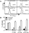
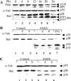
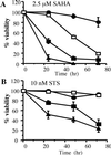
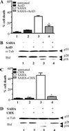
Many chemotherapeutic agents induce mitochondrial-membrane disruption to initiate apoptosis. However, the upstream events leading to drug-induced mitochondrial perturbation have remained poorly defined. We have used a variety of physiological and pharmacological inhibitors of distinct apoptotic pathways to analyze the manner by which suberoylanilide hydroxamic acid (SAHA), a chemotherapeutic agent and histone deacetylase inhibitor, induces cell death. We demonstrate that SAHA initiates cell death by inducing mitochondria-mediated death pathways characterized by cytochrome c release and the production of reactive oxygen species, and does not require the activation of key caspases such as caspase-8 or -3. We provide evidence that mitochondrial disruption is achieved by means of the cleavage of the BH3-only proapoptotic Bcl-2 family member Bid. SAHA-induced Bid cleavage was not blocked by caspase inhibitors or the overexpression of Bcl-2 but did require the transcriptional regulatory activity of SAHA. These data provide evidence of a mechanism of cell death mediated by transcriptional events that result in the cleavage of Bid, disruption of the mitochondrial membrane, and production of reactive oxygen species to induce cell death.
Figures


Similar articles
-
Role of caspases, Bid, and p53 in the apoptotic response triggered by histone deacetylase inhibitors trichostatin-A (TSA) and suberoylanilide hydroxamic acid (SAHA).J Biol Chem. 2003 Apr 4;278(14):12579-89. doi: 10.1074/jbc.M213093200. Epub 2003 Jan 29. J Biol Chem. 2003. PMID: 12556448
-
Suberoylanilide hydroxamic acid (SAHA) overcomes multidrug resistance and induces cell death in P-glycoprotein-expressing cells.Int J Cancer. 2002 May 10;99(2):292-8. doi: 10.1002/ijc.10327. Int J Cancer. 2002. PMID: 11979447
-
Cadmium induces apoptotic cell death in WI 38 cells via caspase-dependent Bid cleavage and calpain-mediated mitochondrial Bax cleavage by Bcl-2-independent pathway.Biochem Pharmacol. 2004 Nov 1;68(9):1845-55. doi: 10.1016/j.bcp.2004.06.021. Biochem Pharmacol. 2004. PMID: 15450950
-
Histone deacetylase inhibitors in programmed cell death and cancer therapy.Cell Cycle. 2005 Apr;4(4):549-51. doi: 10.4161/cc.4.4.1564. Epub 2005 Apr 28. Cell Cycle. 2005. PMID: 15738652 Review.
-
The roles of Bid.Apoptosis. 2002 Oct;7(5):433-40. doi: 10.1023/a:1020035124855. Apoptosis. 2002. PMID: 12207176 Review.
Cited by
-
Valproic acid, an antiepileptic drug with histone deacetylase inhibitory activity, potentiates the cytotoxic effect of Apo2L/TRAIL on cultured thoracic cancer cells through mitochondria-dependent caspase activation.Neoplasia. 2006 Jun;8(6):446-57. doi: 10.1593/neo.05823. Neoplasia. 2006. PMID: 16820090 Free PMC article.
-
Class I HDAC inhibitors enhance YB-1 acetylation and oxidative stress to block sarcoma metastasis.EMBO Rep. 2019 Dec 5;20(12):e48375. doi: 10.15252/embr.201948375. Epub 2019 Oct 31. EMBO Rep. 2019. PMID: 31668005 Free PMC article.
-
Bid mediates anti-apoptotic COX-2 induction through the IKKbeta/NFkappaB pathway due to 5-MCDE exposure.Curr Cancer Drug Targets. 2010 Feb;10(1):96-106. doi: 10.2174/156800910790980160. Curr Cancer Drug Targets. 2010. PMID: 20088789 Free PMC article.
-
MHC class II regulation by epigenetic agents and microRNAs.Immunol Res. 2010 Mar;46(1-3):45-58. doi: 10.1007/s12026-009-8128-3. Immunol Res. 2010. PMID: 19771399 Free PMC article. Review.
-
Differential Mechanisms of Cell Death Induced by HDAC Inhibitor SAHA and MDM2 Inhibitor RG7388 in MCF-7 Cells.Cells. 2018 Dec 22;8(1):8. doi: 10.3390/cells8010008. Cells. 2018. PMID: 30583560 Free PMC article.
References
Publication types
MeSH terms
Substances
LinkOut - more resources
Full Text Sources
Other Literature Sources
Research Materials
Miscellaneous

