. 2001 Sep 3;154(5):937-50.
doi: 10.1083/jcb.200101007.

The nucleoporin Nup60p functions as a Gsp1p-GTP-sensitive tether for Nup2p at the nuclear pore complex

Affiliations

The nucleoporin Nup60p functions as a Gsp1p-GTP-sensitive tether for Nup2p at the nuclear pore complex

D Denning et al. J Cell Biol. .

Abstract

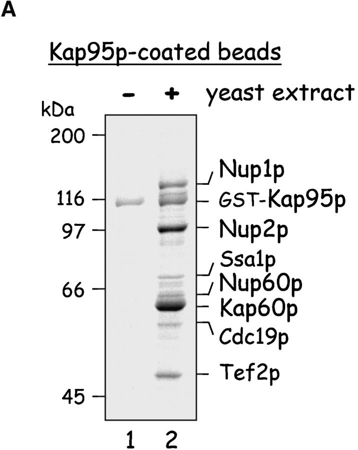
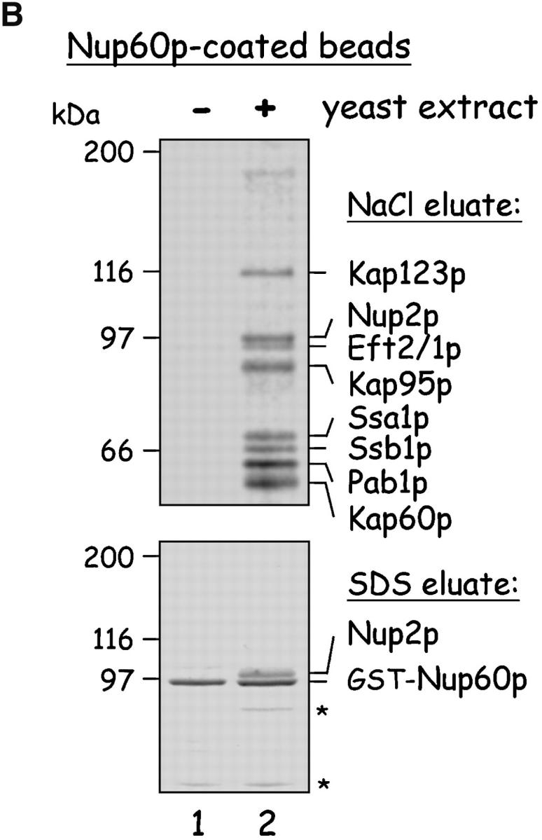
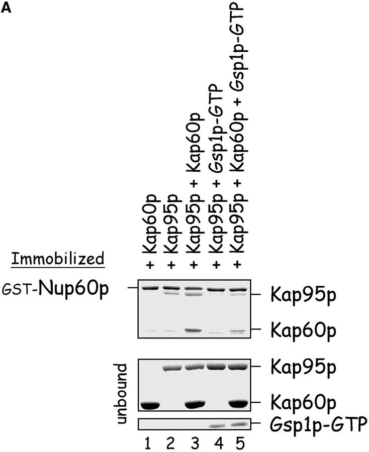
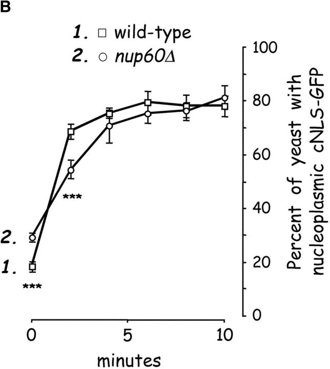
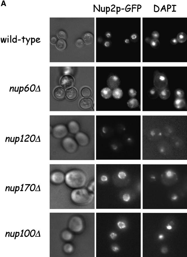
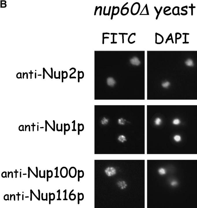
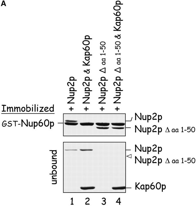
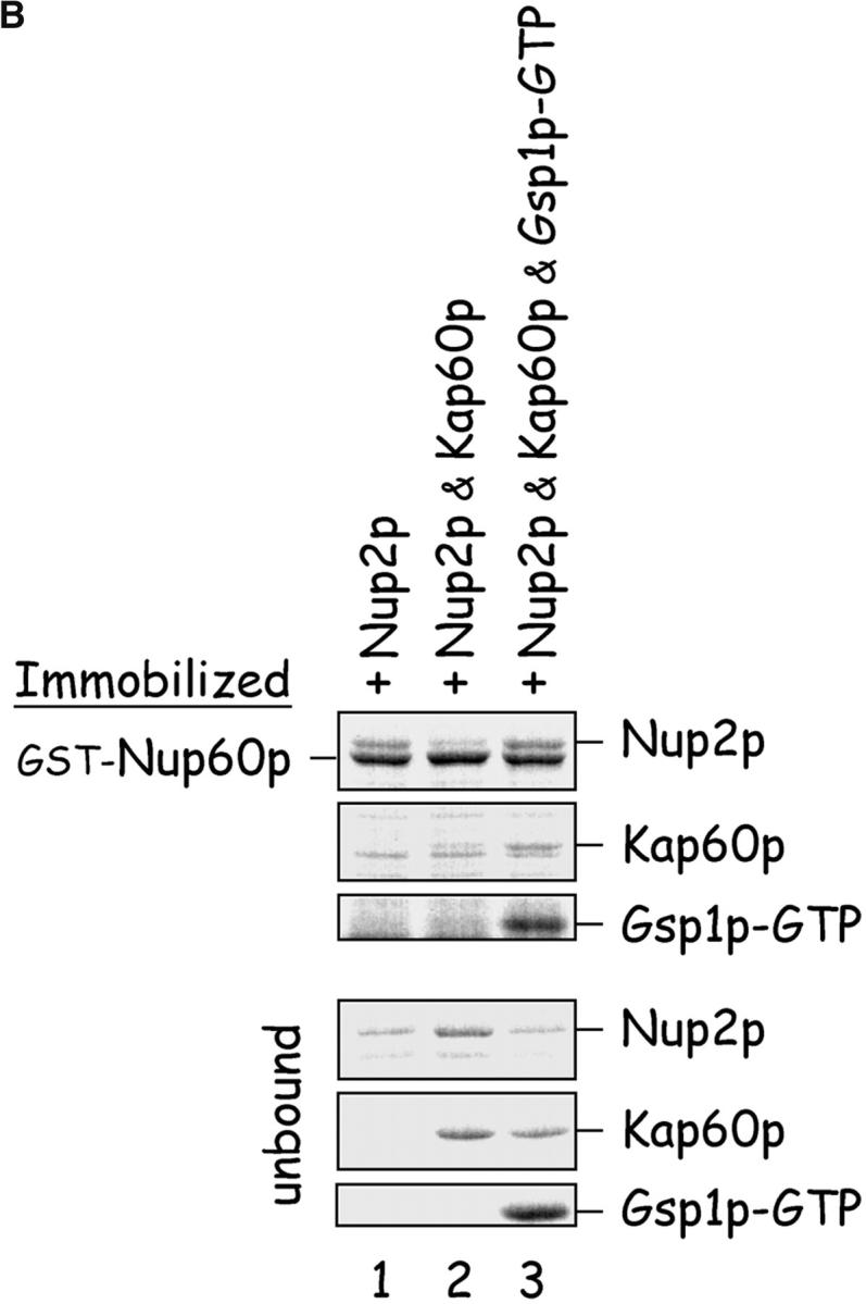
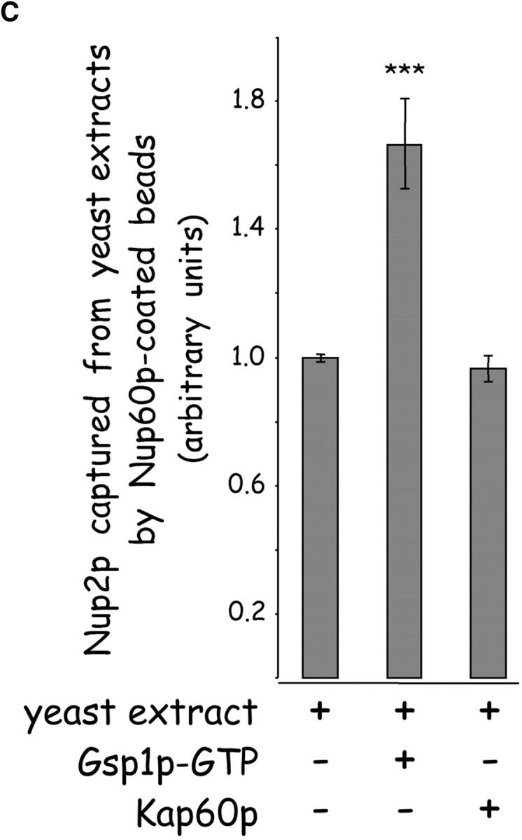
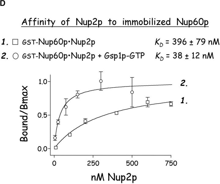
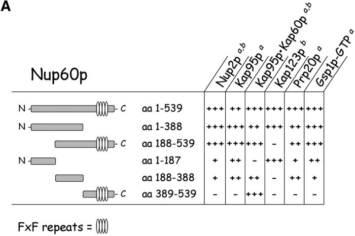
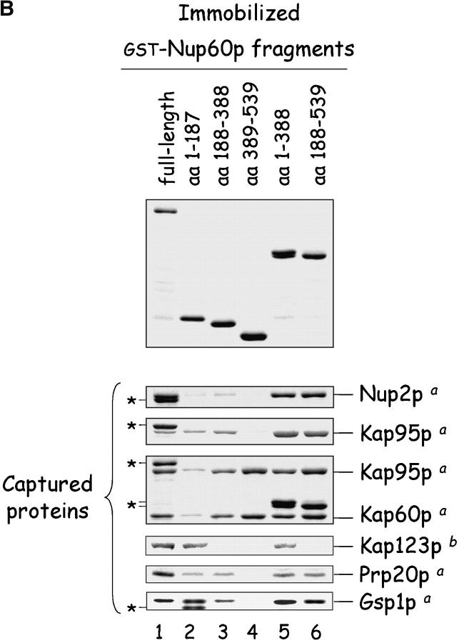
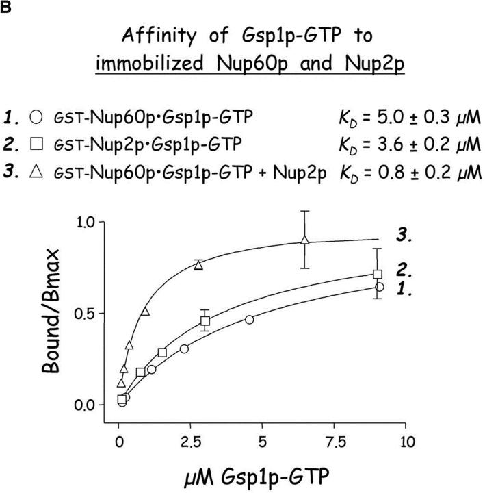
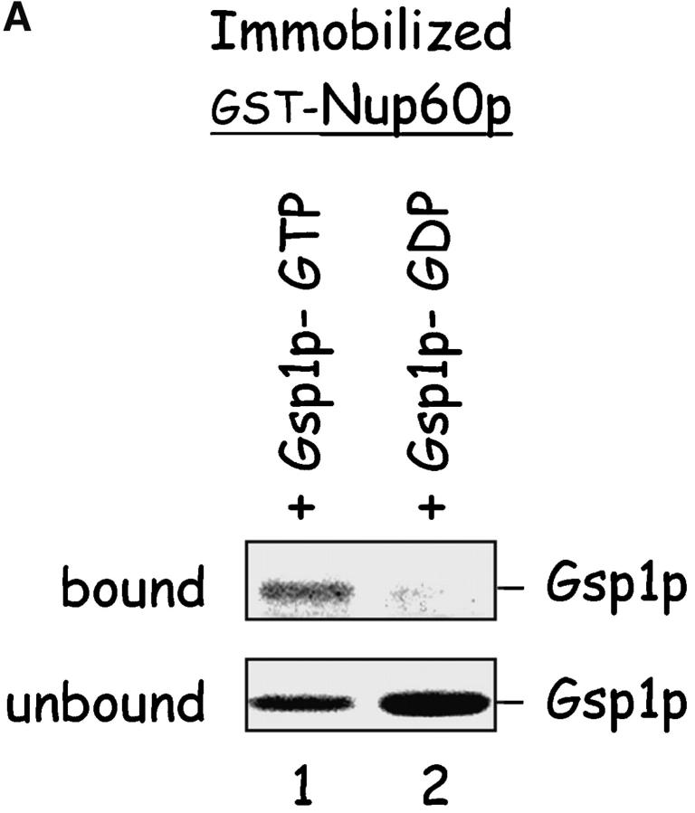
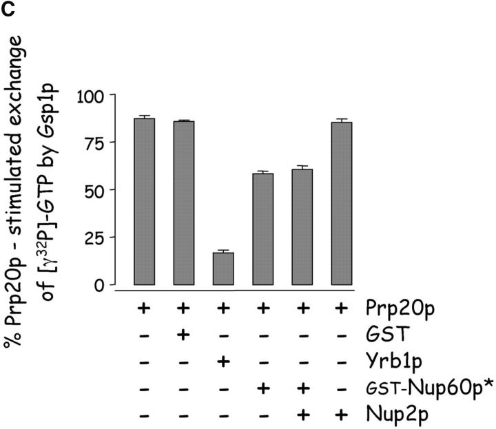
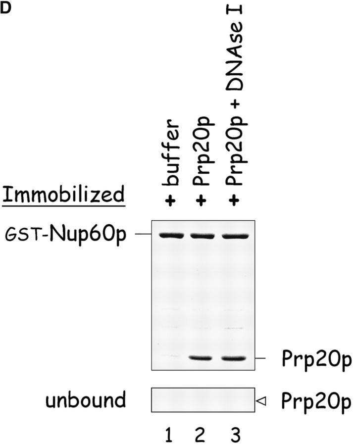
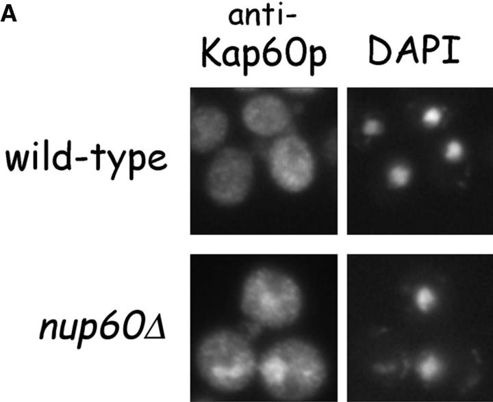
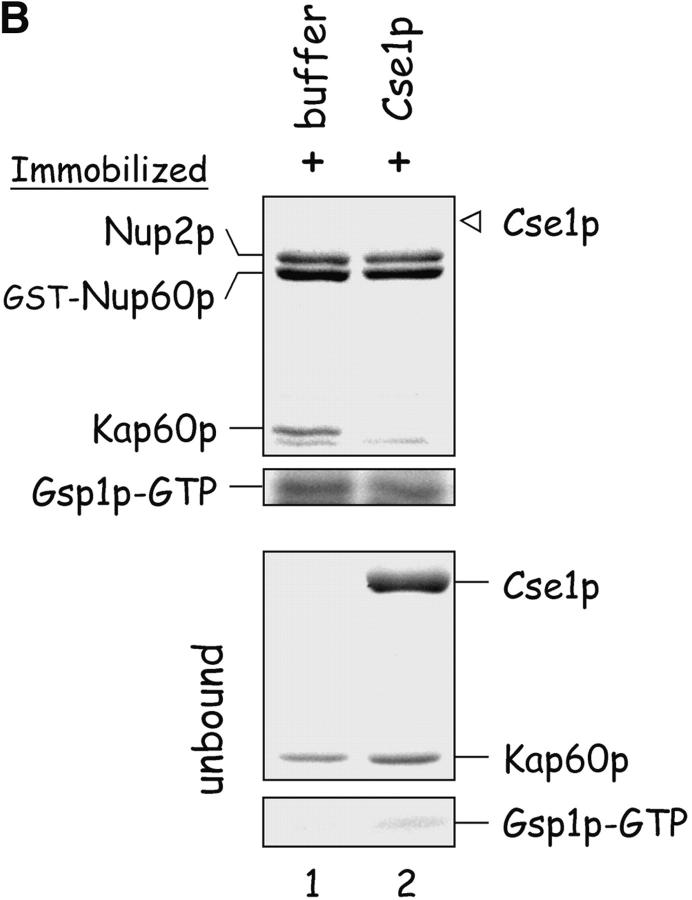
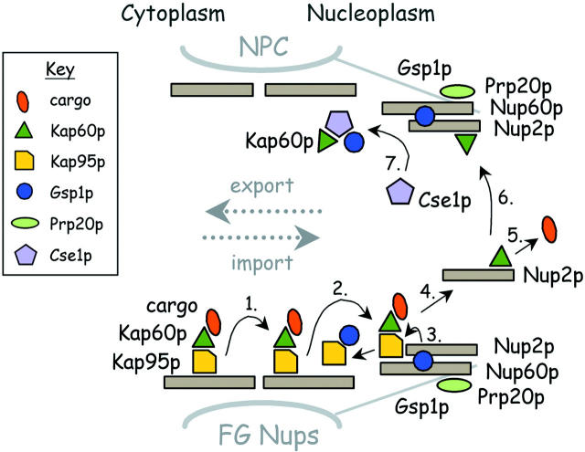
The nucleoporins Nup60p, Nup2p, and Nup1p form part of the nuclear basket structure of the Saccharomyces cerevisiae nuclear pore complex (NPC). Here, we show that these necleoporins can be isolated from yeast extracts by affinity chromatography on karyopherin Kap95p-coated beads. To characterize Nup60p further, Nup60p-coated beads were used to capture its interacting proteins from extracts. We find that Nup60p binds to Nup2p and serves as a docking site for Kap95p-Kap60p heterodimers and Kap123p. Nup60p also binds Gsp1p-GTP and its guanine nucleotide exchange factor Prp20p, and functions as a Gsp1p guanine nucleotide dissociation inhibitor by reducing the activity of Prp20p. Yeast lacking Nup60p exhibit minor defects in nuclear export of Kap60p, nuclear import of Kap95p-Kap60p-dependent cargoes, and diffusion of small proteins across the NPC. Yeast lacking Nup60p also fail to anchor Nup2p at the NPC, resulting in the mislocalization of Nup2p to the nucleoplasm and cytoplasm. Purified Nup60p and Nup2p bind each other directly, but the stability of the complex is compromised when Kap60p binds Nup2p. Gsp1p-GTP enhances by 10-fold the affinity between Nup60p and Nup2p, and restores binding of Nup2p-Kap60p complexes to Nup60p. The results suggest a dynamic interaction, controlled by the nucleoplasmic concentration of Gsp1p-GTP, between Nup60p and Nup2p at the NPC.


Figures

Figure 1.
Figure 1.
Yeast proteins that bind Nup60p and Kap95p. (A) Proteins in yeast extracts captured on Kap95p-coated Sepharose beads. GST-Kap95p (5 μg) was immobilized on glutathione-coated Sepharose beads (beads) and incubated with yeast extract (10 mg protein) or buffer as indicated. After washing beads, bound proteins were eluted with 250 mM MgCl2, collected by precipitation with trichloroacetic acid and deoxycholate, resolved by SDS-PAGE, and stained with Coomassie blue. Visible proteins were identified by mass spectrometry (see Materials and methods). Note that the three Nups captured by Kap95p are components of the nuclear basket structure of the yeast NPC. (B) Proteins in yeast extracts captured on Nup60p-coated beads. GST-Nup60p (1 μg) was immobilized on the beads and incubated with yeast extract (10 mg protein) or buffer as before. Bound proteins were eluted with 1 M NaCl (top) followed by SDS (bottom) and were identified as before. The asterisks mark a degradation product of GST–Nup60p. The identity of the ∼180-kD protein in the top panel could not be determined. Note that a portion of Nup2p remained tightly bound to Nup60p-coated beads even after incubation in 1 M NaCl.
Figure 1.
Figure 1.
Yeast proteins that bind Nup60p and Kap95p. (A) Proteins in yeast extracts captured on Kap95p-coated Sepharose beads. GST-Kap95p (5 μg) was immobilized on glutathione-coated Sepharose beads (beads) and incubated with yeast extract (10 mg protein) or buffer as indicated. After washing beads, bound proteins were eluted with 250 mM MgCl2, collected by precipitation with trichloroacetic acid and deoxycholate, resolved by SDS-PAGE, and stained with Coomassie blue. Visible proteins were identified by mass spectrometry (see Materials and methods). Note that the three Nups captured by Kap95p are components of the nuclear basket structure of the yeast NPC. (B) Proteins in yeast extracts captured on Nup60p-coated beads. GST-Nup60p (1 μg) was immobilized on the beads and incubated with yeast extract (10 mg protein) or buffer as before. Bound proteins were eluted with 1 M NaCl (top) followed by SDS (bottom) and were identified as before. The asterisks mark a degradation product of GST–Nup60p. The identity of the ∼180-kD protein in the top panel could not be determined. Note that a portion of Nup2p remained tightly bound to Nup60p-coated beads even after incubation in 1 M NaCl.
Figure 5.
Figure 5.
Nup60p is a docking site for Kap95p–Kap60p heterodimers. (A) Nup60p binds Kap95p, but only in the absence of Gsp1p–GTP. GST–Nup60p (1 μg) was immobilized on beads and incubated with Kap95p (2 μg), Kap60p (3 μg), and/or Gsp1p–GTP Q71L (0.5 μg). After 1 h at 4°C, unbound and bound proteins were collected, resolved by SDS-PAGE, and stained with Coomassie blue. Note that Kap95p monomers bind to Nup60p, that Kap60p enhances binding of Kap95p to Nup60p, and that Gsp1p–GTP blocks binding of Kap95p and Kap95p–Kap60p heterodimers to Nup60p. (B) Nup60p plays a minor role in Kap95p–Kap60p-dependent import of cNLS-bearing cargo into the nucleus. Wild-type and nup60Δ yeast expressing the SV-40 T-antigen NLS fused to GFP were metabolically poisoned to deplete intracellular ATP and assayed for recovery of nuclear import upon removal of the poison (see Materials and methods). Values plotted at the indicated time points represent the mean fraction of yeast with predominantly nucleoplasmic NLS-GFP from six separate experiments (error bars represent SEM). The asterisks (***) indicate P < 0.01 for comparison of the two values at the indicated time points (unpaired, two-tailed t test). Note that yeast lacking Nup60p exhibit a slower initial rate of cNLS–GFP nuclear import.
Figure 5.
Figure 5.
Nup60p is a docking site for Kap95p–Kap60p heterodimers. (A) Nup60p binds Kap95p, but only in the absence of Gsp1p–GTP. GST–Nup60p (1 μg) was immobilized on beads and incubated with Kap95p (2 μg), Kap60p (3 μg), and/or Gsp1p–GTP Q71L (0.5 μg). After 1 h at 4°C, unbound and bound proteins were collected, resolved by SDS-PAGE, and stained with Coomassie blue. Note that Kap95p monomers bind to Nup60p, that Kap60p enhances binding of Kap95p to Nup60p, and that Gsp1p–GTP blocks binding of Kap95p and Kap95p–Kap60p heterodimers to Nup60p. (B) Nup60p plays a minor role in Kap95p–Kap60p-dependent import of cNLS-bearing cargo into the nucleus. Wild-type and nup60Δ yeast expressing the SV-40 T-antigen NLS fused to GFP were metabolically poisoned to deplete intracellular ATP and assayed for recovery of nuclear import upon removal of the poison (see Materials and methods). Values plotted at the indicated time points represent the mean fraction of yeast with predominantly nucleoplasmic NLS-GFP from six separate experiments (error bars represent SEM). The asterisks (***) indicate P < 0.01 for comparison of the two values at the indicated time points (unpaired, two-tailed t test). Note that yeast lacking Nup60p exhibit a slower initial rate of cNLS–GFP nuclear import.
Figure 2.
Figure 2.
Nup2p is tethered to the NPC via Nup60p. (A) Direct visualization of Nup2p–GFP fusions in yeast. Various yeast strains that express NUP2-GFP from the NUP2 locus were grown in rich media at 30°C and were observed live under a fluorescence microscope. DAPI stain was used to visualize DNA in nuclei and pictures were taken using nuclei as the focal point. Note that in wild-type, nup170Δ, and nup100Δ yeast, Nup2p–GFP fusions accumulate in a punctate pattern at the nuclear periphery, but in nup60Δ yeast Nup2p–GFP is mislocalized to the nucleoplasm and cytoplasm. (B) Indirect immunofluorescence visualization of Nup2p, Nup1p, and Nup100p/Nup116p in nup60Δ yeast. nup60Δ yeast grown to early log phase in rich media at 30°C were fixed in 3.7% formaldehyde for 10 min and processed for immunofluorescence microscopy using affinity-purified anti-Nup antibodies and FITC-labeled secondary antibodies (left). DAPI was used to visualize nuclei (right). Note the mislocalization of Nup2p to the nucleoplasm in nup60Δ yeast in contrast to the normal punctate staining of Nup1p and Nup100p/Nup116p at the nuclear envelope.
Figure 2.
Figure 2.
Nup2p is tethered to the NPC via Nup60p. (A) Direct visualization of Nup2p–GFP fusions in yeast. Various yeast strains that express NUP2-GFP from the NUP2 locus were grown in rich media at 30°C and were observed live under a fluorescence microscope. DAPI stain was used to visualize DNA in nuclei and pictures were taken using nuclei as the focal point. Note that in wild-type, nup170Δ, and nup100Δ yeast, Nup2p–GFP fusions accumulate in a punctate pattern at the nuclear periphery, but in nup60Δ yeast Nup2p–GFP is mislocalized to the nucleoplasm and cytoplasm. (B) Indirect immunofluorescence visualization of Nup2p, Nup1p, and Nup100p/Nup116p in nup60Δ yeast. nup60Δ yeast grown to early log phase in rich media at 30°C were fixed in 3.7% formaldehyde for 10 min and processed for immunofluorescence microscopy using affinity-purified anti-Nup antibodies and FITC-labeled secondary antibodies (left). DAPI was used to visualize nuclei (right). Note the mislocalization of Nup2p to the nucleoplasm in nup60Δ yeast in contrast to the normal punctate staining of Nup1p and Nup100p/Nup116p at the nuclear envelope.
Figure 3.
Figure 3.
Gsp1p–GTP and Kap60p modulate the interaction between Nup60p and Nup2p. (A) The interaction between Nup2p and Nup60p, and the effect of Kap60p. GST–Nup60p (1 μg) was immobilized on beads and incubated with Nup2p (0.5 μg), Nup2pΔ (aa 1–50) (0.5 μg), or Kap60p (1 μg) as indicated. After 1 h at 4°C, unbound and bound proteins were collected, resolved by SDS-PAGE, and visualized with Coomassie blue. Note that Nup2p binds Nup60p, that Kap60p prevents the interaction, and that the NH2 terminus of Nup2p is not required for binding Nup60p. (B) Effect of Gsp1p–GTP on the interaction between Nup2p and Nup60p. GST–Nup60p (1 μg) was immobilized on beads and incubated with Nup2p (0.5 μg), Kap60p (1 μg), or Gsp1p–GTP (His-Gsp1p Q71L) (1 μg) as before. Note that Kap60p interferes with the interaction of Nup2p with Nup60p, but that the presence of Gsp1p–GTP restores binding and promotes formation of Nup60p–Gsp1p–Nup2p–Kap60p complexes. (C) Gsp1p–GTP enhances binding of Nup2p to Nup60p in yeast extracts. GST-Nup60p (1 μg) was immobilized on beads and was incubated with yeast extract (∼1 mg) supplemented with 1.25 μM recombinant Gsp1p–GTP (Q71L), 0.5 μM recombinant Kap60p, or no additional protein. The amount of Nup2p bound to Nup60p-coated beads was determined by quantitative Western blotting as described in Materials and methods. The amount of Nup2p was expressed as the ratio of Nup2p bound per unit of immobilized GST–Nup60p, using the incubation of extract without additions as baseline. Shown are the mean ratios for two samples with error bars representing the SEM; this experiment was performed three times with similar results. The asterisks (***) indicate a P < 0.05 for comparison of mean Nup2p captured from extracts supplemented or not with additional Gsp1p–GTP (unpaired, two-tailed t test). Note that addition of Gsp1p–GTP to yeast extract increases by ∼65% the amount of Nup2p bound to Nup60p-coated beads. (D) Gsp1p–GTP increases the affinity between Nup60p and Nup2p. Nup60p-coated beads were incubated with various concentrations of radiolabeled Nup2p for 2 h at 25°C in binding buffer with 10 mg/ml BSA and protease inhibitors. The concentration of GST–Nup60p within the beads was 25 nM and 150 nM for experiments with or without Gsp1p–GTP, respectively. The dissociation constant (KD) of the Nup60p–Nup2p complex in the presence and absence of 3 μM Gsp1p–GTP Q71L was calculated as described in Materials and methods. To facilitate comparison, the results were plotted as a fraction of maximal Nup2p bound versus Nup2p concentration. Each data point was performed in duplicate and error bars represent SEM. Note the 10-fold higher affinity between Nup60p and Nup2p in the presence of Gsp1p–GTP.
Figure 3.
Figure 3.
Gsp1p–GTP and Kap60p modulate the interaction between Nup60p and Nup2p. (A) The interaction between Nup2p and Nup60p, and the effect of Kap60p. GST–Nup60p (1 μg) was immobilized on beads and incubated with Nup2p (0.5 μg), Nup2pΔ (aa 1–50) (0.5 μg), or Kap60p (1 μg) as indicated. After 1 h at 4°C, unbound and bound proteins were collected, resolved by SDS-PAGE, and visualized with Coomassie blue. Note that Nup2p binds Nup60p, that Kap60p prevents the interaction, and that the NH2 terminus of Nup2p is not required for binding Nup60p. (B) Effect of Gsp1p–GTP on the interaction between Nup2p and Nup60p. GST–Nup60p (1 μg) was immobilized on beads and incubated with Nup2p (0.5 μg), Kap60p (1 μg), or Gsp1p–GTP (His-Gsp1p Q71L) (1 μg) as before. Note that Kap60p interferes with the interaction of Nup2p with Nup60p, but that the presence of Gsp1p–GTP restores binding and promotes formation of Nup60p–Gsp1p–Nup2p–Kap60p complexes. (C) Gsp1p–GTP enhances binding of Nup2p to Nup60p in yeast extracts. GST-Nup60p (1 μg) was immobilized on beads and was incubated with yeast extract (∼1 mg) supplemented with 1.25 μM recombinant Gsp1p–GTP (Q71L), 0.5 μM recombinant Kap60p, or no additional protein. The amount of Nup2p bound to Nup60p-coated beads was determined by quantitative Western blotting as described in Materials and methods. The amount of Nup2p was expressed as the ratio of Nup2p bound per unit of immobilized GST–Nup60p, using the incubation of extract without additions as baseline. Shown are the mean ratios for two samples with error bars representing the SEM; this experiment was performed three times with similar results. The asterisks (***) indicate a P < 0.05 for comparison of mean Nup2p captured from extracts supplemented or not with additional Gsp1p–GTP (unpaired, two-tailed t test). Note that addition of Gsp1p–GTP to yeast extract increases by ∼65% the amount of Nup2p bound to Nup60p-coated beads. (D) Gsp1p–GTP increases the affinity between Nup60p and Nup2p. Nup60p-coated beads were incubated with various concentrations of radiolabeled Nup2p for 2 h at 25°C in binding buffer with 10 mg/ml BSA and protease inhibitors. The concentration of GST–Nup60p within the beads was 25 nM and 150 nM for experiments with or without Gsp1p–GTP, respectively. The dissociation constant (KD) of the Nup60p–Nup2p complex in the presence and absence of 3 μM Gsp1p–GTP Q71L was calculated as described in Materials and methods. To facilitate comparison, the results were plotted as a fraction of maximal Nup2p bound versus Nup2p concentration. Each data point was performed in duplicate and error bars represent SEM. Note the 10-fold higher affinity between Nup60p and Nup2p in the presence of Gsp1p–GTP.
Figure 6.
Figure 6.
Mapping of Nup, karyopherin, Prp20p, and Gsp1p–GTP binding sites on Nup60p. (A) The cartoon depicts the Nup60p fragments used in this study provides a summary of the binding interactions observed in B. The + and − designations provide a qualitative assessment of the binding avidity. The absence of detectable binding is denoted by −, whereas +, ++, and +++ represent relative degrees of binding. (B) Binding of karyopherins, Nup2p, Prp20p, and Gsp1p–GTP to various Nup60p fragments. Each GST–Nup60p fragment (1 μg each) was immobilized on beads and incubated with purified Nup2p, Kap95p, Kap95p–Kap60p heterodimers, Prp20p, or Gsp1p–GTP (1 μg each), as indicated. After 1 h at 4°C, the beads were washed and bound proteins were collected, resolved by SDS-PAGE, and visualized with Coomassie blue stain. Alternatively, each GST–Nup60p fragment (5 μg each) was immobilized on beads and incubated with 10 mg of yeast extract for 2 h at 4°C. After washing the beads, bound proteins were eluted with 1 M NaCl, collected by precipitation with trichloroacetic acid and deoxycholate, resolved by SDS-PAGE, and stained with Coomassie blue. Asterisks designate GST–Nup60p fragments used as bait. The superscripts “a” and “b” denote the source of proteins used in the experiments: “a” marks cases where purified recombinant proteins where used, and “b” marks cases where yeast extracts were used. Note the shift in binding site selection of Kap95p in the presence and absence of Kap60p.
Figure 6.
Figure 6.
Mapping of Nup, karyopherin, Prp20p, and Gsp1p–GTP binding sites on Nup60p. (A) The cartoon depicts the Nup60p fragments used in this study provides a summary of the binding interactions observed in B. The + and − designations provide a qualitative assessment of the binding avidity. The absence of detectable binding is denoted by −, whereas +, ++, and +++ represent relative degrees of binding. (B) Binding of karyopherins, Nup2p, Prp20p, and Gsp1p–GTP to various Nup60p fragments. Each GST–Nup60p fragment (1 μg each) was immobilized on beads and incubated with purified Nup2p, Kap95p, Kap95p–Kap60p heterodimers, Prp20p, or Gsp1p–GTP (1 μg each), as indicated. After 1 h at 4°C, the beads were washed and bound proteins were collected, resolved by SDS-PAGE, and visualized with Coomassie blue stain. Alternatively, each GST–Nup60p fragment (5 μg each) was immobilized on beads and incubated with 10 mg of yeast extract for 2 h at 4°C. After washing the beads, bound proteins were eluted with 1 M NaCl, collected by precipitation with trichloroacetic acid and deoxycholate, resolved by SDS-PAGE, and stained with Coomassie blue. Asterisks designate GST–Nup60p fragments used as bait. The superscripts “a” and “b” denote the source of proteins used in the experiments: “a” marks cases where purified recombinant proteins where used, and “b” marks cases where yeast extracts were used. Note the shift in binding site selection of Kap95p in the presence and absence of Kap60p.
Figure 4.
Figure 4.
Nup60p binds Gsp1p–GTP and Prp20p, and functions as a Gsp1p GDI. (A) Nup60p binds Gsp1p–GTP. GST-Nup60p (1 μg) was immobilized on beads and incubated with His-Gsp1p (2 μg) preloaded with GTP or GDP. After 1 h at 4°C, bound and unbound proteins were resolved by SDS-PAGE and visualized with Coomassie blue. Note that Nup60p binds Gsp1p–GTP, but not Gsp1p–GDP. (B) Affinity of Gsp1p–GTP to Nup60p, Nup2p, and Nup60p–Nup2p complexes. GST–Nup-coated beads were incubated with various concentrations of His-Gsp1p–[γ-32P]GTP for 2 h at 4°C in binding buffer with 10 mg/ml BSA and protease inhibitors. The concentrations of GST–Nup60p and GST–Nup2p within beads were 800 nM and 1.5 μM, respectively. The dissociation constants (KD) of the Nup2p–Gsp1p–GTP complex and the Nup60p–Gsp1p–GTP complex in the presence and absence of 500 nM Nup2p were calculated as described in Materials and methods. Results were plotted as a fraction of maximal Gsp1p–GTP bound versus Gsp1p–GTP concentration. Each data point was performed in duplicate and the error bars represent SEM. Note that Nup60p and Nup2p cooperate to bind Gsp1p–GTP. (C) Nup60p inhibits the Prp20p-stimulated release of GTP from Gsp1p. His-Gsp1p–[γ-32P]GTP immobilized on nickel-coated agarose beads (15 nM Gsp1p–GTP within the beads) was incubated with 0.9 nM Prp20p and 1 mM GDP, plus 4 μM GST–Nup60p (aa 188–539), Yrb1p, Nup2p, Kap95p, or GST. GST–Nup60p (aa 188–539) (indicated by asterisk) was used instead of full-length Nup60p due to its superior solubility and protease resistance. After 10 min, Prp20p activity was stopped with ice-cold buffer, beads were washed, and the [γ-32P]GTP that remained bound to the beads was quantified by scintillation counting. Each data point was performed in duplicate and error bars represent SEM. Note that Nup60p reduces (but does not abolish) the activity of Prp20p. (D) Nup60p binds Prp20p. GST–Nup60p (1 μg) was immobilized on beads and incubated with purified Prp20p (1 μg) in the presence or absence of DNAse I and RNAse I (1 U and 1 μg, respectively). After 1 h at 4°C, unbound and bound proteins were resolved by SDS-PAGE and visualized with Coomassie blue staining. Note that purified Prp20p binds Nup60p.
Figure 4.
Figure 4.
Nup60p binds Gsp1p–GTP and Prp20p, and functions as a Gsp1p GDI. (A) Nup60p binds Gsp1p–GTP. GST-Nup60p (1 μg) was immobilized on beads and incubated with His-Gsp1p (2 μg) preloaded with GTP or GDP. After 1 h at 4°C, bound and unbound proteins were resolved by SDS-PAGE and visualized with Coomassie blue. Note that Nup60p binds Gsp1p–GTP, but not Gsp1p–GDP. (B) Affinity of Gsp1p–GTP to Nup60p, Nup2p, and Nup60p–Nup2p complexes. GST–Nup-coated beads were incubated with various concentrations of His-Gsp1p–[γ-32P]GTP for 2 h at 4°C in binding buffer with 10 mg/ml BSA and protease inhibitors. The concentrations of GST–Nup60p and GST–Nup2p within beads were 800 nM and 1.5 μM, respectively. The dissociation constants (KD) of the Nup2p–Gsp1p–GTP complex and the Nup60p–Gsp1p–GTP complex in the presence and absence of 500 nM Nup2p were calculated as described in Materials and methods. Results were plotted as a fraction of maximal Gsp1p–GTP bound versus Gsp1p–GTP concentration. Each data point was performed in duplicate and the error bars represent SEM. Note that Nup60p and Nup2p cooperate to bind Gsp1p–GTP. (C) Nup60p inhibits the Prp20p-stimulated release of GTP from Gsp1p. His-Gsp1p–[γ-32P]GTP immobilized on nickel-coated agarose beads (15 nM Gsp1p–GTP within the beads) was incubated with 0.9 nM Prp20p and 1 mM GDP, plus 4 μM GST–Nup60p (aa 188–539), Yrb1p, Nup2p, Kap95p, or GST. GST–Nup60p (aa 188–539) (indicated by asterisk) was used instead of full-length Nup60p due to its superior solubility and protease resistance. After 10 min, Prp20p activity was stopped with ice-cold buffer, beads were washed, and the [γ-32P]GTP that remained bound to the beads was quantified by scintillation counting. Each data point was performed in duplicate and error bars represent SEM. Note that Nup60p reduces (but does not abolish) the activity of Prp20p. (D) Nup60p binds Prp20p. GST–Nup60p (1 μg) was immobilized on beads and incubated with purified Prp20p (1 μg) in the presence or absence of DNAse I and RNAse I (1 U and 1 μg, respectively). After 1 h at 4°C, unbound and bound proteins were resolved by SDS-PAGE and visualized with Coomassie blue staining. Note that purified Prp20p binds Nup60p.
Figure 7.
Figure 7.
Nup60p plays a role in the nuclear export of Kap60p. (A) The location of Kap60p in wild-type and nup60Δ yeast was detected by indirect immunofluorescence using affinity-purified anti-Kap60p antibodies. Yeast grown to early log phase at 30°C in rich media were fixed in 3.7% formaldehyde for 1 h and processed for immunofluorescence microscopy (left). Note the moderate accumulation of Kap60p in nuclei of nup60Δ yeast compared with wild-type. (B) Cse1p accepts Kap60p and Gsp1p–GTP from a donor Nup2p–Gsp1p–GTP–Nup2p–Kap60p complex in vitro. GST–Nup60p (1 μg) was immobilized on beads and incubated with Nup2p (2 μg), Gsp1p–GTP (Q71L) (2 μg), and Kap60p (2 μg) for 1 h at 4°C to form the Nup2p–Gsp1p–GTP–Nup2p–Kap60p complex. After washing the beads to remove unbound proteins, the quaternary complex was mixed with buffer of Cse1p (1 μg). After 1 h at 4°C, unbound and bound proteins were collected, resolved by SDS-PAGE, and stained with Coomassie blue. Note that when Cse1p is present, all of the Kap60p and some Gsp1p are lost from the immobilized Nup60p. Also note that Cse1p does not bind to Nup60p–Nup2p complexes.
Figure 7.
Figure 7.
Nup60p plays a role in the nuclear export of Kap60p. (A) The location of Kap60p in wild-type and nup60Δ yeast was detected by indirect immunofluorescence using affinity-purified anti-Kap60p antibodies. Yeast grown to early log phase at 30°C in rich media were fixed in 3.7% formaldehyde for 1 h and processed for immunofluorescence microscopy (left). Note the moderate accumulation of Kap60p in nuclei of nup60Δ yeast compared with wild-type. (B) Cse1p accepts Kap60p and Gsp1p–GTP from a donor Nup2p–Gsp1p–GTP–Nup2p–Kap60p complex in vitro. GST–Nup60p (1 μg) was immobilized on beads and incubated with Nup2p (2 μg), Gsp1p–GTP (Q71L) (2 μg), and Kap60p (2 μg) for 1 h at 4°C to form the Nup2p–Gsp1p–GTP–Nup2p–Kap60p complex. After washing the beads to remove unbound proteins, the quaternary complex was mixed with buffer of Cse1p (1 μg). After 1 h at 4°C, unbound and bound proteins were collected, resolved by SDS-PAGE, and stained with Coomassie blue. Note that when Cse1p is present, all of the Kap60p and some Gsp1p are lost from the immobilized Nup60p. Also note that Cse1p does not bind to Nup60p–Nup2p complexes.
Figure 8.
Figure 8.
A cartoon depicting the NPC and the proposed role of Nup60p in nuclear import and export reactions.

Similar articles

Cited by

References

    1. Allen, N., L. Huang, A. Burlingame, and M. Rexach. 2001. Proteomic analysis of nucleoporin interacting proteins. J. Biol. Chem. 276:29268–29274. - PubMed
    1. Baker, D., L. Hicke, M. Rexach, M. Schleyer, and R. Schekman. 1988. Reconstitution of SEC gene product-dependent intercompartmental protein transport. Cell. 54:335–344. - PubMed
    1. Belanger, K.D., M.A. Kenna, S. Wei, and L.I. Davis. 1994. Genetic and physical interactions between Srp1p and nuclear pore complex proteins Nup1p and Nup2p. J. Cell Biol. 126:619–630. - PMC - PubMed
    1. Booth, J.W., K.D. Belanger, M.I. Sannella, and L.I. Davis. 1999. The yeast nucleoporin Nup2p is involved in nuclear export of importin α/Srp1p. J. Biol. Chem. 274:32360–32367. - PubMed
    1. Brachmann, C.B., A. Davies, G.J. Cost, E. Caputo, J. Li, P. Hieter, and J.D. Boeke. 1998. Designer deletion strains derived from Saccharomyces cerevisiae S288C: a useful set of strains and plasmids for PCR-mediated gene disruption and other applications. Yeast. 14:115–132. - PubMed

Publication types

MeSH terms