. 2001 Aug;108(4):601-9.
doi: 10.1172/JCI12821.

Blocking Smad7 restores TGF-beta1 signaling in chronic inflammatory bowel disease

Affiliations

Blocking Smad7 restores TGF-beta1 signaling in chronic inflammatory bowel disease

G Monteleone et al. J Clin Invest. 2001 Aug.

Abstract

TGF-beta1 functions as a negative regulator of T cell immune responses, signaling to target cells using the Smad family of proteins. We show here that Smad7, an inhibitor of TGF-beta1 signaling, is overexpressed in inflammatory bowel disease (IBD) mucosa and purified mucosal T cells. Both whole tissue and isolated cells exhibit defective signaling through this pathway, as measured by phospho-Smad3 immunoreactivity. Specific antisense oligonucleotides for Smad7 reduce Smad7 protein expression in cells isolated from patients with IBD, permitting the cells to respond to exogenous TGF-beta1. TGF-beta1 cannot inhibit proinflammatory cytokine production in isolated lamina propria mononuclear cells from patients with Crohn disease (CD), but inhibition of Smad7 restores TGF-beta1 signaling and enables TGF-beta1 to inhibit cytokine production. In inflamed mucosal tissue explants from patients with CD, inhibition of Smad7 also restores p-Smad3 and decreases proinflammatory cytokine production, an effect that is partially blocked by anti-TGF-beta1. These results show that Smad7 blockade of TGF-beta1 signaling helps maintain the chronic production of proinflammatory cytokines that drives the inflammatory process in IBD and that inhibition of Smad7 enables endogenous TGF-beta to downregulate this response.


Figures
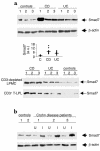
Figure 1
Figure 1
Smad 7 in IBD. (a) Representative expression of Smad7 (upper blot) and β-actin (lower blot) protein in colonic mucosal samples taken from three normal controls, three patients with active CD, and three with UC. The example is representative of five separate experiments analyzing in total mucosal samples from eight patients with CD, eight with UC, and eight normal controls. Quantitative data are shown in the middle panel as measured by densitometry scanning of Western blots. Values are expressed in arbitrary units (a.u.). Each point represents the value (a.u.) of Smad7 in mucosal samples taken from a single subject. Horizontal bars indicate the median value. Shown in the bottom panel is expression of Smad7 protein in both CD3-depleted LPMCs and positively selected CD3+ T lymphocytes (T-LPL) from intestinal mucosal samples taken from three patients with CD, three patients with UC, and three normal controls. The example is representative of three separate experiments analyzing Smad7 in both CD3-depleted LPMCs and CD3+ T-LPLs from five patients with CD, four with UC, and six normal controls. (b) Expression of Smad7 (upper blot) and β-actin (lower blot) protein in CD: effect of disease activity. Colonic mucosal biopsy specimens were taken from two normal controls and from inflamed (I) and uninflamed (U) areas of three patients with CD. Arrows indicate Smad7 and β-actin detected by specific polyclonal antibodies. The example is representative of two separate experiments using in total samples from four controls and five CD patients.
Figure 2
Figure 2
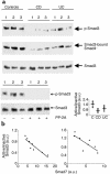
(a) Reduced phosphorylation of Smad3 in IBD tissue. Active (phosphorylated, p-Smad3) and inactive Smad3 in mucosal samples of normal controls and patients with CD or UC. Total proteins were immunoprecipitated with a specific Smad3 antibody and subsequently incubated with a phosphoserine antibody (upper blot). After detection of p-Smad3, the membrane was stripped and incubated with a second Smad3 antibody (lower blot) to ascertain equivalent loading of the lanes. To investigate interactions between Smad3 and Smad4, proteins were immunoprecipitated with an anti-Smad3 antibody and subsequently incubated with a Smad4 antibody (middle blot). The example is representative of three separate experiments analyzing in total mucosal samples from eight patients with CD, eight with UC, and eight normal controls. The bottom right panel of a shows quantitative analysis of active/inactive Smad3 ratio in colonic mucosal samples from eight controls, eight patients with CD, and 8 with UC, as measured by densitometry scanning of Western blots. Values are expressed in arbitrary units (a.u.). Each point represents the value (a.u.) of active/inactive Smad3 ratio in mucosal samples taken from a single subject. Horizontal bars indicate the mean. The bottom left panel of a shows a lack of phosphorylated-Smad3 (p-Smad3) signal after treatment with protein phosphatase-2A (PP-2A). Total protein was extracted from three normal controls immunoprecipitated with a specific Smad3 antibody and incubated in the absence or presence of PP-2A. Proteins were then incubated with a phosphoserine antibody. After detection of p-Smad3, the membrane was stripped and incubated with a second Smad3 antibody to show that PP-2A does not affect the content of total Smad3 proteins. The example is representative of two separate experiments in which mucosal samples from six normal controls were used. (b) Relation between active/inactive Smad3 ratio and Smad7 in mucosal samples taken from patients with CD (left) and UC (right). Both Smad3 and Smad7 levels were measured by densitometry scanning of Western blots and expressed in arbitrary units (a.u.). The expression of Smad7 is inversely related to the active/inactive Smad3 ratio in both CD (r = 0.818; P < 0.003) and UC (r = 0.616; P < 0.03).
Figure 3
Figure 3
Stimulation of CD and UC LPMCs with TGF-β1 does not result in enhanced p-Smad3. LPMCs isolated from the colon of one normal control, one CD patient, or one UC patient were cultured in the presence or absence of TGF-β1 (1 ng/ml) for 1 hour. Phosphorylated (top blot, p-Smad3) and total (bottom blot) Smad3 were analyzed in proteins extracted from LPMCs as indicated in Figure 2. The example is representative of four separate experiments, in each of which, LPMCs from one normal control, one patient with CD, and one patient with UC were analyzed. In all cases, LPMCs from normal patients showed endogenous p-Smad3 that was enhanced by TGF-β1, whereas in all IBD patients, endogenous p-Smad3 was barely detectable after TGF-β1 stimulation. The bottom panel shows quantitative analysis of active/inactive Smad3 ratio in TGF-β1–stimulated LPMCs from four controls, four patients with CD, and four with UC, as measured by densitometry scanning of Western blots. Values are expressed in arbitrary units (a.u.). Each point represents the value (a.u.) of active/inactive Smad3 ratio in LPMC samples taken from a single subject. Horizontal bars indicate the mean.
Figure 4
Figure 4
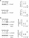
Antisense to Smad7 restores TGF-β1 signaling in both CD and UC LPMCs. (a) Treatment of CD and UC LPMCs with a specific Smad7 antisense but not a sense oligonucleotide inhibits Smad7 expression. CD and UC LPMCs were cultured in the absence (U, unstimulated) or presence of a specific Smad7 antisense (AS) or control sense (S) oligonucleotide for 24 hours. Arrows indicate Smad7 detected by a specific polyclonal antibody. The example is representative of three separate experiments analyzing in total LPMCs from five CD or five UC patients. Quantitative data are shown in the right panel as measured by densitometry scanning of Western blots. Values are expressed in arbitrary units (a.u.). Each point represents the value (a.u.) of Smad7 in LPMCs taken from a single subject. Horizontal bars indicate the median value. (b) Inhibition of Smad7 restores the TGF-β1–induced Smad3 phosphorylation in both CD and UC LPMCs. LPMCs were cultured in the absence (U, unstimulated) or presence of a specific Smad7 antisense (AS) or control sense (S) oligonucleotide, and then stimulated with 1 ng/ml TGF-β1 for 1 hour. The right panel shows quantitative analysis of active/inactive Smad3 ratio in LPMC from five patients with CD and five with UC, as measured by densitometry scanning of Western blots. Values are expressed in arbitrary units (a.u.). Each point represents the value (a.u.) of active/inactive Smad3 ratio in LPMCs taken from a single subject. Horizontal bars indicate the mean.
Figure 5
Figure 5
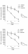
Antisense to Smad7 enables TGF-β1 to inhibit CD LPMC proinflammatory cytokine production. CD LPMCs were cultured in the absence or presence of a specific Smad7 antisense or sense oligonucleotide for 24 hours. The cells were washed with PBS, resuspended in RPMI 1640 supplemented with HL-1 and cultured with or without SEB in the presence or absence of graded doses of TGF-β1 for 12 hours. In CD LPMCs pretreated with the Smad7 antisense oligonucleotide, as little as 0.1 ng/ml of TGF-β1 significantly decreased transcripts for both IFN-γ and TNF-α (P < 0.002). In CD LPMCs precultured with medium or sense Smad7 oligonucleotide, only a modest decrease in IFN-γ and TNF-α transcripts was seen with 10 ng/ml TGF-β1 (P ≤ 0.05). Data are expressed as mean ± SEM of four separate experiments, in each of which, LPMCs from one CD patient were used. The inset shows that, as expected, in LPMCs from normal controls, TGF-β1 dose-dependently inhibited SEB-stimulated cytokine transcripts, regardless of whether LPMCs were pretreated or not with oligonucleotides. Data are expressed as mean ± SEM of three separate experiments, in each of which, LPMCs from one normal control were used.
Figure 6
Figure 6
(a) Inhibition of Smad7 protein by a specific Smad7 antisense oligonucleotide in CD mucosal tissue results in increased p-Smad3 and decreased tissue TNF-α and IFN-γ. The example is representative of four separate experiments, in each of which inflamed mucosal tissue from one CD patient was used. Identical results were seen in each patient. (b) Addition of a neutralizing TGF-β1 antibody decreases but does not abrogate the Smad7 antisense effect on both p-Smad3 and cytokine expression. The example is representative of three separate experiments, in each of which, inflamed mucosal tissue from one CD patient was used. Identical results were seen in each patient.

Comment in

Similar articles

Cited by

References

    1. Fiocchi C. Inflammatory bowel disease: etiology and pathogenesis. Gastroenterology. 1998;115:182–205. - PubMed
    1. Wahl SM. Transforming growth factor β: the good, the bad, and the ugly. J Exp Med. 1994;180:1587–1590. - PMC - PubMed
    1. Letterio JJ, Roberts AB. Regulation of immune responses by TGF-β. Annu Rev Immunol. 1998;16:137–161. - PubMed
    1. Piek E, Heldin C-H, ten Dijke P. Specificity, diversity, and regulation in TGF-β superfamily signaling. FASEB J. 1999;13:2105–2124. - PubMed
    1. Heldin C-H, Kohei M, ten Dijke P. TGF-beta signalling from cell membrane to nucleus through SMAD proteins. Nature. 1997;390:465–471. - PubMed

Publication types

MeSH terms