. 2001 Aug 1;21(15):5501-12.
doi: 10.1523/JNEUROSCI.21-15-05501.2001.

Subunit heterogeneity of cytoplasmic dynein: Differential expression of 14 kDa dynein light chains in rat hippocampus

Affiliations

Subunit heterogeneity of cytoplasmic dynein: Differential expression of 14 kDa dynein light chains in rat hippocampus

J Z Chuang et al. J Neurosci. .

Abstract

Cytoplasmic dynein is a multi-subunit protein complex in which each subunit is encoded by a few genes. How these subunit isoforms are assembled and regulated to mediate the diverse functions of cytoplasmic dynein is unknown. We previously have shown that two highly conserved 14 kDa dynein light chains, Tctex-1 and RP3, have different cargo-binding abilities. In this report, coimmunoprecipitation revealed that Tctex-1 and RP3 were present in mutually exclusive dynein complexes of brain. Two specific antibodies were used to examine the localization of these two dynein light chains in adult rat hippocampal formation and cerebral cortex. By light microscopy, Tctex-1 and RP3 immunoreactivities exhibited distinct and almost complementary distribution patterns in both brain regions. In hippocampal formation, Tctex-1 immunoreactivity was most enriched in somata of newly generated granule cells and scant in the mature granule and pyramidal cell somata. In contrast, RP3 immunoreactivity was abundant in pyramidal and granule cell somata. Ultrastructural analysis of the dentate gyrus revealed both dynein light chains were associated with various membranous organelles that often were affiliated with microtubules. In addition, Tctex-1 and RP3 immunoreactivities were preferentially and highly enriched on membranous organelles and/or vesicles of axon terminals and dendritic spines, respectively. These results suggest that dynein complexes with different subunit composition, and possibly function, are expressed differentially in a spatially and temporally regulated manner. Furthermore, Tctex-1 and RP3 may play important roles in synaptic functions.


Figures
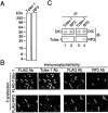
Fig. 1.
Fig. 1.
Specific detection of Tctex-1 and RP3 in rat brain lysate, transfected cells, and immunoprecipitates and assembly of Tctex-1 and RP3 in distinct dynein complexes. A, Rat brain lysate was resolved on SDS-PAGE and immunoblotted with anti-Tctex-1 or anti-RP3 antibody. Both antibodies recognized a single ∼14 kDa molecule, although Tctex-1 migrated slightly faster than RP3 on SDS-PAGE. B, HEK 293 cells transfected with FLAG–Tctex-1 or FLAG–RP3 expression vector were double labeled with anti-FLAG monoclonal antibody and anti-Tctex-1 or anti-RP3 antibody (as indicated). Although the anti-FLAG antibody recognized both transfected FLAG–Tctex-1 and FLAG–RP3, Tctex-1 antibody recognized only FLAG–Tctex-1, but not FLAG–RP3. Arrows point to examples of transfected cells. Likewise, anti-RP3 antibody immunolabeled only the FLAG–RP3, but not FLAG–Tctex-1, transfected cells. Note that anti-Tctex-1 antibody also lightly labeled the endogenous Tctex-1 present in the nontransfected cells (left panels). There is no detectable endogenous RP3 in HEK 293 cells. C, Cytoplasmic dynein complexes were immunoprecipitated from brain lysates with anti-Tctex-1 or anti-RP3 antibody. The immunoprecipitates were separated on SDS-PAGE and transferred to nitrocellulose. The top half of the filter was immunoblotted with anti-dynein intermediate chain (DIC) to demonstrate that approximately equal amounts of the dynein complex were precipitated by each antibody (top panel). The bottom half of the nitrocellulose filter was immunoblotted with anti-Tctex-1 or anti-RP3 antibody. Anti-Tctex-1 antibody detected Tctex-1 in the anti-Tctex-1 precipitate, but not in the anti-RP3 precipitate. Likewise, anti-RP3 detected RP3 in the anti-RP3 precipitate, but not in the anti-Tctex-1 precipitate. This result suggests that the dynein complex precipitated by Tctex-1 antibody did not contain RP3, and vice versa. Furthermore, Tctex-1 antibody and RP3 antibody did not cross-react with the antigens of each other.
Fig. 2.
Fig. 2.
Distinct distributions of RP3 and Tctex-1 in the hippocampal formation and cerebral cortex. Coronal sections of adult Sprague Dawley rat brain were immunolabeled for Tctex-1 (A–C) or RP3 (D–F), using affinity-purified antibodies, followed by peroxidase color development with diaminobenzidine. A, Perikarya exhibiting intense Tctex-1 immunoreactivity were found in the subgranular zone (arrows) of the dentate gyrus. Neurons containing Tctex-1 labeling also were scattered throughout the entire hippocampal formation. Moderate levels of diffusive Tctex-1 labeling were found in the inner molecular layer (iml) and hilus of the dentate gyrus (h) and stratum lucidum of CA3 region (open arrow). B, Higher magnification of the boxed area in Ashows that dendritic processes of Tctex-1 labeled somata in the subgranular zone often penetrated (arrowheads) through the granule cell layer (gcl). Both Tctex-1-labeled perikarya (open arrow) and varicose processes (arrows) were found throughout the hilus and CA3 regions. C, A low-magnification view of the cerebral cortex (layers 1–6) shows Tctex-1-immunoreactive neurons scattered throughout all layers. A higher magnification micrograph of theboxed area in layer 5 (inset) revealed perikaryal labeling of Tctex-1 in a small subset of pyramidal cells in cerebral cortex. In addition, many varicose processes were also Tctex-1-immunoreactive. D, Diffuse RP3 immunoreactivity was concentrated in the perikarya of the CA1 and CA3 pyramidal cells and granule cells. E, A higher magnification micrograph of the boxed area in D demonstrates that RP3 immunoreactivity also was found in many small puncta over the granule cell layer (gcl), hilar interneurons (arrow), and CA3 neurons. F, The majority of cerebral cortical neurons was RP3-immunoreactive. Theinset reveals the diffuse as well as the grainy perikarya labeling of RP3 in these pyramidal cortical neurons. Scale bars: A, C, D, F, 500 μm; B, E, 100 μm.
Fig. 3.
Fig. 3.
Fluorescent micrographs of Tctex-1 immunoreactivity in TOAD-64-containing neurons and a subset of parvalbumin-containing interneurons. A, B, Consecutive coronal sections of the rat brains were singly labeled with either Tctex-1 (A) or TOAD-64 (B) antibodies, followed by Alexa-488-conjugated goat anti-rabbit IgG. Neurons labeled for Tctex-1 (A) had an almost identical distribution to neurons labeled with TOAD-64 (B) in the subgranular zone. The dendritic processes (arrows) of Tctex-1 and TOAD-64 perikarya penetrated through the granule cell layer. C, D, Confocal images of hippocampal slices were double labeled with Tctex-1 rabbit antibody (C) and parvalbumin monoclonal antibody (D), followed by corresponding secondary antibodies. Tctex-1 labeling sometimes colocalized with parvalbumin-labeled cells (arrows) in the hilus of the dentate gyrus. Scale bars, 50 μm.
Fig. 4.
Fig. 4.
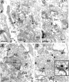
Electron micrograph of the immunoperoxidase labeling of Tctex-1 in the central hilus. Patches of Tctex-1 immunoprecipitate were associated primarily with the small synaptic vesicle clusters (open arrows) and mitochondria (mit; arrows) in a mossy fiber terminal (Mt) and small axon terminals (At). In one mossy fiber terminal (Mt1) some intensely labeled synaptic vesicles were closely apposed to the synaptic specialization. Near the synaptic contact of mossy fiber terminal 2 (Mt2) a large pleomorphic vesicle also had Tctex-1 immunoreaction products affiliated with it (curved arrow). Almost all of the spines contacted by Tctex-1-positive terminals are unlabeled. USp, Unlabeled spine;UAt, unlabeled axon terminal. Scale bars, 0.5 μm.
Fig. 5.
Fig. 5.
Electron micrographs of Tctex-1 immunogold labeling in the hilus (A, B, D) and in the inner molecular layer (C) of the dentate gyrus.A, A Tctex-1-labeled axon terminal (At) synapsed on an unlabeled dendritic spine (uSp), emanating from the shaft of an labeled dendrite (Den). In axon terminals, immunogold-silver particles indicative of Tctex-1 were associated with small synaptic vesicles and larger pleomorphic vesicles (curved arrow). These organelles often were affiliated with the active zone (filled arrow). In the dendritic shaft, Tctex-1 immunoreactivity (open arrow) was affiliated with MTs (m), smooth endoplasmic reticulum (sr), mitochondria (mit), and multivesicular bodies (mvb).B, A large Tctex-1-labeled mossy fiber terminal forms multiple asymmetric synapses on dendritic spines that invaginate or indent the terminal. In the labeled terminal, Tctex-1-derived immunogold particles often were found in vesicles that were larger than small synaptic vesicles (curved arrows) and that often were found in the vicinity of the active zone (arrows). Occasionally, Tctex-1 immunogold was seen on smooth endoplasmic reticulum (sr). C, Three small axon terminals (At1, At2, At3) containing Tctex-1 labeling that is affiliated with synaptic vesicles near active zones (arrows) are shown. Tctex-1 immunoreactivity also was associated with smooth endoplasmic reticulum (sr;open arrow) and various membrane profiles (arrowheads) in the neighboring axon (Ax). D, Immunogold particles indicative of Tctex-1 were associated with synaptic vesicles. Some Tctex-1-immunoreactive synaptic vesicles were larger than the small synaptic vesicles (curvedarrows) and were located in the vicinity of the active zone (arrows), which is enlarged in the inset. Scale bars, 0.5 μm.
Fig. 6.
Fig. 6.
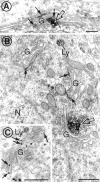
The Golgi localization of RP3 in the granule cells. A, In a granule cell perikaryon RP3 immunoreactivity was associated with Golgi cisternae (arrows) and clusters of vesicles likely to be in thetrans-Golgi network (open arrow).B, RP3-derived immunoperoxidase precipitate was associated with the Golgi apparatus (G) near the membrane stacks (arrows) as well as the slightly dilated ends. Prominent patches of RP3 immunoreactivity were found in the clusters of vacuolar/tubular membrane profiles on thetrans (concave) face of the Golgi complex (open arrow). An unlabeled lysosome (Ly), which contains multiple electron-dense grains, also was found.C, High density of RP3 immunogold particles was observed on the Golgi membrane stacks and the tubular vesicles on thetrans side of the Golgi apparatus. Scale bars, 0.5 μm.
Fig. 7.
Fig. 7.
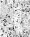
Electron micrographs of the RP3 localization in the hilus proper of the dentate gyrus. A, A low-magnification electron micrograph shows that RP3 immunoperoxidase precipitate was found primarily in dendritic spines (filled arrows). RP3-immunoreactive spines frequently originated from the same branched protuberance (open arrows). The inset shows a high-magnification view of a branched spine in which RP3 was present on only two spine heads and absent from another. USp, Unlabeled spine. The PSDs (small arrows) usually were labeled heavily, in contrast to the corresponding PSDs (arrowheads) in an unlabeled spine. B, Several RP3-containing dendritic spines (arrows) invaginated into a few large mossy fiber terminals (Mt), which essentially have no reaction products. C, In a longitudinally sectioned dendrite (Den) a spine extends and branches into two spine heads, one RP3-positive and one RP3-negative (the branching is marked with anasterisk). The classic spine apparatus (sa), which has flattened sacs of smooth endoplasmic reticulum, was decorated by RP3 immunoreactivity. Moreover, intense RP3 labeling was seen routinely on PSDs and surrounding the multivesicular body (curved arrow) within the labeled spine head. In the dendritic shaft, RP3-derived immunoperoxidase precipitate was associated with tubulovesicular smooth endoplasmic reticulum (open arrow) and vesicles of various sizes (arrows) that often were arrayed on MTs (m). Patches of RP3 immunoprecipitates (arrowheads) sometimes were associated closely with the MTs themselves. Scale bars, 0.5 μm.
Fig. 8.
Fig. 8.
MT and membrane organelle association of RP3 in the postsynaptic elements. A, RP3 immunoperoxidase precipitate was concentrated near MT (m) tracks (arrows) within a dendritic profile. The outer membranes of mitochondria (mit) and vesicular profiles (curved arrow) within the dendrite also were RP3-immunoreactive. RP3 immunoreactivity was notably enriched in dendritic spines, as demonstrated by the high-level RP3 immunoreaction product associated with membranes of vesicles (v), multivesicular body (mvb), and PSD (open arrow) within spines. No RP3 immunoreactivity was observed in several mossy fiber terminals that were in direct contact with RP3-positive dendrites/spines.B, A high-magnification view of three RP3-positive spine heads invaginated into an RP3-negative mossy fiber terminal. The cytosolic face of multiple spine organelles/structures, such as multivesicular body (mvb), large vesicles (V), and PSD (open arrows), was decorated by the RP3 immunoreaction product. Scale bars, 0.5 μm.
Fig. 9.
Fig. 9.
Ultrastructural study of RP3 in the dental hilus, using immunogold labeling. A, Two large dendrites (Den) were contacted by multiple axon terminals. In the dendrite cytoplasm RP3-derived immunogold particles were associated with the tubular-like smooth endoplasmic reticulum (open arrows). Many of these RP3-positive membranes (arrows) also were located near the synaptic contacts to axon terminals (At). A few axon terminals shown in this micrograph lacked RP3 labeling. B, RP3 immunoreactivity was associated with the smooth endoplasmic reticulum (arrow) and the PSD (open arrow) of two spines arising from separate dendrites (Den).C, In a dendritic profile the RP3-derived immunogold particles (open arrows) were clustered on the postsynaptic element apposing from an axon terminal. Smooth endoplasmic reticulum (curved arrows) also was labeled by RP3 immunogold particles D, In a dendritic profile several RP3-derived immunogold particles (open arrow) were concentrated at the postsynaptic elements that were in direct contact with a large axon terminal (At). E, In a spine head the gold particles indicative of RP3 were localized to a typical multi-cisternae spine apparatus (sa; open arrow) and smooth endoplasmic reticulum (arrow).mvb, Multivesicular body; Mt, mossy fiber terminal. Scale bars, 0.5 μm.

Similar articles

Cited by

References

    1. Allen MJ, Shan X, Caruccio P, Froggett SJ, Moffat KG, Murphey RK. Targeted expression of truncated glued disrupts giant fiber synapse formation in Drosophila. J Neurosci. 1999;19:9374–9384. - PMC - PubMed
    1. Altman J, Das GD. Autoradiographic and histological evidence of postnatal hippocampal neurogenesis in rats. J Comp Neurol. 1965;124:319–335. - PubMed
    1. Amaral DG. A Golgi study of cell types in the hilar region of the hippocampus in the rat. J Comp Neurol. 1978;182:851–914. - PubMed
    1. Aniento F, Emans N, Griffiths G, Gruenberg J. Cytoplasmic dynein-dependent vesicular transport from early to late endosomes. J Cell Biol. 1993;123:1373–1387. - PMC - PubMed
    1. Baas PW, Deitch JS, Black MM, Banker GA. Polarity orientation of microtubules in hippocampal neurons: uniformity in the axon and nonuniformity in the dendrite. Proc Natl Acad Sci USA. 1988;85:8335–8339. - PMC - PubMed

Publication types

MeSH terms

LinkOut - more resources