

Host range of small-ruminant lentivirus cytopathic variants determined with a selectable caprine arthritis- encephalitis virus pseudotype system
- PMID: 11462010
- PMCID: PMC114973
- DOI: 10.1128/JVI.75.16.7384-7391.2001
Host range of small-ruminant lentivirus cytopathic variants determined with a selectable caprine arthritis- encephalitis virus pseudotype system
Abstract
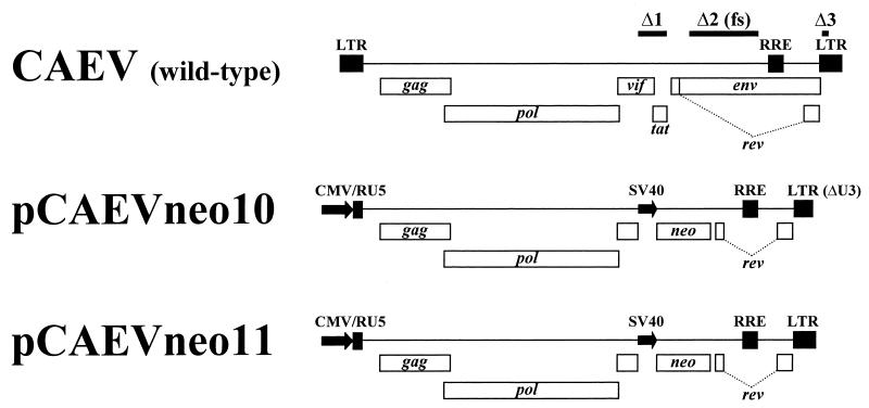
The small-ruminant lentiviruses ovine maedi-visna virus (MVV) and caprine arthritis-encephalitis virus (CAEV) cause encephalitis, progressive pneumonia, arthritis, and mastitis in sheep and goats. Icelandic MVV strains, which are lytic in tissue culture, have a wide species distribution of functional receptors, which includes human cells. In contrast, functional receptors for the nonlytic CAEV CO are absent from human cells. To determine if the wide species distribution of functional receptors is a common property of MVV strains or related to cytopathic phenotype, we tested the infectivity of viruses pseudotyped with the envelope glycoproteins of MVV K1514, CAEV CO, and lytic and nonlytic North American MVV strains to cells of different species. Replication-defective CAEV proviral constructs lacking the env, tat, and vif genes and carrying the neomycin phosphotransferase gene in the vif-tat region were developed for the infectivity assays. Cotransfection of human 293T cells with these proviral constructs and plasmids expressing CAEV, MVV, or vesicular stomatitis virus envelope glycoproteins produced infectious pseudotyped virus which induced resistance of infected cells to G418. Using these pseudotypes, we confirmed the wide species distribution of Icelandic MVV receptors and the narrow host range of CAEV. However, functional receptors for the two North American MVV strains tested, unlike the Icelandic MVV and similar to CAEV, were limited to cells of ruminant species, regardless of cytopathic phenotype. The results indicate a differential receptor recognition by MVV strains which is unrelated to cytopathic phenotype.
Figures
Similar articles
-
Differential receptor usage of small ruminant lentiviruses in ovine and caprine cells: host range but not cytopathic phenotype is determined by receptor usage.Virology. 2002 Sep 15;301(1):21-31. doi: 10.1006/viro.2002.1575. Virology. 2002. PMID: 12359443
-
Genetic characterization of two phenotypically distinct North American ovine lentiviruses and their possible origin from caprine arthritis-encephalitis virus.Virology. 1996 Nov 1;225(1):1-10. doi: 10.1006/viro.1996.0569. Virology. 1996. PMID: 8918528
-
Restrictive type of replication of ovine/caprine lentiviruses in ovine fibroblast cell cultures.Virology. 1996 Aug 1;222(1):21-30. doi: 10.1006/viro.1996.0394. Virology. 1996. PMID: 8806484
-
Maedi-visna virus and its relationship to human immunodeficiency virus.AIDS Rev. 2005 Oct-Dec;7(4):233-45. AIDS Rev. 2005. PMID: 16425963 Review.
-
Small ruminant lentiviruses (SRLVs) break the species barrier to acquire new host range.Viruses. 2013 Jul 23;5(7):1867-84. doi: 10.3390/v5071867. Viruses. 2013. PMID: 23881276 Free PMC article. Review.
Cited by
-
Immunization against small ruminant lentiviruses.Viruses. 2013 Aug 2;5(8):1948-63. doi: 10.3390/v5081948. Viruses. 2013. PMID: 23917352 Free PMC article. Review.
-
Caprine arthritis-encephalitis virus envelope surface glycoprotein regions interacting with the transmembrane glycoprotein: structural and functional parallels with human immunodeficiency virus type 1 gp120.J Virol. 2003 Nov;77(21):11578-87. doi: 10.1128/jvi.77.21.11578-11587.2003. J Virol. 2003. PMID: 14557643 Free PMC article.
-
Multiplex assay for the simultaneous detection of antibodies against small ruminant lentivirus, Mycobacterium avium subsp. paratuberculosis, and Brucella melitensis in goats.Vet World. 2023 Apr;16(4):704-710. doi: 10.14202/vetworld.2023.704-710. Epub 2023 Apr 8. Vet World. 2023. PMID: 37235146 Free PMC article.
-
Small ruminant lentiviruses: genetic variability, tropism and diagnosis.Viruses. 2013 Apr 23;5(4):1175-207. doi: 10.3390/v5041175. Viruses. 2013. PMID: 23611847 Free PMC article. Review.
-
Retroviral infections in sheep and goats: small ruminant lentiviruses and host interaction.Viruses. 2013 Aug 19;5(8):2043-61. doi: 10.3390/v5082043. Viruses. 2013. PMID: 23965529 Free PMC article. Review.
References
-
- August M J, Harter D H. Visna virus-induced fusion of continuous simian kidney cells. Arch Ges Virusforsch. 1974;44:92–101. - PubMed
-
- Berkowitz R D, Ilves H, Plavec I, Veres G. Gene transfer systems derived from visna virus: analysis of virus production and infectivity. Virology. 2001;279:116–129. - PubMed
-
- Beyer J C, Chebloune Y, Mselli-Lakhal L, Hötzel I, Kumpula-McWhirter N, Cheevers W P. Immunization with plasmid DNA expressing the caprine arthritis-encephalitis virus envelope gene: quantitative and qualitative aspects of antibody response to viral surface glycoproteins. Vaccine. 2001;19:1643–1651. - PubMed
-
- Brown H R, Thormar H. Persistence of visna virus in murine and hamster cell cultures without the appearance of cell transformation. Microbios. 1975;13:51–60.
-
- Chebloune Y, Sheffer D, Karr B M, Stephens E, Narayan O. Restrictive type of replication of ovine/caprine lentiviruses in ovine fibroblast cell cultures. Virology. 1996;222:21–30. - PubMed
Publication types
MeSH terms
Substances
Grants and funding
LinkOut - more resources
Full Text Sources
