Functional analysis of cyclin-dependent kinase inhibitors of Arabidopsis
- PMID: 11449057
- PMCID: PMC139548
- DOI: 10.1105/tpc.010087
Functional analysis of cyclin-dependent kinase inhibitors of Arabidopsis
Abstract
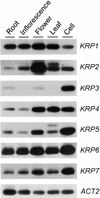
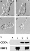
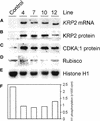
Cyclin-dependent kinase inhibitors, such as the mammalian p27(Kip1) protein, regulate correct cell cycle progression and the integration of developmental signals with the core cell cycle machinery. These inhibitors have been described in plants, but their function remains unresolved. We have isolated seven genes from Arabidopsis that encode proteins with distant sequence homology with p27(Kip1), designated Kip-related proteins (KRPs). The KRPs were characterized by their domain organization and transcript profiles. With the exception of KRP5, all presented the same cyclin-dependent kinase binding specificity. When overproduced, KRP2 dramatically inhibited cell cycle progression in leaf primordia cells without affecting the temporal pattern of cell division and differentiation. Mature transgenic leaves were serrated and consisted of enlarged cells. Although the ploidy levels in young leaves were unaffected, endoreduplication was suppressed in older leaves. We conclude that KRP2 exerts a plant growth inhibitory activity by reducing cell proliferation in leaves, but, in contrast to its mammalian counterparts, it may not control the timing of cell cycle exit and differentiation.
Figures


Similar articles
-
The cyclin-dependent kinase inhibitor KRP2 controls the onset of the endoreduplication cycle during Arabidopsis leaf development through inhibition of mitotic CDKA;1 kinase complexes.Plant Cell. 2005 Jun;17(6):1723-36. doi: 10.1105/tpc.105.032383. Epub 2005 Apr 29. Plant Cell. 2005. PMID: 15863515 Free PMC article.
-
Downregulation of multiple CDK inhibitor ICK/KRP genes upregulates the E2F pathway and increases cell proliferation, and organ and seed sizes in Arabidopsis.Plant J. 2013 Aug;75(4):642-55. doi: 10.1111/tpj.12228. Epub 2013 May 30. Plant J. 2013. PMID: 23647236
-
Arabidopsis cyclin-dependent kinase inhibitors are nuclear-localized and show different localization patterns within the nucleoplasm.Plant Cell Rep. 2007 Jul;26(7):861-72. doi: 10.1007/s00299-006-0294-3. Epub 2007 Jan 26. Plant Cell Rep. 2007. PMID: 17253089
-
Plant Cyclin-Dependent Kinase Inhibitors of the KRP Family: Potent Inhibitors of Root-Knot Nematode Feeding Sites in Plant Roots.Front Plant Sci. 2017 Sep 8;8:1514. doi: 10.3389/fpls.2017.01514. eCollection 2017. Front Plant Sci. 2017. PMID: 28943880 Free PMC article. Review.
-
Modifying plant growth and development using the CDK inhibitor ICK1.Cell Biol Int. 2003;27(3):297-9. doi: 10.1016/s1065-6995(02)00310-4. Cell Biol Int. 2003. PMID: 12681342 Review. No abstract available.
Cited by
-
Overexpression of Orysa;KRP4 drastically reduces grain filling in rice.Planta. 2024 Aug 22;260(4):78. doi: 10.1007/s00425-024-04512-0. Planta. 2024. PMID: 39172243
-
Precocious cell differentiation occurs in proliferating cells in leaf primordia in Arabidopsis angustifolia3 mutant.Front Plant Sci. 2024 Apr 16;15:1322223. doi: 10.3389/fpls.2024.1322223. eCollection 2024. Front Plant Sci. 2024. PMID: 38689848 Free PMC article.
-
Sugar signals pedal the cell cycle!Front Plant Sci. 2024 Mar 18;15:1354561. doi: 10.3389/fpls.2024.1354561. eCollection 2024. Front Plant Sci. 2024. PMID: 38562561 Free PMC article. Review.
-
The SIAMESE family of cell-cycle inhibitors in the response of plants to environmental stresses.Front Plant Sci. 2024 Feb 16;15:1362460. doi: 10.3389/fpls.2024.1362460. eCollection 2024. Front Plant Sci. 2024. PMID: 38434440 Free PMC article. Review.
-
Anatomical Mechanisms of Leaf Blade Morphogenesis in Sasaella kogasensis 'Aureostriatus'.Plants (Basel). 2024 Jan 23;13(3):332. doi: 10.3390/plants13030332. Plants (Basel). 2024. PMID: 38337866 Free PMC article.
References
-
- Beeckman, T., and Viane, R. (2000). Embedding thin plant specimens for oriented sectioning. Biotech. Histochem. 75, 23–26. - PubMed
-
- Bell, M.H., Halford, N.G., Ormrod, J.C., and Francis, D. (1993). Tobacco plants transformed with cdc25, a mitotic inducer gene from fission yeast. Plant Mol. Biol. 23, 445–451. - PubMed
-
- Burssens, S., de Almeida Engler, J., Beeckman, T., Richard, C., Shaul, O., Ferreira, P., Van Montagu, M., and Inzé, D. (2000). Developmental expression of the Arabidopsis thaliana CycA2;1 gene. Planta 211, 623–631. - PubMed
-
- Chen, J., Jackson, P.K., Kirschner, M.W., and Dutta, A. (1995). Separate domains of p21 involved in the inhibition of Cdk kinase and PCNA. Nature 374, 386–388. - PubMed
Publication types
MeSH terms
Substances
Associated data
- Actions
- Actions
- Actions
- Actions
- Actions
- Actions
- Actions
- Actions
LinkOut - more resources
Full Text Sources
Other Literature Sources
Molecular Biology Databases
Miscellaneous

