. 2001 Jul 16;20(14):3728-37.
doi: 10.1093/emboj/20.14.3728.

Heterologous dimerization domains functionally substitute for the double-stranded RNA binding domains of the kinase PKR

Affiliations

Heterologous dimerization domains functionally substitute for the double-stranded RNA binding domains of the kinase PKR

T L Ung et al. EMBO J. .

Abstract

The protein kinase PKR (dsRNA-dependent protein kinase) phosphorylates the eukaryotic translation initiation factor eIF2alpha to downregulate protein synthesis in virus-infected cells. Two double-stranded RNA binding domains (dsRBDs) in the N-terminal half of PKR are thought to bind the activator double-stranded RNA, mediate dimerization of the protein and target PKR to the ribosome. To investigate further the importance of dimerization for PKR activity, fusion proteins were generated linking the PKR kinase domain to heterologous dimerization domains. Whereas the isolated PKR kinase domain (KD) was non-functional in vivo, expression of a glutathione S-transferase-KD fusion, or co-expression of KD fusions containing the heterodimerization domains of the Xlim-1 and Ldb1 proteins, restored PKR activity in yeast cells. Finally, coumermycin-mediated dimerization of a GyrB-KD fusion protein increased eIF2alpha phosphorylation and inhibited reporter gene translation in mammalian cells. These results demonstrate the critical importance of dimerization for PKR activity in vivo, and suggest that a primary function of double-stranded RNA binding to the dsRBDs of native PKR is to promote dimerization and activation of the kinase domain.


Figures
None
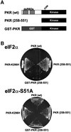
Fig. 1. Expression of a GST–PKR kinase domain fusion protein, but not the isolated kinase domain alone, is toxic in yeast. (A) Schematics of wild-type human PKR [PKR (wt)] showing two dsRNA binding domains (dsRBDs) in the N-terminal half of the protein and the kinase domain (residues ∼263–551) in the C-terminal half of the protein; the isolated PKR kinase domain [PKR (258–551)]; and a GST–PKR kinase domain fusion protein (GST–PKR). (B) Plasmids expressing wild-type PKR (p1420), PKR-K296H (p1421), the isolated PKR kinase domain [PKR (258–551); pC681], or the GST–PKR kinase domain fusion protein (pC661), as indicated, under the control of a yeast GAL-CYC1 hybrid promoter were introduced into strain J80 containing wild-type eIF2α and strain J82 containing eIF2α-S51A, as indicated. Trans formants were streaked on SGal medium (synthetic minimal medium containing 10% galactose) supplemented with essential nutrients, and the plates were incubated at 30°C for 6 days.
None
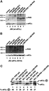
Fig. 2. Immunoblot analysis of PKR expression and isoelectric focusing analysis of eIF2α phosphorylation in yeast strains expressing various forms of PKR. (A and B) Immunoblot analysis of PKR expression. The yeast strains J80 [(A), wild-type eIF2α] and J82 [(B), eIF2α-S51A] were transformed with plasmids to express the indicated forms of PKR or empty vector as follows: lane 1, empty vector (pEMBLyex4); lane 2, wild-type PKR (p1420); lane 3, PKR (258–551) (pC681); lane 4, GST–PKR (pC661); lane 5, Ldb–PKR (pC903) plus vector (pEMBLyex4); lane 6, Lim–PKR (pC901) plus vector (p2444); lane 7, Ldb–PKR (pC903) plus Lim–PKR (pC901). The various PKR proteins were expressed under the control of a yeast GAL-CYC1 promoter. Transformants were grown to exponential phase in SD medium, and then shifted to inducing conditions (SGR medium containing 10% galactose plus 2% raffinose) for ∼18 h. Whole-cell extracts were prepared and 100 µg aliquots were subjected to SDS–PAGE followed by immunoblot analysis using polyclonal antisera against GST–PKR or eIF2α, as indicated. Immune complexes were detected by enhanced chemiluminescence. The molecular mass of SDS size standards are shown on the left. The black or white dots identify the relevant PKR protein(s). (C) Isoelectric focusing analysis of eIF2α phosphorylation. The yeast strains J80 (S51) or J82 (S51A) were transformed with plasmids expressing the indicated PKR proteins and grown as described above. Whole-cell extracts were prepared and 20 µg aliquots were resolved by isoelectric focusing PAGE and then subjected to immunoblot analysis using polyclonal anti-eIF2α antiserum as described previously (Dever et al., 1992). Lane 7 is a darker and higher contrast exposure of lane 4. The positions of basally phosphorylated (eIF2α) and eIF2α phosphorylated on Ser51 (eIF2α–P) are indicated on the right. The percentage of total eIF2α that is phosphorylated on Ser51 was determined by quantitative densitometry and NIH Image software and is indicated below the lanes.
None
Fig. 3. Ribosome association of wild-type PKR, but not the GST–PKR fusion protein. Transformants of strain J82 (expressing eIF2α-S51A) containing the PKR plasmid p1420 or the GST–PKR plasmid pC661 were grown in SGal medium to an OD600 ∼1.5. Whole-cell extracts were prepared in the presence of cycloheximide (50 mg/ml) and MgCl2 (10 mM), and then subjected to velocity sedimentation on 5–47% sucrose gradients as described previously (Zhu et al., 1997; Romano et al., 1998b). The gradients were fractionated while monitoring absorbance at 254 nm to identify the positions of free 40 and 60S subunits, and 80S monosomes (as indicated by the arrows). The distribution of PKR and GST–PKR along the gradients was visualized by SDS–PAGE and immunoblot analysis using polyclonal antiserum raised against the GST–PKR fusion protein. The first lane in each panel was loaded with 1/50 of the input (I) extracts fractionated on the gradients.
None
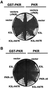
Fig. 4. GST–PKR and PKR (wt) show different sensitivities to the PKR inhibitors encoded by the vaccinia virus K3L and E3L genes. (A) Pseudosubstrate inhibition of PKR and GST–PKR. Yeast strain H1894 was transformed with a URA3 plasmid expressing GST–PKR (pC661), wild-type PKR (p1419), or the empty vector (pEMBLyex4) plus a LEU2 plasmid expressing the vaccinia virus K3L (pC365) or K3L-H47R (pC366) protein or the empty vector (pRS425). (B) Inhibition of PKR, but not GST–PKR, by the dsRNA-binding proteins E3L and PKR-ΔK. Yeast strain H1894 was transformed with a URA3 plasmid expressing GST–PKR (pC661) or wild-type PKR (p1419) plus a LEU2 plasmid expressing the vaccinia virus E3L (pC1315) or K3L-H47R (pC366) protein or the empty vector (pRS425), or a TRP1 plasmid expressing a truncated version of PKR lacking the kinase domain (PKR-ΔK, pC1316). All proteins in both (A) and (B) were expressed under the control of a yeast GAL-CYC1 promoter. Transformants were streaked on SGal minimal complete medium (synthetic minimal medium containing 10% galactose and supplemented with all amino acids), and the plates were incubated at 30°C for 8 days.
None
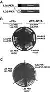
Fig. 5. Reconstitution of PKR activity in yeast by co-expression of Lim–PKR and Ldb–PKR fusion proteins. (A) Schematics of Lim–PKR and Ldb–PKR. In Lim–PKR the first 56 amino acids of the Xenopus Xlim-1 protein (black box) are fused to the PKR kinase domain residues 258–551 (gray box). In Ldb–PKR residues 290–350 of the 375 amino acid Xenopus Ldb1 protein (cross-hatched box) are fused to the PKR kinase domain residues 258–551 (gray box). As indicated by the double-headed arrow, the Xenopus Lim and Ldb domains are known to heterodimerize. (B) The yeast strains J80 (eIF2α) and J82 (eIF2α-S51A) were transformed with the Lim–PKR expression plasmid (pC901) plus plasmids expressing Ldb–PKR (pC903) or Lim–PKR (pC944) or the empty vector (p2444), as indicated. (C) Yeast strain J80 was co-transformed with the Ldb–PKR plasmid (pC903) plus the Lim–PKR (pC901) or Lim–PKR-K296H (pC1097) plasmid or empty vector (pEMBLyex4), as indicated. All Lim–PKR and Ldb–PKR fusion proteins in (B) and (C) were expressed under the control of a yeast GAL-CYC1 promoter. Transformants were streaked on SGal minimal complete medium (synthetic minimal medium containing 10% galactose and supplemented with all amino acids), and the plates were incubated at 30°C for 7 days.
None
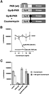
Fig. 6. Coumermycin-induced activation of GyrB–PKR fusion proteins in mammalian cells. (A) Schematics of wild-type PKR [PKR(wt)]; GyrB–PKR fusion protein consisting of the N-terminal 220 amino acids of E.coli GyrB fused to the human PKR kinase domain residues 258–551 (GyrB–PKR); and coumermycin (black dumbbell)-mediated dimerization of GyrB–PKR fusion proteins (GyrB–PKR + coumermycin). (B) NIH 3T3 cells were co-transfected with the luciferase reporter plasmid pGL3-Control (Promega) and either empty vector (vector, pC869), or plasmids to express either wild-type PKR [PKR (wt), pC882] or the GyrB–PKR fusion protein [GyrB–PKR (258–551), pC939], as indicated. Twenty-four hours following transfection, cells were treated with dimethylsulfoxide (DMSO) alone or the indicated concentration of coumermycin dissolved in DMSO. After another 24 h, cells were harvested, lysed and samples of the whole-cell extracts were assayed for luciferase activity. The results are the average and standard deviation from three independent experiments. (C) NIH 3T3 cells were co-transfected with the luciferase reporter plasmid pGL3-Control (Promega) and either empty vector (vector, pC869), or plasmids to express wild-type PKR [PKR (wt), pC882], GyrB–PKR (pC939), or GyrB–PKR-K296H (pC940), as indicated. Twenty-four hours following transfection, cells were treated with DMSO alone (–coumermycin) or with 100 ng/ml coumermycin dissolved in DMSO. Following 24 h stimulation, cells were harvested, lysed and samples of the whole-cell extracts were assayed for luciferase activity. The results are the average and standard deviation from three independent experiments.
None
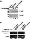
Fig. 7. Coumermycin treatment does not alter GyrB–PKR expression or luciferase reporter mRNA levels in NIH 3T3 cell transfectants. (A) Analysis of PKR expression. NIH 3T3 cells were co-transfected with the luciferase reporter plasmid pGL3-Control (Promega) and either empty vector (lane 1, vector, pC869), or plasmids to express GyrB–PKR (lanes 2 and 3, pC939) or GyrB–PKR-K296H (lanes 4 and 5, pC940), as indicated. Twenty-four hours following transfection, cells were treated with DMSO alone (– coumermycin) or with 100 ng/ml coumermycin dissolved in DMSO (+ coumermycin). Following 24 h stimulation with the drug, cells were harvested, lysed and samples of the whole-cell extracts were subjected to SDS–PAGE and then immunoblotted with anti-PKR (upper panel) or anti-TFIIB (lower panel) antisera as indicated. (B) Analysis of luciferase mRNA levels. NIH 3T3 cells were co-transfected with the luciferase reporter plasmid pGL3-Control (pGL3-Luc) or empty vector (pC869), as indicated, and either empty vector (no label, pC869) or plasmids to express wild-type PKR [PKR (1–551), pC882], GyrB–PKR (pC939), or GyrB–PKR-K296H (pC940), as indicated. Twenty-four hours following transfection, cells were treated with DMSO alone (– coumermycin) or with 100 ng/ml coumermycin dissolved in DMSO (+ coumermycin). Following 24 h stimulation, cells were harvested, lysed and the amount of luciferase reporter and β-actin mRNAs was determined by RT–PCR, as described previously (Kawagishi-Kobayashi et al., 2000). Lanes 1 and 2 are control experiments for the RT–PCR analysis in which either cellular RNA was omitted (lane 1) or the RNA was obtained from non-transfected cells (lane 2).
None
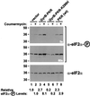
Fig. 8. Increased phosphorylation of eIF2α on Ser51 in NIH 3T3 cells expressing GyrB–PKR and treated with coumermycin. NIH 3T3 cells were co-transfected with the luciferase reporter plasmid pGL3-Control and either empty vector (lanes 1 and 2, vector, pC869), or plasmids to express GyrB–PKR (lanes 3 and 4, pC939), GyrB–PKR-K296H (lanes 5 and 6, pC940), or wild-type PKR [lanes 7 and 8, PKR (wt), pC882], as indicated. Twenty-four hours following transfection, cells were treated with DMSO alone (– coumermycin) or with 100 ng/ml coumermycin dissolved in DMSO (+ coumermycin). Following 24 h stimulation, cells were harvested, lysed and samples of the whole-cell extracts were subjected to SDS–PAGE and then immunoblotted with phosphospecific antibodies raised against an eIF2α peptide containing phosphoserine-51 (DeGracia et al., 1997) (upper and middle panel). Subsequently, the blot was stripped and probed with anti-eIF2α monoclonal antibodies (Scorsone et al., 1987) (lower panel). As indicated, the middle panel is a longer exposure (60 s) of the same blot presented in the top panel (5 s exposure). The relative level of eIF2α phosphorylation in comparison with the untreated vector transfectant (lane 1) was determined by quantitative densitometry and NIH Image software, and is indicated below the lanes.

Similar articles

Cited by

References

    1. Barber G.N., Wambach,M., Wong,M.L., Dever,T.E., Hinnebusch,A.G. and Katze,M.G. (1993) Translational regulation by the interferon-induced double-stranded RNA-activated 68-kDa protein kinase. Proc. Natl Acad. Sci. USA, 90, 4621–4625. - PMC - PubMed
    1. Bertolotti A., Zhang,Y., Hendershot,L., Harding,H. and Ron,D. (2000) Dynamic interaction of BiP and ER stress transducers in the unfolded-protein response. Nature Cell Biol., 2, 326–332. - PubMed
    1. Breen J.J., Agulnick,A.D., Westphal,H. and Dawid,I.B. (1998) Interactions between LIM domains and the LIM domain-binding protein Ldb1. J. Biol. Chem., 273, 4712–4717. - PubMed
    1. Carpick B.W., Graziano,V., Schneider,D., Maitra,R.K., Lee,X. and Williams,B.R.G. (1997) Characterization of the solution complex between the interferon-induced, double-stranded RNA-activated protein kinase and HIV-1 trans-activating region RNA. J. Biol. Chem., 272, 9510–9516. - PubMed
    1. Cesareni G. and Murray,J.A.H. (1987) Plasmid vectors carrying the replication origin of filamentous single-stranded phages. In Setlow,J.K. and Hollaender,A. (eds), Genetic Engineering: Principles and Methods. Vol. 9. Plenum Press, New York, NY, pp. 135–154.