. 2001 Jul 2;20(13):3495-505.
doi: 10.1093/emboj/20.13.3495.

PML mediates the interferon-induced antiviral state against a complex retrovirus via its association with the viral transactivator

Affiliations

PML mediates the interferon-induced antiviral state against a complex retrovirus via its association with the viral transactivator

T Regad et al. EMBO J. .

Abstract

The promyelocytic leukaemia (PML) protein localizes in the nucleus both in the nucleoplasm and in matrix-associated multiprotein complexes known as nuclear bodies (NBs). The number and the intensity of PML NBs increase in response to interferon (IFN). Overexpression of PML affects the replication of vesicular stomatitis virus and influenza virus. However, PML has a less powerful antiviral activity against these viruses than the IFN mediator MxA. Here, we show that overexpression of PML, but not that of Mx1 or MxA, leads to a drastic decrease of a complex retrovirus, the human foamy virus (HFV), gene expression. PML represses HFV transcription by complexing the HFV transactivator, Tas, preventing its direct binding to viral DNA. This physical interaction requires the N-terminal region of Tas and the RING finger of PML, but does not necessitate PML localization in NBs. Finally, we show that IFN treatment inhibits HFV replication in wild-type but not in PML-/- cells. These findings point to a role for PML in transcriptional repression and suggest that PML could play a key role in mediating an IFN-induced antiviral state against a complex retrovirus.


Figures

None
Fig. 1. Overexpression of PML confers resistance to infection by HFV. U373 MG neo, U373 MG-PML, U373 MG untreated or treated with 1000 U/ml IFNα for 24 h were infected with HFV at a m.o.i. of 0.1 for 3 days. (A) Immunofluorescence microscopy was performed with mouse anti-PML antibodies visualized with Texas red and rabbit anti-HFV antibodies followed by FITC labelling. (B) Twenty micrograms of protein extracts from infected U373 MG control, IFN-treated, U373 MG neo and U373 MG-PML cells were analysed by western blotting and revealed by polyclonal anti-Gag antibodies. The same blots were reprobed with monoclonal anti-Bet and polyclonal anti-Actin antibodies. (C) GP+E-86 hygro and GP+E-86-PML cells were infected at a m.o.i. of 0.1 or 1 for 3 days, 20 µg of protein extracts were analysed by western blotting and revealed by anti-HFV antibodies. The lower panel shows the Coomassie Blue-stained proteins. (D) Expression of Mx1 or MxA, two other IFN-induced mediators, did not confer resistance to HFV. NIH 3T3 neo, overexpressing Mx1 or MxA, were infected with HFV at a m.o.i. of 0.1 for 3 days; 20 µg of protein extracts were analysed by western blotting and revealed by anti-HFV antibodies. The lower panel shows the Coomassie Blue-stained proteins.
None
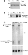
Fig. 2. Viral DNA and mRNA in PML-expressing or in IFN-treated cells. (A) U373 MG neo and U373 MG-PML cells were infected with HFV for 3 h at a m.o.i. of 1. After extensive washing with cold PBS, cultures were treated with acetic acid (0.2 M, pH 2.5) containing 0.5 M NaCl for 6 min at 4°C in order to remove HFV on cell surface membrane. Similar amounts of DNA from Hirt supernatant were analysed by Southern blotting and hybridization with full-length HFV DNA. (B) Similar amounts of Hirt DNA from U373 MG neo, U373 MG-PML and U373 MG pretreated or not with 1000 U/ml IFNα, all infected with HFV for 30 h at a m.o.i. of 0.1, were analysed by Southern blotting using a full-length HFV probe. (C) Total cellular RNA was extracted from U373 MG controls, U373 MG-PML and U373 MG pretreated for 24 h with 1000 U/ml IFNα, all infected with HFV at a m.o.i. of 0.1 for 30 h. Twenty micrograms of RNA were analysed with HFV tas and GAPDH probes.
None
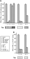
Fig. 3. Inhibition by PML of Tas-mediated transactivation of the HFV LTR and IP. CHO cells transiently co-transfected with LTR-luc and tas or with IP-CAT and tas and a plasmid expressing the empty vector (hygro), wild-type PML, PML mutant PML Δ(216–333), the RING finger PML mutants Q59C60 and C57,60, the C-terminal PML mutant (Stop381), PML 3K mutant or U373 MG. In all cases, pCMV βgal was used to monitor transfection efficiency. Twelve hours later, U373 MG cells were untreated or treated with 1000 U/ml IFNα. All cells were harvested 48 h post-transfection. Luciferase (A) and CAT (B) activities were measured. The graphs show the fold luciferase or CAT activity exhibited by the reporter constructs upon Tas transactivation in the absence or presence of PML. The data represent the average calculated from triplicates of two independent experiments and are expressed as a percentage of control cells (transfected with tas and LTR or IP) with the standard deviation indicated. Equivalent aliquots from all samples were analysed by western blotting for Tas expression.
None
Fig. 4. (A) Tas co-localizes with PML on to NBs. Tas and PML were transiently transfected in CHO cells. After fixation, PML was detected using monoclonal anti-PML antibodies and visualized with Texas red labelling. Tas was detected with rabbit anti-HFV antibodies, followed by anti-rabbit FITC. The right panel shows two-colour overlay results, demonstrating co-localization of Tas and PML proteins (yellow). (B) Tas interacts with PML. CHO cells expressing the empty vector (hygro) or stably expressing PML (CHO-PML) were transfected with tas, lysed and fractionated into cytoplasmic (C) and nuclear (N) components. One portion of these fractions was immunoprecipitated with polyclonal anti-PML antibodies and subsequent western blot probed with anti-HFV antibodies (left panel). The second portion was immunoprecipitated with anti-HFV antibodies and subsequent westerns probed with serum anti-PML (right panel). (C) Implication of the PML RING domain in Tas–PML interaction. CHO cells stably expressing the empty vector (hygro), PML, the PML coiled-coil mutant Δ(216–333), the RING finger PML mutants Q59C60 and Cys57,60 were transfected with tas. CHO cells were co-transfected with tas and PML or the PML 3K mutant (*). Two days later, cells were lysed and nuclear fractions were immunoprecipitated with anti-PML antibodies and subsequent western blot probed with anti-HFV antibodies. (D) Immunoprecipitation of PML and Tas from transcribed–translated proteins. Expression vectors encoding Tas, PML or a mixture of both were in vitro translated in the presence of [35S]methionine using the TnT rabbit-coupled reticulocyte lysate system (Promega) and analysed by 10% SDS–PAGE (INPUTS). Equimolar amounts of labelled proteins were immunoprecipitated by anti-HFV antibodies or anti-PML antibodies. Bound proteins were analysed by 13% SDS–PAGE followed by autoradiography.
None
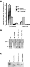
Fig. 5. (A) LTR transactivation by Tas mutants in the absence or presence of PML. CHO cells expressing the empty vector (hygro) or CHO-PML were transfected with LTR and Tas, TasΔN(8–69), TasΔDB(102–161) or TasΔC(235–294). In each case, pCMV βgal was used to monitor transfection efficiency. All cells were harvested 48 h post-transfection and luciferase activity was measured. The data represent averages calculated from triplicates of three independent experiments and are expressed as a percentage of control cells (transfected with tas and LTR) with the standard deviation indicated. (B) The N-terminal region of Tas is necessary for Tas–PML interaction. CHO-PML were transfected with Tas, TasΔN(8–69), TasΔDB(102–161) or TasΔC(235–294). Two days later, cells were lysed and nuclear fractions were immunoprecipitated with polyclonal anti-PML antibodies and subsequent western blot probed with anti-HFV antibodies. (C) Expression of Tas mutants corresponding to the INPUTS of (B).
None
Fig. 6. (A) Binding of Tas to the LTR or IP sequences in the absence or presence of PML. EMSA was performed with extracts from CHO-hygro, CHO-PML, CHO-PML Q59C60 and CHO-PML Cys57,60 transfected with tas. Radiolabelled sequences derived from HFV LTR or IP were used and super-shifting was performed with mouse polyclonal anti-Tas antibodies. (B) Tas expression in EMSA samples. Equivalent aliquots from EMSA samples were analysed by western blotting and revealed by anti-HFV antibodies. (C) Expression of Tas corresponding to the inputs of (A) and (B).
None
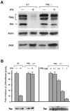
Fig. 7. (A) Response to HFV infection after IFN treatment in wild-type and PML–/– MEF. Wild-type and PML–/– MEF untreated or treated with 1000 U/ml mouse IFNα for 24 h were infected with HFV at a m.o.i. of 0.1. Two days post-infection, western blots were performed using polyclonal anti-HFV, anti-Actin or anti-PKR antibodies. (B) Effect of IFN or PML on Tas-mediated transactivation of LTR in MEF cells. Left panel: wild-type and PML–/– MEF were transfected with LTR–luc and tas. Twelve hours later, cells were untreated or treated with 1000 U/ml IFNα. Right panel: PML–/– MEF were transfected with LTR–luc and tas, and increasing concentrations of PML. In all cases, pCMV βgal was used to monitor transfection efficiency. Cells were harvested 48 h post-transfection and luciferase activity was measured. The data represent averages calculated from triplicates of three independent experiments and are expressed as a percentage of control cells (transfected with tas and LTR) with the standard deviation indicated. Equivalent aliquots from all samples were analysed by western blotting for Tas expression.

Similar articles

Cited by

References

    1. Alcalay M., Tomassoni,L., Colombo,E., Stoldt,S., Grignani,F., Fagioli,M., Szekely,L., Helin,K. and Pelicci,P.G. (1998) The promyelocytic leukemia gene product (PML) forms stable complexes with the retinoblastoma protein. Mol. Cell. Biol., 18, 1084–1093. - PMC - PubMed
    1. Chelbi-Alix M. and de Thé,H. (1999) Herpes virus induces proteasome-dependent degradation of the nuclear bodies-associated PML and Sp100. Oncogene, 18, 935–941. - PubMed
    1. Chelbi-Alix M.K., Pelicano,L., Quignon,F., Koken,M.H.M., Venturini,L., Stadler,M., Pavlovic,J., Degos,L. and de Thé,H. (1995) Induction of the PML protein by interferons in normal and APL cells. Leukemia, 9, 2027–2033. - PubMed
    1. Chelbi-Alix M.K., Pelicano,L., Quignon,F., Koken,M.H.M. and de Thé,H. (1996) PML is a primary target gene of interferon and could mediate some of its biological activities. Tumor Biol., 99, 17–27.
    1. Chelbi-Alix M.K., Quignon,F., Pelicano,L., Koken,M.H.M. and de Thé,H. (1998) Resistance to virus infection conferred by the interferon-induced promyelocytic leukemia protein. J. Virol., 72, 1043–1051. - PMC - PubMed

Publication types

MeSH terms