. 2001 Jun 25;153(7):1465-78.
doi: 10.1083/jcb.153.7.1465.

Nup2p dynamically associates with the distal regions of the yeast nuclear pore complex

Affiliations

Nup2p dynamically associates with the distal regions of the yeast nuclear pore complex

D J Dilworth et al. J Cell Biol. .

Abstract

Nucleocytoplasmic transport is mediated by the interplay between soluble transport factors and nucleoporins resident within the nuclear pore complex (NPC). Understanding this process demands knowledge of components of both the soluble and stationary phases and the interface between them. Here, we provide evidence that Nup2p, previously considered to be a typical yeast nucleoporin that binds import- and export-bound karyopherins, dynamically associates with the NPC in a Ran-facilitated manner. When bound to the NPC, Nup2p associates with regions corresponding to the nuclear basket and cytoplasmic fibrils. On the nucleoplasmic face, where the Ran--GTP levels are predicted to be high, Nup2p binds to Nup60p. Deletion of NUP60 renders Nup2p nucleoplasmic and compromises Nup2p-mediated recycling of Kap60p/Srp1p. Depletion of Ran--GTP by metabolic poisoning, disruption of the Ran cycle, or in vitro by cell lysis, results in a shift of Nup2p from the nucleoplasm to the cytoplasmic face of the NPC. This mobility of Nup2p was also detected using heterokaryons where, unlike nucleoporins, Nup2p was observed to move from one nucleus to the other. Together, our data support a model in which Nup2p movement facilitates the transition between the import and export phases of nucleocytoplasmic transport.


Figures

Figure 1
Figure 1
Nup2p localizes to multiple sites within the NPC. Nup2p–pA was localized in purified NEs (A) and nuclei (B) by IEM using gold-conjugated antibodies. Montages of 20 NPCs were prepared with the cytoplasmic side of the NPCs oriented up (determined by the presence of ribosomes on the NE). In NEs, Nup2p–pA localized solely to the nuclear face of the NPC ∼63 nm from the midplane of the NPC. In intact nuclei, Nup2p–pA was detected on both faces of the NPC ∼36 nm from the midplane. For comparison, Nup159p–pA and Nup1p–pA (not shown) were also localized under these conditions, demonstrating that the relative localization of Nup1p–pA, Nup159p–pA, and nuclear Nup2p–pA did not vary significantly between the different preparations. The positions of Nup2p–pA, Nup159p–pA, and Nup1p–pA in each montage is summarized in C.
Figure 3
Figure 3
Nup2p–GFP and Nup49p–GFP expression levels and turnover rates are equivalent. (A) Relative nucleoporin levels quantified by analytical flow cytometry of GFP-tagged nucleoporins. Shown is a histogram of the background-normalized mean fluorescent intensity for Nup2p–GFP, Nup49p–GFP, Nsp1p–GFP, Nup60p–GFP, and Nup159p–GFP. These data establish that Nup2p–GFP and Nup49p–GFP are expressed at roughly equal levels with respect to other nucleoporins. Error bars represent the standard deviation over four independent experiments. (B) Quantitation of nucleoporin turnover rates using a photobleach/recovery assay. Nup2p–GFP or Nup49p–GFP-expressing cells were photobleached in an area encompassing the nucleus or the entire cell and then imaged at 20, 40, and 60 min after the photobleach. The average nuclear fluorescent intensity of bleached cells as a percentage of the initial fluorescence is plotted over time. Data points are the average of five cells, and the error bars represent the standard deviation. The recovery rate of Nup2p–GFP is equivalent to that of Nup49p–GFP and there was no significant difference between whole cell bleaching and bleaching of only the nuclear signal. Thus, the appearance of Nup2p–GFP in the recipient nucleus in heterokaryon experiments is due to movement of Nup2p–GFP rather than de novo protein synthesis or a cytoplasmic pool of Nup2p–GFP.
Figure 2
Figure 2
Nup2p is mobile. (A) Confocal images of bright field and GFP fluorescence acquired 15–45 min after heterokaryon formation. Nup2p–GFP signal is detectable in the recipient kar1-1 nucleus during this time (Nup2p–GFP), whereas Nup49p–GFP remained in the donor nucleus (Nup49p–GFP). Other control nucleoporins, Nup60p and Nsp1p, were also detected only in the donor nucleus over these time courses (data not shown). (B) Summary of nucleoporin mobility assay results. Heterokaryons were scored as mobile if GFP signal was detected in two well-separated nuclei; static, if only one fluorescing nucleus could be seen or inconclusive in situations where it was difficult to distinguish between a dividing donor nucleus or adjacent donor/recipient nuclei. In the time interval tested, >60% of Nup2p–GFP heterokaryons exhibited GFP signal in a clearly distinct recipient nucleus, whereas we detected no movement in Nup60p–GFP and Nup49p–GFP heterokaryons over this time course. The percentage of inconclusive heterokaryons reported for Nup2p–GFP was significantly higher than that for control nucleoporins, which likely reflects the conservative nature of the classification. (C) Gain of signal in the recipient nucleus of Nup2p–GFP heterokaryons is concomitant with a loss of fluorescence in the donor nucleus. Nup2p–GFP heterokaryons were monitored for 1 h, and the percentage of the total initial nuclear fluorescence for the donor and recipient nuclei are plotted over time. Shown below are representative image slices at each time point. The fluorescence of a nonmating Nup2p–GFP cell in the same field of view was also determined to show sample acquisition bleaching. All plots are the average of two heterokaryons/cells, and error bars represent the standard deviation of the two measurements.
Figure 4
Figure 4
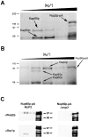
Nup2p docks at the NPC through an interaction with Nup60p and facilitates the formation of a tetrameric complex between Nup60p, Kap60p, Kap95p, and Nup2p. (A) Nup2p–pA from whole cell lysates was bound to IgG–Sepharose. Coprecipitating proteins were eluted with a MgCl2 gradient, separated by SDS-PAGE, and detected by Coomassie blue staining. Shown, from left to right, are molecular mass standards (kD) followed by the fractions eluted by treatment with 0.2, 0.5, 1.0, 2.0, and 4.0 M MgCl2. The two abundantly copurifying proteins were identified by mass spectrometry as Kap60p and Kap95p. No copurifying nucleoporins were detected. (B) Nup60p was immunoprecipitated from yeast whole cell lysates and analyzed as in A. Immunoprecipitation of Nup60p–pA from whole cell lysates coprecipitated Kap60p, Kap95p, and Nup2p, suggesting that Nup60p is the nucleoporin that anchors Nup2p to the nuclear face of the NPC. We detected no coprecipitating proteins by Coomassie staining when the same immunoprecipitation was performed from a strain lacking Nup2p (data not shown). (C) Immunoblot analysis of Nup60p–pA immunoprecipitations in wild-type and Δnup2 strains confirms the absence of both Nup2p and Kap60p in the Δnup2 strain. The 0.5, 1.0, 2.0, and 4.0 M MgCl2 elution fractions from Nup60–pA immunoprecipitations in wild-type and Δnup2 strains were probed using anti-Kap60p (anti-SRP1) and anti-Nup2p antibodies. The absence of Kap60p in the immunoprecipitation from strains lacking Nup2p indicates that Nup2p facilitates the interaction between Nup60p and Kap60p and suggests that the interaction between Nup2p and Nup60p is direct. The two closely migrating bands recognized by the anti-Nup2p antibody are specific to Nup2p as neither band is present in strains lacking Nup2p. The signal observed in the 4,000 mM elution fraction represents Nup60–pA and Nup60p–pA breakdown products that bound to the rabbit polyclonal antibodies.
Figure 7
Figure 7
Genetic interactions between NUP2, NUP60, and KAP60. (A) Δnup2 and Δnup60 strains were crossed and sporulated with the covering plasmid, pLDB60 (NUP2 URA3 CEN). Progeny of the indicated genotypes were assayed for the ability to grow without the NUP2-covering plasmid by growth on fluoroorotic acid. As shown by serial dilution of logarithmically growing cultures, only the double mutant fails to grow on fluororotic acid, indicating that NUP2 and NUP60 are synthetically lethal. (B) To assess any genetic interaction between NUP60 and KAP60, we mated a strain harboring a temperature-sensitive KAP60 allele, srp1-31, with a nup60 null strain. Wild-type and single and double mutant spores were isolated and assayed for their ability to grow at 23°C, 30°C, and 37°C. A genetic interaction was observed between KAP60 and NUP60, as the Δnup60,srp1-31 strain failed to grow at 30°C, whereas the srp1-31 single mutant grew at 30°C but not at 37°C. (C) Expression of a Nup2p mutant lacking the RBD of Nup2p rescues the synthetic lethality observed between Nup2 and Nup60, but conveys a temperature-sensitive phenotype. A plasmid encoding amino acid residues 1–546 of Nup2p (pLDB690) was able to partially rescue the synthetic lethality observed between NUP2 and NUP60. Δnup2nup60 cells carrying the pLDB690 plasmid grew slowly at 23°C and normal at 30°C, but failed to grow at 37°C. Thus, the RBD of Nup2p performs a function that becomes essential in strains lacking Nup60p.
Figure 5
Figure 5
Nup60p facilitates the binding of Nup2p to the NPC. (Top) Nup2p–GFP in wild-type backgrounds exhibits punctate peripheral nuclear rim staining characteristic of a nucleoporin. Like other nucleoporins, Nup2p–GFP clusters to one face of the nuclear rim in cells lacking Nup120p. Consistent with our in vitro binding data, deletion of NUP1 has no effect on the localization of Nup2p–GFP. (Middle) Deletion of NUP60 results in the nuclear accumulation of Nup2p–GFP, but has no effect on the localization of the control nucleoporins, Nsp1p and Nup49p. (Bottom) In strains lacking Nup60p, Nup2p–GFP signal returned to the nuclear rim upon expression of NUP60 from a galactose-inducible promoter.
Figure 6
Figure 6
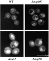
Nup60p is involved in the export of Kap60p from the nucleus. In agreement with previous reports, we observed a nuclear accumulation of Kap60p–GFP in cells lacking Nup2p. This defect was also observed, but was less severe, in strains lacking Nup60p, which suggests that Nup2p docking to Nup60p plays a role in Cse1p-mediated export of Kap60p. The localization of Kap60p–GFP was unaffected by deletion of NUP100, indicating that the redistribution observed in cells lacking Nup2p or Nup60p is specific.
Figure 8
Figure 8
Deletion of the RBD of Nup2p affects its ability to bind to the NPC. (Top) Images of Nup2p–GFP in wild-type and Δnup60 cells as well as for Nup2ΔRBDp–GFP in an otherwise wild-type background. (Middle) Plots of the fluorescent intensity across a nuclear bisect for 15 cells, each of the strains above. (Bottom) Plots of the mean (thick line) and standard deviation (shaded region) of data presented above. Comparison reveals an increased nuclear signal of Nup2ΔRBDp–GFP relative to Nup2p–GFP. However, relative to Nup2p–GFP in strains lacking Nup60p, there remained a significant portion of Nup2ΔRBDp–GFP present at the nuclear rim.
Figure 9
Figure 9
Perturbations in the Ran cycle can return Nup2p–GFP to the NPC in strains lacking Nup60p. (Ai) After initial image acquisition (START), the indicated strains were metabolically poisoned by azide/deoxyglucose treatment for 45 min (POISON). No change in the localization of Nup2p–GFP was observed. In contrast, Nup2p–GFP accumulated at the nuclear rim in strains lacking Nup60p after the 45-min poisoning treatment. The localization of Kap60p–GFP similarly accumulated at the nuclear rim under these conditions. Furthermore, a 10-min recovery period in glucose-containing media resulted in the return of Nup2p–GFP and Kap60p–GFP to their respective steady-state locales (RECOVERY). (Aii) Inactivation of the Ran-GTP exchange factor, Prp20p, enhances binding of Nup2p to alternative sites within the NPC. Nup2p–GFP was expressed in Δnup60,prp20-7 cells, grown at 23°C (START), shifted to 37°C for 90 min (TEMP. SHIFT), and then recovered at room temperature for 60 min (RECOVERY). Kap60p–GFP accumulated at the nuclear rim at the nonpermissive temperature, indicating a block in nuclear transport. Neither Nup49p–GFP nor Nup2p–GFP in otherwise wild-type cells appeared affected by this treatment; however, Nup2p–GFP signal returned to the nuclear rim and accumulated in the cytoplasm in a strain lacking Nup60p. This effect was reversible as growth at the permissive temperature restored the steady-state nuclear localization of Nup2p–GFP in the Δnup60 strain. (B) The alternative NPC docking site of Nup2p is likely at the cytoplasmic face of the NPC. Nup2p–pA in cells lacking Nup60p was localized by IEM of purified nuclei. We could find no signal was detected on the nuclear face of the NPC; however, there remained a pool of Nup2p–pA that remained associated with the NPC on the cytoplasmic face.

Similar articles

Cited by

References

    1. Aitchison J.D., Blobel G., Rout M.P. Nup120pa yeast nucleoporin required for NPC distribution and mRNA transport J. Cell Biol 131 1995. 1659 7165a - PMC - PubMed
    1. Aitchison J.D., Rout M.P., Marelli M., Blobel G., Wozniak R.W. Two novel related yeast nucleoporins Nup170p and Nup157pcomplementation with the vertebrate homologue Nup155p and functional interactions with the yeast nuclear pore-membrane protein Pom152p J. Cell Biol 131 1995. 1133 1148b - PMC - PubMed
    1. Aitchison J.D., Blobel G., Rout M.P. Kap104pa karyopherin involved in the nuclear transport of messenger RNA binding proteins. Science. 1996;274:624–627. - PubMed
    1. Akey C.W. Structural plasticity of the nuclear pore complex. J. Mol. Biol. 1995;248:273–293. - PubMed
    1. Allen T.D., Cronshaw J.M., Bagley S., Kiseleva E., Goldberg M.W. The nuclear pore complexmediator of translocation between nucleus and cytoplasm. J. Cell Sci. 2000;113:1651–1659. - PubMed

Publication types

MeSH terms