. 2001 Jun 15;20(12):3145-55.
doi: 10.1093/emboj/20.12.3145.

Vac8p release from the SNARE complex and its palmitoylation are coupled and essential for vacuole fusion

Affiliations

Vac8p release from the SNARE complex and its palmitoylation are coupled and essential for vacuole fusion

M Veit et al. EMBO J. .

Abstract

Activated fatty acids stimulate budding and fusion in several cell-free assays for vesicular transport. This stimulation is thought to be due to protein palmitoylation, but relevant substrates have not yet been identified. We now report that Vac8p, a protein known to be required for vacuole inheritance, becomes palmitoylated when isolated yeast vacuoles are incubated under conditions that allow membrane fusion. Similar requirements for Vac8p palmitoylation and vacuole fusion, the inhibition of vacuole fusion by antibodies to Vac8p and the strongly reduced fusion of vacuoles lacking Vac8p suggest that palmitoylated Vac8p is essential for homotypic vacuole fusion. Strikingly, palmitoylation of Vac8p is blocked by the addition of antibodies to Sec18p (yeast NSF) only. Consistent with this, a portion of Vac8p is associated with the SNARE complex on vacuoles, which is lost during Sec18p- and ATP-dependent priming. During or after SNARE complex disassembly, palmitoylation occurs and anchors Vac8p to the vacuolar membrane. We propose that palmitoylation of Vac8p is regulated by the same machinery that controls membrane fusion.


Figures

None
Fig. 1. Identification of Vac8p as a target of palmitoylation on isolated vacuoles. (A) Vacuole fusion depends on palmitoylation. Vacuoles (6 µg) from yeast strains BJ3505 and DKY6281 were incubated in a 30 µl reaction in the presence of ATP for 90 min at 26°C. Where indicated, cytosol (15 µg), CoA (10 µM), palmitate (200 µM) or Br-Pal (200 µM) was added to the reaction. Then, fusion activity was measured (Haas et al., 1994). (B) Vac8p is palmitoylated during the fusion reaction. Vacuoles from DKY6281 (60 µg) were labeled with [3H]palmitate (150 µCi) in a 300 µl volume at 30°C in the absence or presence of ATP (1 mM), cytosol (0.5 µg/µl) and CoA (10 µM). After 90 min, vacuoles were isolated by centrifugation (5 min, 4°C, 12 000 g), washed with 500 µl of PS buffer and resuspended in SDS sample buffer without 2-mercaptoethanol (lanes 1–4). Lane 5 shows an immuno precipitation of one complete reaction. The sample shown in lane 6 was labeled in parallel to lane 5, but analyzed without immunoprecipitation. Palmitoylated proteins were identified by SDS–PAGE and fluorography. After film exposure, the gel shown in lanes 5 and 6 was rehydrated, treated with 1 M hydroxylamine pH 6.8 overnight, dried and analyzed by fluorography (lanes 7 and 8) as before. (C) Vac8p palmitoylation is sensitive to reducing agents. Vacuoles were incubated for 90 min in the presence of ATP, cytosol, CoA and [3H]palmitate as in (B). After reisolation, three identical vacuole pellets were resuspended in SDS sample buffer without (lane 1), with 5% (lane 2) or with 10% (lane 3) 2-mercaptoethanol. Samples were boiled for 4 min at 95°C prior to SDS–PAGE analysis and fluorography.
None
Fig. 2. Requirements for Vac8p palmitoylation and vacuole fusion coincide. (A) Vac8p palmitoylation requires an optimal CoA concentration. A fusion reaction (300 µl) containing 60 µg of vacuoles from DKY6281 was incubated for 60 min in the presence of cytosol, ATP, [3H]palmitate and increasing amounts of CoA. Palmitoylated Vac8p was identified by SDS–PAGE and fluorography as described in Figure 1. (B) Vacuoles from both tester strains were incubated in a 30 µl reaction volume under similar conditions with varying CoA concentrations to determine fusion activity. (C) Addition of Br-Pal blocks Vac8p palmitoylation. Vacuoles (60 µg) were incubated in a 300 µl volume for 60 min in the presence of ATP, cytosol, CoA and [3H]palmitate. Br-Pal was added at the indicated concentrations to the fusion reaction. Palmitoylated Vac8p was analyzed as in (A). (D) Fusion is blocked by Br-Pal. Vacuoles from both tester strains were incubated at 26°C in the presence of ATP, CoA, cytosol and Br-Pal or palmitate at the indicated concentrations. Fusion was determined after 90 min incubation. Note that palmitate has no stimulatory effect on vacuole fusion if cytosol is added to the assay (see also lanes 8 and 10 in Figure 1A).
None
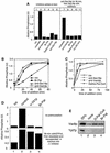
Fig. 3. Vac8p is labeled in the beginning of the fusion reaction. (A) Kinetics of Vac8p palmitoylation. Vacuole fusion reactions (300 µl) containing ATP, cytosol and CoA (10 µM) were labeled with [3H]palmitate for 0, 10, 30, 45 or 90 min. Palmitoylation of Vac8p was then analyzed as described in Figure 1B. Normalized intensities of labeled bands including standard deviations (n = 3): 100% (10 min), 79 ± 8.5% (30 min), 85 ± 10.5% (45 min), 68 ± 14% (90 min). (B) Vac8p palmitoylation cannot be chased by the addition of cold palmitate. Vacuolar fusion reactions containing ATP, cytosol and CoA (10 µM) were labeled with [3H]palmitate for 10 min. Palmitoylation was chased by addition of excess unlabeled palmitate (30 µM) and incubated for 0, 15, 30 or 60 min. Normalized intensities of labeled bands including standard deviations (n = 3): 100% (0 min), 88 ± 5.5% (15 min), 86 ± 4.5% (30 min), 79 ± 9% (60 min). (C) Vac8p is palmitoylated only within the first 10 min of the fusion reaction. Vacuolar fusion reactions containing ATP, cytosol and CoA (10 µM) were labeled with [3H]palmitate at 0, 10, 30 or 60 min after the start of the reaction and incubations were continued for 15 min. (D) Synthesis of Pal-CoA is restricted to the start of the reaction. Vacuoles from DKY6281 were incubated at 30°C in the presence of cytosol, ATP and CoA. [3H]palmitate was added at the indicated times and labeling was carried out for 15 min. In lane 5, additional CoA (10 µM) and ATP (1 mM) were added together with [3H]palmitate. In lane 7, vacuoles received anti-Sec18p to block priming. Lane 6 is the control without antibodies. Labeling with [3H]palmitate was carried out for 15 min. Reactions were then placed on ice, and lipids were extracted and analyzed by TLC and fluorography as described in Materials and methods. The identity of bands was verified with marker lipids run on the same TLC plate. FS = fatty acids and neutral lipids, PS = phosphatidylserine, PI = phosphatidylinositol. (E) [3H]Pal-CoA was added at 0, 10, 20 or 45 min after the start to fusion reactions without CoA and the incubations were continued for 15 min. (F) CoA addition stimulates fusion only within the first 10 min. Vacuoles from both tester strains were incubated in a 30 µl volume in the presence of ATP. At the indicated time points, 10 µM CoA was added and the incubation was continued to a total of 90 min. Then fusion was determined.
None
Fig. 4. Palmitoylation inhibitors block vacuole docking. (A) Requirement for palmitoylation cannot be fulfilled before Sec18p action. Vacuoles were incubated for 30 min at 26°C in the presence of ATP, cytosol and CoA and in the absence (lanes 1–6) or presence (lanes 7–11) of antibodies to Sec18p. The indicated inhibitors were added to the reactions shown in lanes 1–6 before the start. After 30 min, reactions 7–11 were placed on ice, diluted with 300 µl of PS buffer, centrifuged (8000 g, 4 min, 4°C) and resuspended in 30 µl of reaction buffer containing ATP, cytosol, CoA and 100 ng of Sec18p. Br-Pal (200 µM), dethio-Pal-CoA (30 µM) or antibodies to Vam3p were added where indicated. Incubations were for 90 min at 26°C. Then fusion was assayed. (B) Time-of-addition experiment with Br-Pal. A 30× scale fusion reaction was started in the presence of ATP, CoA (10 µM) and cytosol at 26°C. Aliquots (30 µl) were removed at the indicated time, antibodies (200 ng/µl) to Sec18p, Vam3p or Br-Pal (200 µM) were added and samples were incubated further at 26°C for a total of 90 min before being assayed for fusion activity. Ice = samples were placed on ice at the indicated times. (C) As for (B) except that dethio-Pal-CoA (30 µM) was added instead of Br-Pal. (D) The fusion step is insensitive to inhibitors of palmitoylation. Upper panel: fusion reactions (30 µl) containing ATP, cytosol and inhibitors [antibodies to Vti1p (200 ng/µl) or Br-Pal (200 µM) as indicated] were incubated for 90 min at 26°C or on ice. Lower panel: fusion reactions (30 µl) containing ATP, cytosol and 2 mM Mg-GTPγS were incubated at 26°C for 30 min. Samples were then diluted with 150 µl of PS buffer containing 150 mM KCl, and vacuoles were reisolated (3 min, 8000 g, 4°C) and resuspended in 30 µl of reaction buffer containing ATP, cytosol and the inhibitors as indicated. Fusion was determined after an additional 60 min incubation at 26°C or on ice. (E) A block in palmitoylation causes release of Vac8p from the vacuole. BJ3505 vacuoles (15 µg) were incubated in a 75 µl reaction for 30 min at 26°C in the presence of ATP, and Br-Pal (200 µM) or dethio Pal-CoA (30 µM) where indicated. Vacuoles were then reisolated (10 min, 8000 g, 4°C) and proteins in the reaction supernatant were precipitated by 13% trichloroacetic acid (v/v). Proteins were then analyzed by SDS–PAGE and immunoblotting with antibodies to Vac8p and Ypt7p. Bands were quantified by laser densitometry. In lanes 2–4, release to the supernatant was 4.3, 12 and 11% for Vac8p, and 2.6, 3.1 and 2.7% for Ypt7p, respectively.
None
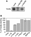
Fig. 5. Vac8p palmitoylation depends on Sec18p. (A) Fusion reactions were labeled with [3H]palmitate in the presence or absence of antibodies to Sec18p or Vam3p (200 ng/µl) for 15 min as described in Figure 1B. Then, vacuoles were reisolated and analyzed by SDS–PAGE and fluorography. (B) Quantification of the inhibition of Vac8p palmitoylation from independent experiments. Labeling of vacuoles with [3H]palmitate was carried out as in (A). Control incubation was set to 100%. Inhibition of Vac8 palmitoylation with antibodies (200 ng/µl) to Sec18p (n = 3), Vam3p (n = 3), Vti1p (n = 1), Nyv1p (n = 1), 5 mM BAPTA (n = 1) and 300–500 µM neomycin (n = 4) was quantified by laser densitometry. Control incubation was set to 100%. All inhibitors blocked the fusion reaction completely (not shown).
None
Fig. 6. Vac8p is part of the SNARE complex. (A) Co-immunoprecipitation of Vac8p with anti-Vti1p antiserum.Vacuoles (50 µg) were incubated in a 250 µl volume with or without ATP and Br-Pal (200 µM) for 15 min at 26°C, pelleted (5 min, 8000 g, 4°C) and solubilized. The detergent extract was incubated overnight with pre-immune IgGs, and then immunoprecipitated for 2 h with antibodies to Vti1p. Retained proteins were eluted and analyzed (see Materials and methods). Immunoblots were decorated with antibodies to Vam3p, Sec17p, Vac8p and Vti1p. Whereas all Vti1p is immunoprecipitated in this experiment, 30% of Vam3p and Sec17p and 2% of Vac8p is found in association with Vti1p (lane 1) as quantified by laser densitometry. (B) Immunoprecipitation of Vam3p with anti-Vac8p antiserum. Vacuoles (50 µg) from DKY6281 containing Vac8p with a C-terminal GFP tag (lane 1) or DKY vacuoles lacking Vac8p (lane 2) were detergent solubilized as in (A) and immunoprecipitation was carried out overnight with protein A–Sepharose-linked antibodies to Vac8p. Western blots were decorated with antibodies to Vac8p and Vam3p. Tagged Vac8p was used in this experiment because of occasional cross-reactivity of the anti-Vac8p antibody with the 60 kDa region on the blot. (C) Cross-linking of Vac8p and Vam3p. Vacuoles from BJ3505 containing a Vac8 protein with a C-terminal TAP tag (see Materials and methods) were incubated in 250 µl of reaction buffer for 30 min on ice with or without 200 µM dithiobis-succinimidyl propionate (DSP, Pierce). Then, 20 mM Tris pH 7.9 was added, vacuoles were reisolated (5 min, 8000 g, 4°C) and detergent solubilized in 1 ml of 1% Triton X-100, 20 mM HEPES–KOH pH 7.9, 300 mM NaCl, 1 mM PMSF, 0.5× PIC. Immunoprecipitation with 30 µl of IgG–Sepharose (Amersham Pharmacia), washing and elution of bound proteins are described in Materials and methods. Blots were decorated with antibodies to Vac8p and Vam3p. Due to high salt washing of the beads, association of Vam3p with Vac8p was lost without DSP pre-treatment. Some Vac8p signal in lane 3 was lost due to cross-linking into high molecular weight complexes.
None
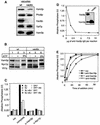
Fig. 7. Vac8p is essential for fusion. (A) Protein composition of vacuoles from wild-type or vac8Δ mutants. Vacuoles (10 µg) from DKY6281 and the corresponding deletion mutant were solubilized in SDS sample buffer, boiled for 4 min at 95°C and analyzed by SDS–PAGE and western blotting. The immunoblot was decorated with antibodies to Vam3p, Vac8p, Pho8p, Vam2p and Ypt7p. (B) Deletion of Vac8p does not influence the integrity of the SNARE complex. Vacuoles from BJ3505 or the corresponding strain lacking Vac8p were purified, incubated for 10 min in the presence or absence of ATP and processed for co-immunoprecipitation with anti-Vti1p antibodies as described in Figure 6A. (C) Fusion of vac8Δ vacuoles. Vacuoles from the tester strains and the respective vac8Δ strains were incubated for 90 min at 26°C in the presence of cytosol, Sec18p (20 ng), CoA (10 µM) and ATP in the combinations shown. Inhibitors such as IgGs to Vam3p or Nyv1p (both at 200 ng/µl) were added where indicated. After the incubation, fusion was assayed as described. v8Δ stands for vac8Δ. (D) Sensitivity of vacuole fusion to antibodies against Vac8p. Fusion reactions (30 µl) containing vacuoles from both tester strains, ATP and cytosol were incubated for 90 min at 26°C. Increasing amounts of purified IgGs against full-length Vac8p (see Materials and methods) were added where indicated. After 90 min, fusion was measured. The inset shows an immunoblot of vacuoles (wt = 5 µg, vac8Δ = 50 µg) decorated with Vac8p (new) and Vam3p antibodies. (E) Vac8p antibodies inhibit at priming. Reactions were incubated and aliquots removed as described in Figure 4B, except that IgGs to Sec18, Vam3p and Vac8p (200 ng/µl) were added as inhibitors.

Similar articles

Cited by

References

    1. Bano M.C., Jackson,C.S. and Magee,A.I. (1998) Pseudo-enzymatic S-acylation of a myristoylated Yes protein kinase peptide in vitro may reflect non enzymatic S-acylation in vivo. Biochem. J., 330, 723–731. - PMC - PubMed
    1. Berger M. and Schmidt,M.F. (1984) Identification of acyl donors and acceptor proteins for fatty acid acylation in BHK cells infected with Semliki Forest virus. EMBO J., 3, 713–719. - PMC - PubMed
    1. Berthiaume L. and Resh,M.D. (1995) Biochemical characterization of a palmitoyl acyltransferase activity that palmitoylates myristoylated proteins. J. Biol. Chem., 270, 22399–22405. - PubMed
    1. Choi J.Y. and Martin,C.E. (1999) The Saccharomyces cerevisiae FAT1 gene encodes an acyl-CoA synthetase that is required for maintenance of very long chain fatty acid levels. J. Biol. Chem., 274, 4671–4683. - PubMed
    1. Conradt B., Shaw,J., Vida,T., Emr,S. and Wickner,W. (1992) In vitro reactions of vacuole inheritance in Saccharomyces cerevisiae. J. Cell Biol., 119, 1469–1479. - PMC - PubMed

Publication types

MeSH terms