. 2001 Jun 1;20(11):2954-65.
doi: 10.1093/emboj/20.11.2954.

A subcomplex of three eIF3 subunits binds eIF1 and eIF5 and stimulates ribosome binding of mRNA and tRNA(i)Met

Affiliations

A subcomplex of three eIF3 subunits binds eIF1 and eIF5 and stimulates ribosome binding of mRNA and tRNA(i)Met

L Phan et al. EMBO J. .

Abstract

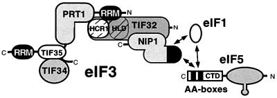
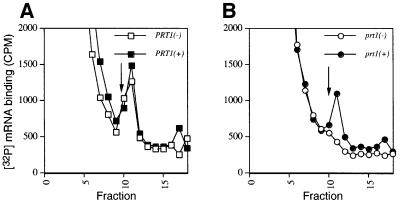
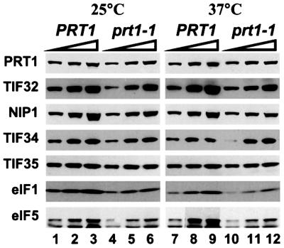
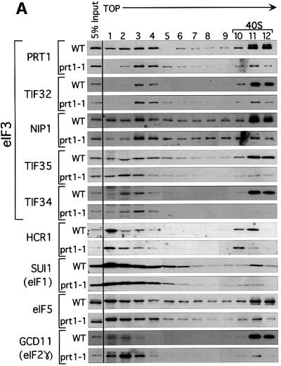
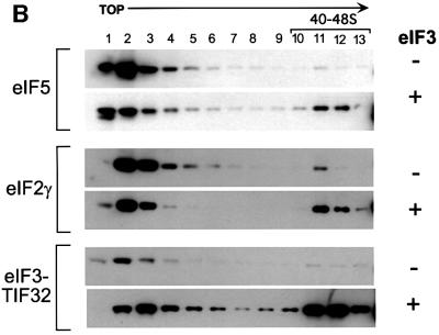
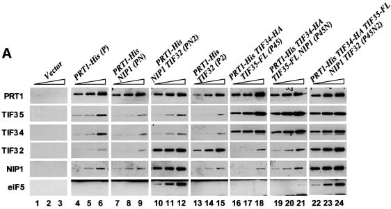
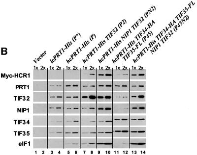
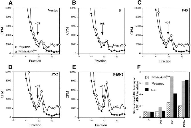
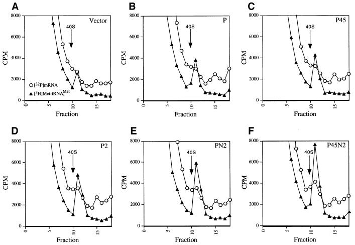
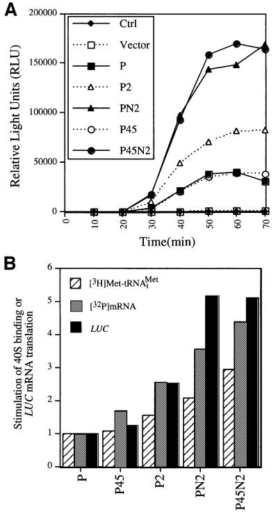
Yeast translation initiation factor 3 contains five core subunits (known as TIF32, PRT1, NIP1, TIF34 and TIF35) and a less tightly associated component known as HCR1. We found that a stable subcomplex of His8-PRT1, NIP1 and TIF32 (PN2 subcomplex) could be affinity purified from a strain overexpressing these eIF3 subunits. eIF5, eIF1 and HCR1 co-purified with this subcomplex, but not with distinct His8-PRT1- TIF34-TIF35 (P45) or His8-PRT1-TIF32 (P2) sub complexes. His8-PRT1 and NIP1 did not form a stable binary subcomplex. These results provide in vivo evidence that TIF32 bridges PRT1 and NIP1, and that eIFs 1 and 5 bind to NIP1, in native eIF3. Heat-treated prt1-1 extracts are defective for Met-tRNA(i)Met binding to 40S subunits, and we also observed defective 40S binding of mRNA, eIFs 1 and 5 and eIF3 itself in these extracts. We could rescue 40S binding of Met- tRNA(i)Met and mRNA, and translation of luciferase mRNA, in a prt1-1 extract almost as well with purified PN2 subcomplex as with five-subunit eIF3, whereas the P45 subcomplex was nearly inactive. Thus, several key functions of eIF3 can be carried out by the PRT1-TIF32-NIP1 subcomplex.


Figures

None
Fig. 1. Predicted interactions among yeast eIF3 subunits, eIF5, eIF1 and HCR1. Yeast eIF3 contains five core subunits (TIF32, PRT1, NIP1, TIF34 and TIF35) and a less tightly associated component known as HCR1. PRT1 and TIF35 contain RRMs, and TIF32 contains an HCR1-like domain (HLD). Interactions among these proteins detected by yeast two-hybrid or in vitro binding assays are depicted schematically as points of contact between the representative shapes. The CTD of eIF5 contains a conserved bipartite motif (AA-boxes) required for its interaction with eIF3–NIP1. NIP1 additionally interacts with eIF1, and eIF1 interacts with the eIF5-CTD in these binding assays. The N-terminal portion of eIF5 contains a zinc-finger motif depicted as a prong. (See text for references.)
None
Fig. 2. Rescue of [32P]mRNA binding to the 40S ribosome in heat-inactivated prt1-1 extract by purified eIF3. Twenty microliters (∼300 µg) of extract prepared from PRT1 strain LPY200 (A) or prt1-1 strain H1676 (B) were heat treated at 37°C for 5 min and incubated in a 40 µl reaction containing ∼2 pmol of [32P]MFA2 mRNA (∼200 000 c.p.m.), 1× translation buffer, 1.2 mM GMPPNP, and either 1.5 pmol purified eIF3 (+) or buffer alone (–), at 26°C for 20 min. The reactions were stopped by adding formaldehyde to 0.3% and chilled on ice for 10 min before loading on a 7.5–30% sucrose gradient and centrifuging for 5 h at 41 000 r.p.m. in an SW41 rotor at 4°C. Fractions of 0.6 ml were collected using an ISCO gradient fraction collector and assayed for [32P]mRNA by mixing 0.2 ml of each fraction with 1 ml of water and 10 ml of scintillation fluid, and counting in a scintillation counter. The arrow in each panel marks the migration position of 40S ribosomes.
None
Fig. 3. Ni2+ affinity purification of intact eIF3 complexes containing eIF1 and eIF5 from heat-treated extract containing His8-prt1-1. Translation extracts were prepared from strains LPY201 (PRT1-His) and LPY202 (prt1-1-His) grown in YPD medium (Sherman et al., 1974) at 26°C to an OD600 of 1.0. The extracts were supplemented with imidazole to a final concentration of 20 mM, incubated at 25 or 37°C for 5 min, as indicated, and subjected to Ni2+ chelation chromatography using buffer A (see Materials and methods). Equivalent aliquots (2.5, 5 and 10 µl) of the Ni2+-NTA–silica eluates were separated by SDS–PAGE using 4–20% gels and subjected to immunoblot analysis using rabbit polyclonal antibodies against the proteins shown on the left, at the following dilutions: PRT1, 1:3000; TIF32, 1:3000; NIP1, 1:1000; TIF34, 1:500; TIF35, 1:5000; eIF5, 1:10000; SUI1/eIF1, 1:1000. Immune complexes were detected by chemiluminescence (ECL™, Amersham Pharmacia Biotech) using horseradish peroxidase-conjugated secondary antibodies (Amersham Pharmacia Biotech).
None
Fig. 4. Binding of eIF3, eIF2, eIF5 and eIF1 to 40S ribosomes is defective in the heat-treated prt1-1 extract. (A) Twenty microliters (∼300 µg) of translation extracts prepared from PRT1 strain LPY200 or prt1-1 strain H1676 were heat treated at 37°C for 5 min and incubated in a 40 µl reaction containing 1× translation buffer and 1.2 mM GMPPNP at 26°C for 20 min. The reactions were stopped by adding formaldehyde to 0.3% and incubating on ice for 10 min. A portion of each reaction (5%) was removed (input samples) and the remainder was separated on a 7.5–30% sucrose gradient as described in Figure 2. Fractions (0.6 ml) were collected and precipitated with 1.0 ml of ethanol at –20°C. The precipitates were washed once with ethanol, dried and resuspended in 50 µl of loading buffer, and separated by SDS–PAGE using 4–20% gradient gels. The separated proteins were subjected to immunoblot analysis using antibodies against the proteins indicated on the left. Antibodies were used at the same dilutions described in Figure 3, with the addition of antibodies against HCR1 (1:500) (Valášek et al., 2001) and GCD11 (1:10000). The position of 40S ribosomes in the gradients is indicated over fractions 10–12. (B) Heat-treated PRT1 and prt1-1 extracts were analyzed as in (A) except that 4.5 pmol of highly purified eIF3 (+) or buffer alone (–) were added to each reaction, as indicated on the right, prior to incubation at 26°C for 20 min. The reactions were stopped and analyzed as described in (A).
None
Fig. 5. eIF5 and eIF1 co-purify with eIF3 subcomplexes containing His8-PRT1, NIP1 and TIF32. (A) His8-PRT1 and associated proteins were purified from the PRS by nickel chelation chromatography in buffer containing 350 mM KCl (buffer B; see Materials and methods) from the following strains overexpressing different combinations of eIF3 subunits: LPY60 (empty vectors), LPY65 (P), LPY66 (PN), LPY67 (PN2), LPY68 (P2), LPY85 (P45), LPY86 (P45N) and LPY87(P45N2). The letters and numbers in parentheses designate overexpression of His8-PRT1 (P), HA-TIF34 (4), FLAG-TIF35 (5), NIP1(N) and TIF32 (2). Three serial dilutions of the Ni2+-NTA–silica eluates for each preparation were resolved by 4–20% SDS–PAGE and subjected to immunoblot analysis as described in Figures 3 and 4. Samples of the control extract loaded in lanes 1–3 (Vector) contained 0.5, 1 and 2 µg of total protein, respectively. Samples of the P preparation containing overexpressed His8-PRT1 alone in lanes 4–6 contained 0.38, 0.75 and 1.5 µg total protein, respectively. For the remaining preparations, the amounts loaded were predetermined to contain the same quantities of His8-PRT1 as in lanes 4–6. (B) Yeast strains LPY60, LPY65, LPY67, LPY68, LPY85 and LPY87, described in (A), were transformed with low-copy-number plasmid YCpLVHM-T encoding c-myc-tagged HCR1 to create strains LPY142 (Vector), LPY134 (P), LPY136 (PN2), LPY137 (P2), LPY138 (P45) and LPY140 (P45N2), respectively. In addition, strain LPY191 (P*) was constructed containing YCpLVHM-T and a low-copy plasmid bearing PRT1-His. His8-PRT1 and associated proteins were purified from PRS by nickel chelation chromatography in a buffer containing 100 mM KCl (buffer A; see Materials and methods). Two dilutions of the Ni2+-NTA–silica eluates for each preparation were resolved by 4–20% SDS–PAGE and subjected to immunoblot analysis as described in Figures 3 and 4, except that anti-c-Myc monoclonal antibodies (Boehringer-Mannheim; 1:2000) were used to probe for c-Myc-tagged HCR1. Samples of the control preparation in lanes 1 and 2 (Vector) contained 0.25 (1×) and 0.5 µg (2×) of total protein, respectively, as did the P* preparation in lanes 3 and 4. Samples of the P preparation in lanes 5 and 6 contained 0.125 (1×) and 0.25 µg (2×) of total protein. For the remaining preparations, the samples were predetermined to contain approximately the same amounts of His8-PRT1 as in lanes 5 and 6.
None
Fig. 6. Rescue of 40S binding of [32P]mRNA and [3H]Met-tRNAiMet and LUC mRNA translation in the prt1-1 extract by the PN2 subcomplex in a PRS. (AE) For each panel, 20 µl (∼300 µg) of heat-treated translation extract from the prt1-1 strain were incubated in a 40 µl reaction containing 1× translation buffer, 1.2 mM GMPPNP, 1.2 pmol of [3H]Met-tRNAiMet (∼90 000 c.p.m.), 2 pmol of [32P]MFA2 mRNA (∼200 000 c.p.m.) and 15 µg of PRS (prepared as described in Materials and methods using buffer A) from the strains described in Figure 5A containing different combinations of overexpressed eIF3 subunits. The identity of the PRS added to the reaction is indicated in the upper right corner of the panel by the designations defined in Figure 5A. The reactions were carried out at 26°C for 20 min, stopped by addition of formaldehyde to 0.3% on ice for 10 min, and then separated on sucrose gradients. An aliquot (0.2 ml) of each fraction was assayed for [3H]Met-tRNAiMet and [32P]MFA2 mRNA by liquid scintillation counting, as described in Figure 2. The arrow in each panel marks the position of the 40S ribosomes in the gradient. (FIn vitro translation reactions were carried out using 35 µl of heat-treated prt1-1 extract in a 70 µl reaction containing 1× translation buffer, 1.2 mM GTP, 4 µg of capped LUC mRNA, 10 U of RNAsin (Promega), all 20 amino acids at 0.1 mM and 15 µg of the PRS fractions. The reactions were incubated at 26°C, and 10 µl aliquots were removed every 10 min and assayed for luciferase production by adding 100 µl of pre-mixed luciferase assay reagents (Promega) in an automated injection luminometer (Analytical Luminescence Laboratory) and measuring the emitted light. The relative stimulation of LUC mRNA translation by the P45, PN2 and P45N2 PRS fractions was calculated by normalizing the relative light units (RLU) measured after a 70 min incubation for the amount of His8-PRT1 present in the PRS fraction as determined by immunoblot analysis (data not shown). The normalized RLU values for the P45, PN2 and P45N2 reactions were divided by the normalized RLU value for the P reaction and plotted graphically (LUC, black bars). The stimulation of [3H]Met-tRNAiMet and [32P]MFA2 mRNA binding to 40S subunits in reactions (C–E) relative to that observed in (B) was also depicted graphically as hatched and gray bars, respectively. The relative amount of [3H]Met-tRNAiMet or [32P]MFA2 mRNA bound to 40S ribosomes in each experiment was determined by measuring the total area under the c.p.m. peak in the 40S region of the gradient using NIH Image software 1.61. The peak area measured for the Vector sample (A) was subtracted from the peak areas measured in (B–E), and the remainders were normalized for the quantity of Prt1-His8 in the PRS fraction added to the reaction. The resulting values for (C–E) were depicted graphically relative to that obtained for the P complex (B), which was set to 1.0.
None
Fig. 7. Rescue of [32P]mRNA and [3H]Met-tRNAiMet binding to 40S ribosomes in prt1-1 extract by affinity-purified PN2 subcomplex. (AF) For each panel, 20 µl (∼300 µg) of heat-treated translation extract from prt1-1 strain H1676 were incubated in a 40 µl reaction containing 1× translation buffer, 1.2 mM GMPPNP, 1.2 pmol of [3H]Met-tRNAiMet (∼90 000 c.p.m.), 2 pmol of [32P]MFA2 mRNA (∼200 000 c.p.m.) and an aliquot of the Ni2+-affinity-purified preparations containing His8-PRT1 described in Figure 5A. Two to three picomoles of the P45N2 preparation were added to the reaction shown in (F). The amounts of the other His8-PRT1-containing complexes added in (B–E) contained the same amount of His8-PRT1 used in (F), as judged by immunoblot analysis (Figure 5A) and Coomassie Blue staining (data not shown) of the different preparations. The aliquot of Vector preparation used in (A) was identical in volume to that of the P preparation used in (B). The reactions were incubated at 26°C for 20 min, and analyzed for the amounts of [32P]mRNA and [3H]Met-tRNAiMet bound to 40S subunits as described in Figures 2 and 6.
None
Fig. 8. Rescue of LUC mRNA translation in prt1-1 extract by affinity-purified PN2 subcomplex. (AIn vitro translation reactions were carried out using 35 µl of heat-treated translation extract from prt1-1 strain in a reaction volume of 70 µl containing 1× translation buffer, 1.2 mM GTP, 4 µg of capped LUC mRNA, 10 U of RNAsin (Promega), each of the 20 amino acids at 0.1 mM, and an aliquot of one of the Ni2+-affinity-purified preparations containing His8-PRT1 described in Figure 5A. The latter were added in the same relative proportions described in Figure 7A–F, using 1.5 pmol of the P45N2 preparation. The reactions were incubated at 26°C and assayed for luciferase production as described in Figure 6. (B) Quantification of the relative activities of eIF3 subcomplexes in stimulating 40S binding of [3H]Met-tRNAiMet and [32P]MFA2 mRNA and of LUC mRNA translation in the prt1-1 extract. The stimulation of LUC mRNA translation by the P2, P45, PN2 and P45N2 complexes observed in (A) was quantified by dividing each RLU value obtained after a 70 min incubation by the corresponding RLU value for the P reaction and plotted graphically (LUC, black bars). The relative amount of [3H]Met-tRNAiMet or [32P]MFA2 mRNA bound to 40S ribosomes in each experiment shown in Figure 7 was determined as described in Figure 6. The peak area measured for the Vector sample (Figure 7A) was subtracted from the peak areas measured in Figure 7B–F. The remainders for the P45N2, PN2, P2 and P45 complexes were depicted graphically relative to the value obtained for the P complex, which was set to 1.0.

Similar articles

Cited by

References

    1. Asano K., Phan,L., Anderson,J. and Hinnebusch,A.G. (1998) Complex formation by all five homologues of mammalian translation initiation factor 3 subunits from yeast Saccharomyces cerevisiae. J. Biol. Chem., 273, 18573–18585. - PubMed
    1. Asano K., Krishnamoorthy,T., Phan,L., Pavitt,G.D. and Hinnebusch,A.G. (1999) Conserved bipartite motifs in yeast eIF5 and eIF2Bε, GTPase-activating and GDP–GTP exchange factors in translation initiation, mediate binding to their common substrate eIF2. EMBO J., 18, 1673–1688. - PMC - PubMed
    1. Asano K., Clayton,J., Shalev,A. and Hinnebusch,A.G. (2000) A multifactor complex of eukaryotic initiation factors eIF1, eIF2, eIF3, eIF5 and initiator tRNAMet is an important translation initiation intermediate in vivo. Genes Dev., 14, 2534–2546. - PMC - PubMed
    1. Asano K., Shalev,A., Phan,L., Nielsen,K., Clayton,J., Valášek,L., Donahue,T.F. and Hinnebusch,A.G. (2001) Multiple roles for the C-terminal domain of eIF5 in initiation complex assembly and GTPase activation. EMBO J., 20, 2326–2337. - PMC - PubMed
    1. Bandyopadhyay A. and Maitra,U. (1999) Cloning and characterization of the p42 subunit of mammalian translation initiation factor 3 (eIF3): demonstration that eIF3 interacts with eIF5 in mammalian cells. Nucleic Acids Res., 27, 1331–1337. - PMC - PubMed

MeSH terms