

Maintenance of TCR clonality in T cells expressing genes for two TCR heterodimers
- PMID: 11381132
- PMCID: PMC34437
- DOI: 10.1073/pnas.121179998
Maintenance of TCR clonality in T cells expressing genes for two TCR heterodimers
Abstract
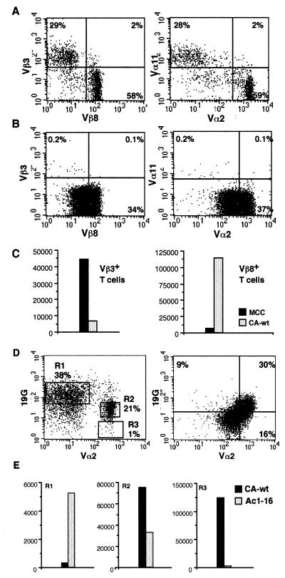
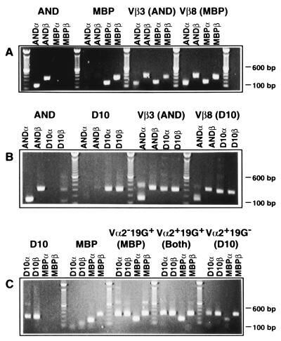
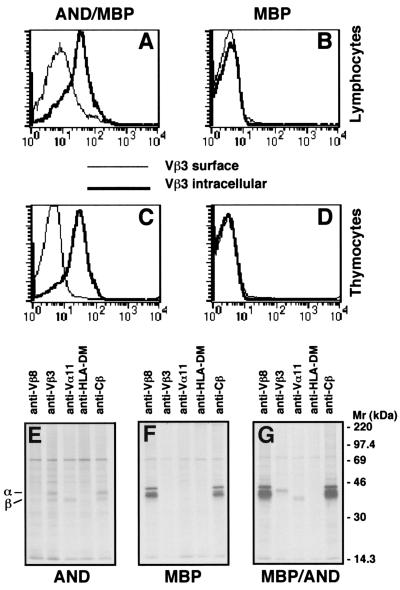
T cell receptor (TCR) allelic exclusion is believed to be primarily mediated by suppression of further recombination at the TCR locus after the expression of a functional TCR protein. Genetic allelic exclusion has been shown to be leaky for the beta chain and, more commonly, for the alpha chain. Here, we demonstrate an additional mechanism by which T cells can maintain monoclonality. T cells from double TCR transgenic mice express only one or the other of the two available TCRs at the cell surface. This "functional allelic exclusion" is apparently due to control of the TCR assembly process because these T cells express RNA and protein for all four transgenic TCR proteins. Lack of cell surface expression of the second TCR may be controlled by a failure to assemble the TCR heterodimer.
Figures

Similar articles
-
Allelic exclusion in pTalpha-deficient mice: no evidence for cell surface expression of two T cell receptor (TCR)-beta chains, but less efficient inhibition of endogeneous Vbeta--> (D)Jbeta rearrangements in the presence of a functional TCR-beta transgene.J Exp Med. 1997 Aug 29;186(5):767-75. doi: 10.1084/jem.186.5.767. J Exp Med. 1997. PMID: 9271592 Free PMC article.
-
Essential role of the pre-T cell receptor in allelic exclusion of the T cell receptor beta locus.Immunity. 1997 Nov;7(5):601-7. doi: 10.1016/s1074-7613(00)80381-7. Immunity. 1997. PMID: 9390684
-
Exclusion and inclusion of TCR alpha proteins during T cell development in TCR-transgenic and normal mice.J Immunol. 2004 Nov 1;173(9):5591-600. doi: 10.4049/jimmunol.173.9.5591. J Immunol. 2004. PMID: 15494509
-
Regulation of TCR alpha and beta gene allelic exclusion during T-cell development.Immunol Today. 1992 Aug;13(8):315-22. doi: 10.1016/0167-5699(92)90044-8. Immunol Today. 1992. PMID: 1324691 Review.
-
Accessibility control of variable region gene assembly during T-cell development.Immunol Rev. 1998 Oct;165:121-30. doi: 10.1111/j.1600-065x.1998.tb01235.x. Immunol Rev. 1998. PMID: 9850857 Review.
Cited by
-
Dual TCR-Expressing T Cells in Cancer: How Single-Cell Technologies Enable New Investigation.Immunohorizons. 2023 May 1;7(5):299-306. doi: 10.4049/immunohorizons.2200062. Immunohorizons. 2023. PMID: 37129560 Free PMC article.
-
Enhanced antitumor activity of T cells engineered to express T-cell receptors with a second disulfide bond.Cancer Res. 2007 Apr 15;67(8):3898-903. doi: 10.1158/0008-5472.CAN-06-3986. Cancer Res. 2007. PMID: 17440104 Free PMC article.
-
Enhanced antitumor activity of murine-human hybrid T-cell receptor (TCR) in human lymphocytes is associated with improved pairing and TCR/CD3 stability.Cancer Res. 2006 Sep 1;66(17):8878-86. doi: 10.1158/0008-5472.CAN-06-1450. Cancer Res. 2006. PMID: 16951205 Free PMC article.
-
Monogenic TCRβ Assembly and Expression Are Paramount for Uniform Antigen Receptor Specificity of Individual αβ T Lymphocytes.J Immunol. 2022 Jul 1;209(1):93-98. doi: 10.4049/jimmunol.2200176. Epub 2022 Jun 13. J Immunol. 2022. PMID: 35697383 Free PMC article.
-
Heterozygous OT-I mice reveal that antigen-specific CD8+ T cells shift from apoptotic to necrotic killers in the elderly.Aging Cell. 2023 Jun;22(6):e13824. doi: 10.1111/acel.13824. Epub 2023 Mar 22. Aging Cell. 2023. PMID: 36947105 Free PMC article.
References
-
- Malissen M, Trucy J, Jouvin-Marche E, Cazenave P A, Scollay R, Malissen B. Immunol Today. 1992;13:315–322. - PubMed
-
- Von Boehmer H. Ann NY Acad Sci. 1995;766:52–61. - PubMed
-
- Mallick C A, Dudley E C, Viney J L, Owen M J, Hayday A C. Cell. 1993;73:513–519. - PubMed
-
- Dudley E C, Girardi M, Owen M J, Hayday A C. Curr Biol. 1995;5:659–669. - PubMed
MeSH terms
Substances
Grants and funding
LinkOut - more resources
Full Text Sources
