. 2001 May 28;153(5):1085-96.
doi: 10.1083/jcb.153.5.1085.

Mba1, a novel component of the mitochondrial protein export machinery of the yeast Saccharomyces cerevisiae

Affiliations

Mba1, a novel component of the mitochondrial protein export machinery of the yeast Saccharomyces cerevisiae

M Preuss et al. J Cell Biol. .

Abstract

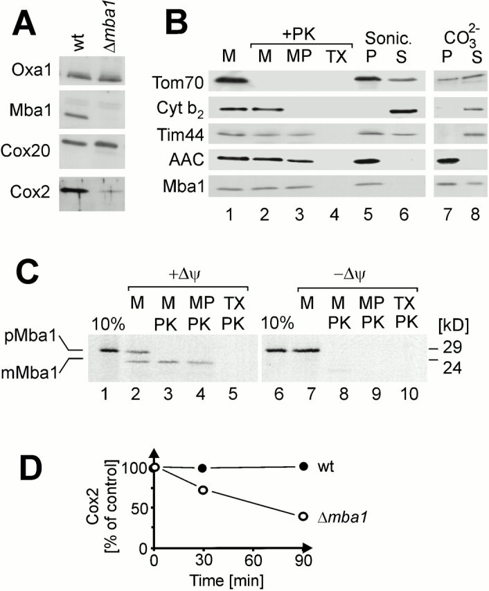
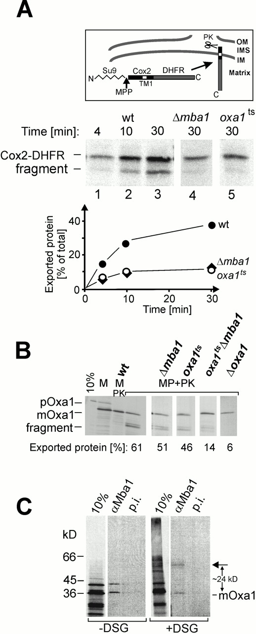
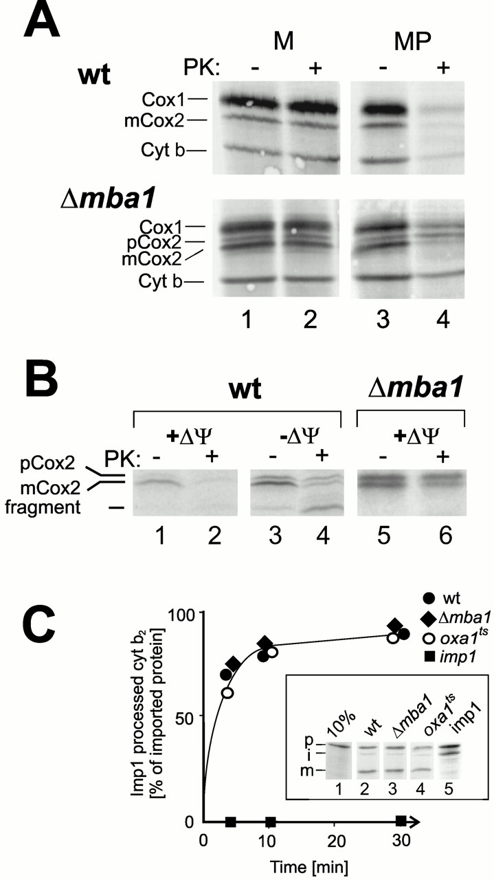
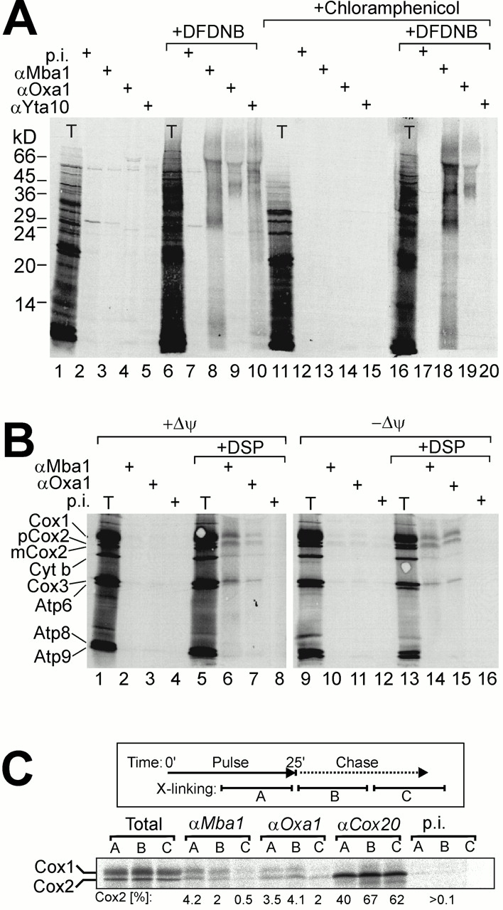
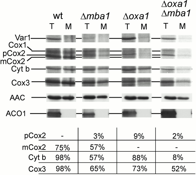
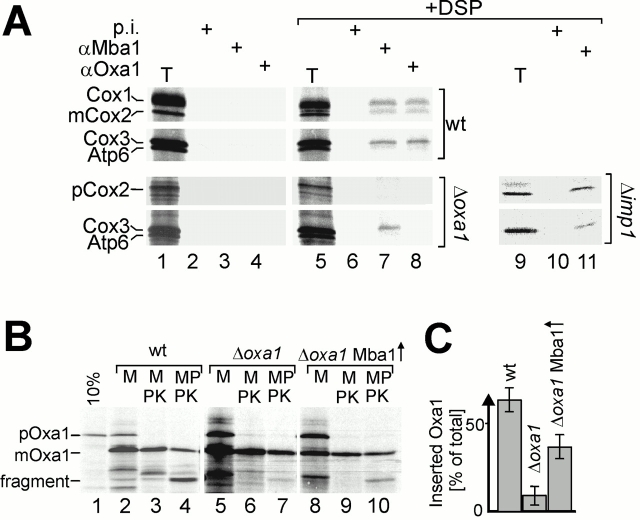
The biogenesis of mitochondria requires the integration of many proteins into the inner membrane from the matrix side. The inner membrane protein Oxa1 plays an important role in this process. We identified Mba1 as a second mitochondrial component that is required for efficient protein insertion. Like Oxa1, Mba1 specifically interacts both with mitochondrial translation products and with conservatively sorted, nuclear-encoded proteins during their integration into the inner membrane. Oxa1 and Mba1 overlap in function and substrate specificity, but both can act independently of each other. We conclude that Mba1 is part of the mitochondrial protein export machinery and represents the first component of a novel Oxa1-independent insertion pathway into the mitochondrial inner membrane.


Figures

Figure 1
Figure 1
Localization of Mba1 on the matrix side of the mitochondrial inner membrane. (A) Western blots of extracts of wild-type (wt) and Δmba1 mitochondria (50 μg) using antisera against Oxa1, Mba1, Cox20, and Cox2. (B) Submitochondrial fractionation. Lane 1, mitochondria (M); lane 2, proteinase K–treated mitochondria; lane 3, proteinase K–treated mitoplasts (MP); lane 4, proteinase K–treated Triton X-100 extract of mitochondria (TX); lane 5, membrane-associated proteins (P); lane 6, soluble protein fraction after sonication of mitochondria (S); lane 7, membrane proteins; and lane 8, soluble protein fraction after carbonate extraction of mitochondria. Marker proteins for the different mitochondrial subcompartments were Tom70 for the outer membrane, cytochrome b2 (Cyt b2) for the intermembrane space, Tim44 for a matrix protein, and ADP/ATP carrier (AAC) for the inner membrane. (C) Import of in vitro–synthesized Mba1 precursor (pMba1) into isolated mitochondria. Mba1 precursor protein was synthesized in reticulocyte lysate and incubated for 20 min at 25°C with wild-type mitochondria. Equal aliquots were either mock treated (lanes 2 and 7), treated with proteinase K (lanes 3 and 8), converted to mitoplasts and treated with proteinase K (lanes 4 and 9), or lysed with 1% Triton X-100 before proteinase K treatment (lanes 5 and 10). For the samples shown in lanes 7–10, the membrane potential (Δψ) was dissipated by addition of 5 μM valinomycin during the import reaction. For comparison, lanes 1 and 6 show 10% of the precursor protein used per import reaction. Proteins were separated by SDS-PAGE and transferred to nitrocellulose. Efficiencies of mitoplast formation and protease treatment were controlled by Western blotting. Mba1 signals were detected by autoradiography. (D) Newly synthesized Cox2 is unstable in Δmba1 mitochondria. Mitochondrial translation products were radiolabeled in wild-type and Δmba1 cells for 30 min. Then the cells were washed twice and reincubated in the presence of 2.5 mg/ml chloramphenicol and 5 mM cold methionine at 30°C for 0, 30, or 90 min. Proteins were separated by SDS-PAGE, and signals of radioactive Cox2 were quantified. The graph shows the amounts of Cox2 relative to the material present directly after the labeling reaction.
Figure 4
Figure 4
Mba1 interacts with nuclear-encoded proteins. (A) Posttranslational insertion of the Cox2 NH2 terminus. In the inset, the export of the Cox2 NH2 terminus after import of pSu9(1-66)pCox2(1-74)-DHFR is depicted. N, NH2 terminus; C, COOH terminus; OM, outer membrane; IMS, intermembrane space; IM, inner membrane; MPP, cleavage site of the mitochondrial processing peptidase; Su9, subunit 9 presequence; TM1, first transmembrane domain of Cox2. (Middle and bottom panels) Wild-type, Δmba1, and oxa1ts mitochondria were pretreated for 10 min at 37°C and incubated with pSu9(1-66)pCox2(1-74)-DHFR for 4, 10, and 30 min at 25°C. The mitochondria were converted into mitoplasts and treated with proteinase K (50 μg/ml) to digest the NH2 terminus of inserted Cox2. Lanes 1–3 show the generation of the fragment in wild-type mitochondria; lanes 4 and 5 show the 30 min reactions of Δmba1 and oxa1ts mitochondria, respectively. The graph shows a quantification of the fraction of inserted Cox2 relative to total imported protein at various times of import. The numbers were corrected for the methionine residues contained in the different Cox2 species. (B) Oxa1 precursor (pOxa1) was synthesized in reticulocyte lysate and incubated for 20 min at 25°C with wild-type, Δmba1, oxa1ts, oxa1ts Δmba1, or Δoxa1 mitochondria. Mitochondria were then converted to mitoplasts (MP) and treated with 50 μg/ml proteinase K (PK) as indicated to convert inserted Oxa1 into a characteristic fragment. The ratio of fragment to total imported Oxa1 was determined by densitometry. (C) Radiolabeled Oxa1 precursor was imported into wild-type mitochondria for 10 min. Then the reaction was split and one half was mock treated, and the other treated with 100 μM DSG for 20 min. The mitochondria were reisolated, lysed, and either directly analyzed by SDS-PAGE (10%) or used for immunoprecipitation with Mba1 antiserum or with preimmune serum. The cross-link product specifically precipitated with Mba1 serum is indicated by an arrow.
Figure 2
Figure 2
Mba1 is required for efficient insertion of Cox2. (A) Wild-type (wt; top) or Δmba1 mitochondria (bottom) were mock treated or converted to mitoplasts. Mitochondrial translation products were radiolabeled for 20 min at 25°C. The samples were split and treated without or with 50 μg/ml proteinase K (PK) as indicated. pCox2, Cox2 precursor; mCox2, mature Cox2; Cyt b, cytochrome b. (B) Translation products were radiolabeled in the presence (−Δψ, lanes 3 and 4) or absence (residual lanes) of 1 μM valinomycin in wild-type or Δmba1 mitoplasts and treated with or without protease K as depicted. The mitoplasts were reisolated and lysed. Radiolabeled Cox2 was visualized by autoradiography after immunoprecipitation with a COOH-terminal Cox2 antiserum. (C) Imp1 processing of cytochrome b2 precursor. Cytochrome b2(1-185)-DHFR precursor protein was synthesized in reticulocyte lysate and incubated for the times indicated at 25°C with wild-type, Δmba1, imp1, or oxa1ts mitochondria. The latter had been preexposed to 37°C to induce the phenotype. Mitochondria were then treated with 50 μg/ml proteinase K to remove nonimported protein. After SDS-PAGE and autoradiography, the fraction of Imp1 processed in relation to total imported protein was quantified by densitometry. The inset shows the signals obtained after import for 20 min in the strains indicated, and 10% of the precursor protein used for each reaction for comparison (lane 1). Complete swelling of the samples was controlled by Western blotting. p, precursor; i, intermediate; m, mature cytochrome b2(1-185)-DHFR.
Figure 5
Figure 5
Mba1 can function independently of Oxa1. (A) Mutations in mba1 and oxa1 cause synthetic growth defects. The strains indicated were grown on YPD medium to log phase and serial 10-fold dilutions of the cultures were spotted on YP plates containing 2% glucose (YPD; top) or 2% glycerol and 2% ethanol (YPEG; bottom). The plates were incubated at the temperatures indicated for 2 (top) or 4 d (bottom). (B) Oxa1 and Mba1 do not cofractionate upon gel filtration. A digitonin extract of wild-type mitochondria was separated on a Superose 12 column. Resulting fractions were analyzed by Western blotting and the signals for Oxa1 and Mba1 were quantified. (C and D) The activities of the bc1 and the Cox complex are decreased in oxa1 and mba1 mutants. Enzyme activities of wild-type, Δmba1, Δoxa1, and Δoxa1 Δmba1 mitochondria were measured in three experiments. Error bars show the standard deviation. (E) ATPase activities of wild-type, Δmba1, Δoxa1, Δoxa1 Δmba1, and Δfzo1 were measured in the presence or absence of oligomycin. The percentage of oligomycin-sensitive ATPase activity is shown. The rho0 mutant Δfzo1 was used as a control for mitochondria lacking functional F0-ATPase.
Figure 3
Figure 3
Mba1 is in direct proximity to mitochondrial translation intermediates. (A) Mitochondrial translation products were radiolabeled for 10 min at 25°C. The reaction was split and treated without (lanes 1–10) or with (lanes 11–20) 2 mg/ml chloramphenicol. After 1 min the samples were either mock treated (lanes 1–5 and 11–15) or treated with 400 μM DFDNB (other lanes) for 20 min. Cross-linking was quenched by the addition of 100 mM glycine. The mitochondria were reisolated, washed, and incubated in 1% SDS for 30 s at 96°C. The extract was centrifuged for 5 min at 15,000 g and either directly applied to SDS-PAGE (T, total) or used for immunoprecipitation with preimmune serum (p.i.) or antiserum against Mba1, Oxa1, and Yta10 as indicated. (B) Mitochondrial translation products were radiolabeled for 15 min in the absence (+Δψ, lanes 1–8) or the presence (−Δψ, lanes 9–16) of 1 μM valinomycin. Then the samples were mock treated (lanes 1–4 and 9–12) or treated with 200 μM DSP (other lanes) for 30 min. Cross-linking and translation were stopped by addition of glycine and unlabeled methionine. Mitochondria were reisolated, washed, and further treated as outlined in A. Lanes labeled T show 10% of the extract used for immunoprecipitation. (C) Transient interaction of Mba1 and Oxa1 with newly synthesized Cox2. Mitochondrial translation products were radiolabeled in three separate reactions for 25 min (Pulse). The labeling was stopped by the addition of 25 μg/ml puromycin and 5 mM methionine, and the samples were further incubated for 30 min (Chase). During this procedure 400 μM of the cross-linker DSP was present for 15 min and then quenched with 100 μM glycine. As depicted in the insert, sample A was cross-linked during the labeling period, B directly afterwards, and C 15 min after the beginning of the chase reaction. The mitochondria were reisolated, lysed, and either directly applied to SDS-PAGE (Total) or used for immunoprecipitation with preimmune serum (p.i.) or antiserum against Mba1, Oxa1, and Cox20 as indicated. Lanes labeled “Total” show 10% of the extract used for immunoprecipitation. The amounts of immunoprecipitated Cox2 were quantified and are shown as a percentage of total Cox2.
Figure 6
Figure 6
oxa1 and mba1 mutants show synthetic protein insertion defects. Translation products were radiolabeled for 20 min at 25°C in mitochondria of the strains indicated. Mitochondria were reisolated and treated with 0.1 M Na2CO3. Samples were split. Proteins were either directly precipitated with TCA (T, total) or after flotation through a sucrose gradient (M, membranes), separated by SDS-PAGE, and transferred to nitrocellulose. Var1, Cox2, cytochrome b, and Cox3 signals were detected by autoradiography, quantified, and the proportions of floatable material are indicated. For control, the soluble protein Aco1 and the membrane protein ATP/ADP carrier (AAC) were detected by Western blotting.
Figure 7
Figure 7
Mba1 can function in the absence of Oxa1. (A) Translation reactions in wild-type, Δoxa1, or Δimp1 mitochondria were incubated with the cleavable cross-linker DSP and analyzed by immunoprecipitation as described in the legend to Fig. 3 B. mCox2, mature Cox2. (B) 35S-labeled Oxa1 precursor was imported for 15 min at 25°C into wild-type, Δoxa1, and Δoxa1Mba1↑ mitochondria. The samples were divided into three parts. Mitochondria (M) were then either mock treated, treated with proteinase K (PK), or converted to mitoplasts (MP) and proteinase K treated. Lane 1 shows 10% of the preprotein used per reaction. (C) The ratio of protease-accessible Oxa1 to total imported Oxa1 was quantified from four independent experiments and depicted in the diagram. Error bars show the standard deviation (n = 4).

Similar articles

Cited by

References

    1. Ackerman S.H., Tzagoloff A. ATP10, a yeast nuclear gene required for the assembly of the mitochondrial F1-F0 complex. J. Biol. Chem. 1990;265:9952–9959. - PubMed
    1. Bauer M., Behrens M., Esser K., Michaelis G., Pratje E. PET1402, a nuclear gene required for proteolytic processing of cytochrome oxidase subunit 2 in yeast. Mol. Gen. Genet. 1994;245:272–278. - PubMed
    1. Bonnefoy N., Chalvet F., Hamel P., Slominski P.P., Dujardin G. OXA1, a Saccharomyces cerevisiae nuclear gene whose sequence is conserved from prokaryotes to eukaryotes controls cytochrome oxidase biogenesis. J. Mol. Biol. 1994;239:201–212. - PubMed
    1. Borst P., Grivell L.A. The mitochondrial genome of yeast. Cell. 1978;15:705–723. - PubMed
    1. Hamel P., Lemaire C., Bonnefoy N., Brivet-Chevillotte P., Dujardin G. Mutations in the membrane anchor of yeast cytochrome c1 compensate for the absence of Oxa1p and generate carbonate-extractable forms of cytochrome c1 . Genetics. 1998;150:601–611. - PMC - PubMed

Publication types

MeSH terms