. 2001 Jun 1;21(11):3839-48.
doi: 10.1523/JNEUROSCI.21-11-03839.2001.

The exocyst complex associates with microtubules to mediate vesicle targeting and neurite outgrowth

Affiliations

The exocyst complex associates with microtubules to mediate vesicle targeting and neurite outgrowth

I E Vega et al. J Neurosci. .

Abstract

During neuronal development, vesicles are targeted to the growth cone to promote neurite outgrowth and synaptogenesis. The Exocyst complex is an essential macromolecule in the secretory pathway that may play a role in vesicle targeting. Although it has been shown that this complex is enriched in rat brain, the molecular mechanism underlying its function is largely unknown. Here, we report that the Exocyst complex coimmunoprecipitates with microtubules from total rat brain lysate. Additionally, the Exocyst complex subcellular localization changes on neuronal differentiation. In undifferentiated pheochromocytoma (PC12) cells, this complex is associated with microtubules at the microtubule organizing center. However, in differentiated PC12 cells and cultured hippocampal neurons, the Exocyst complex and microtubules extend to the growing neurite and colocalize at the growth cone with synaptotagmin. Inhibition of the NGF-activated MAP kinase pathway blocks the Exocyst complex and microtubule redistribution, abolishing neurite outgrowth and promoting cytosolic accumulation of secretory vesicles. Consistently, the overexpression of Exocyst sec10 subunit mutant blocks neurite outgrowth. These results indicate that the Exocyst complex targets secretory vesicles to specific domains of the plasma membrane through its association with the microtubules, promoting neurite outgrowth.


Figures
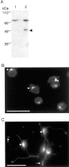
Fig. 1.
Fig. 1.
The Exocyst complex exhibits distinct subcellular localization on neuronal differentiation. Monoclonal antibody against the Exocyst complex subunit Exo70 was produced as explained in Materials and Methods. A, Western blot analysis using the anti-Exo70 monoclonal antibody 70X13F3 showed that it recognized a single band in brain lysate (lane 1). Recombinant Exo70 was used as a molecular weight control (lane 2). Thearrow indicates a degradation product of the recombinant Exo70. B, C, The subcellular localization of the Exocyst complex in both undifferentiated (B) and differentiated (C; NGF; 50 ng/ml) PC12 cells was determined by immunofluorescence microscopy. PC12 cells were fixed with 100% methanol, and the anti-Exo70 antibody was visualized with anti-mouse antibodies conjugated to FITC.B, The Exocyst complex exhibited a perinuclear localization in undifferentiated PC12 cells (arrowheads). C, In differentiated PC12 cells, the Exocyst complex is found in the cell body (arrowheads), along the neurite, and at the growth cone (arrows). Scale bars, 50 μm.
Fig. 2.
Fig. 2.
Brefeldin A does not affect the Exocyst complex perinuclear localization. The localization of Exo70 (A,D, G, J) and the Golgi marker GM130 protein (B, E,H, K) in undifferentiated (A–F) and differentiated (G–L) PC12 cells was determined by immunofluorescence microscopy. PC12 cells were treated with methanol (0.5%, Control) (A–C,G–I) or brefeldin A (5 μg/ml in methanol) (D–F, J–L) as explained in Materials and Methods. The anti-Exo70 antibody was visualized using anti-mouse antibodies conjugated to FITC. Antibodies against GM130 were visualized with anti-rabbit antibodies conjugated to TRITC. Arrowsin G, I, J, andL show the Exocyst complex at the growth cone. Scale bars, 50 μm.
Fig. 3.
Fig. 3.
Microtubule-disrupting drugs affect the Exocyst complex perinuclear localization. Undifferentiated PC12 cells were treated with DMSO (0.5%; Control) (A, D, G), cytochalasin D (CytoD; 10 μm) (B,E, H), or Nocodazole (Ncd; 5 μg/ml) (C, F,I) as described in Materials and Methods. After the drug treatment, cells were fixed in methanol, and the localization of Exo70 (A–C), tubulin (D–F), and actin (G–I) was determined. The arrow in D indicates the location of the MTOC. Anti-Exo70 and anti-acetylated α-tubulin antibodies were visualized using anti-mouse antibodies conjugated to FITC, and anti-actin polyclonal antibodies were visualized with anti-rabbit antibodies conjugated to TRITC.
Fig. 4.
Fig. 4.
The Exocyst complex is associated with MTs. Subcellular fractionation in a 17.5% Percoll self-generating gradient (A) and immunoprecipitation (B–C) studies were performed to determine the subcellular localization and microtubule association of the Exocyst complex. A, Subcellular fractionation of differentiated PC12 cells on a 17.5% Percoll gradient was performed as described in Materials and Methods. Gradient fractions were analyzed by Western blot (see Materials and Methods). B, C, Western blot analysis of PC12 cell (B, lane 1) and rat brain lysates (C, lane 1) were performed after immunoprecipitation with nonspecific mouse immunoglobulin (B, C, lane 2) or anti-Sec8 monoclonal antibody 2E12 (B,C, lane 3). PC12 cell and rat brain lysates were prepared as described in Materials and Methods.
Fig. 5.
Fig. 5.
The redistribution of the Exocyst complex on neuronal differentiation is blocked by inhibition of the MAP kinase pathway. Undifferentiated PC12 cells were preincubated for 1 hr with 30 μm MAP kinase kinase inhibitor PD98059 in DMSO (DF,JL) or with DMSO alone (AC,GI) (0.001%), followed by coincubation with NGF (50 ng/ml) for 3 d. The subcellular localization of MTs (A, D), the Exocyst complex (G, J), and the vesicle marker synaptotagmin (Sytg) (B,E, H, K) was determined (see Materials and Methods). The arrowsindicate the growth cones of differentiated PC12 cells inA–C and G–I. Thearrowheads indicate the perinuclear localization of MTs (D) and the Exocyst complex (J) after the drug treatment.
Fig. 6.
Fig. 6.
The Exocyst complex function is important for neurite outgrowth. A, Recombinant GST-Sec8 (lanes 1–3) and GST-Sec8ΔNT(lanes 4–6) were incubated with Sec6 (lanes 1, 4), Exo70 (lanes 2, 5), and Sec10 (lanes 3,6). GST-Sec10 (lanes 7–9) and GST-Sec10ΔCT (lanes 10–12) were incubated with Sec8 (lanes 7,10), Sec6 (lanes 8, 11), and Exo70 (lanes 9, 12). GST was used as a negative control (lanes 13–16). Binding experiments were performed and analyzed as explained in Materials and Methods. B, PC12 cells were transfected with pIRES2-EGFP (1, 2), pIRES2-EGFP:: sec8ΔNT(3, 4), or pIRES2-EGFP:: sec10ΔCT(5, 6). NGF (50 ng/ml) was added 48 hr after transfection. The EGFP expression and cell morphology were monitored under fluorescence (1, 3,5) or bright field (2, 4,6) 3 d after NGF addition (see Materials and Methods). Arrows identify the same cells observed in fluorescence and bright fields.
Fig. 7.
Fig. 7.
A model of the Exocyst complex function in targeting vesicles toward the growth cone to promote neurite outgrowth. In response to signals that promote cell differentiation, the Exocyst complex coordinates the assembly and redirects the MTs toward specific domains of the plasma membrane. A, The Exocyst complex associates with MTs near or at the MTOC before cell differentiation.B, On the activation of the cell differentiation process, the Exocyst complex is upregulated. The activated complex functions as a scaffold to coordinate the assembly of MTs and to redirect them to specific areas of the plasma membrane, such as the growth cone. Then, the MTs radiate outward from the MTOC and pave the road by which secretory vesicles are delivered to the vicinity of the plasma membrane via microtubule-associated motors, such as kinesins.C, The Exocyst complex associates with the radiating MTs in the extending neurite and the growth cone. Once vesicles are delivered to the growth cone, they are released from the MTs to the plasma membrane. Finally, docking and fusion of these vesicles with the plasma membrane result in membrane addition and/or secretion.

Similar articles

Cited by

References

    1. Adam JC, Pringle JR, Peifer M. Evidence for functional differentiation among Drosophila septins in cytokinesis and cellularization. Mol Biol Cell. 2000;11:3123–3135. - PMC - PubMed
    1. Bennett MK, Calakos N, Scheller RH. Syntaxin: a synaptic protein implicated in docking of synaptic vesicles at presynaptic active zones. Science. 1992;257:255–259. - PubMed
    1. Bock JB, Klumperman J, Davanger S, Scheller RH. Syntaxin 6 functions in trans-Golgi network vesicle trafficking. Mol Biol Cell. 1997;8:1261–1271. - PMC - PubMed
    1. Bowser R, Muller H, Govindan B, Novick P. Sec8p and Sec15p are components of a plasma membrane-associated 19.5S particle that may function downstream of Sec4p to control exocytosis. J Cell Biol. 1992;118:1041–1056. - PMC - PubMed
    1. Brinkeley BR. Microtubule organizing centers. Annu Rev Cell Biol. 1985;1:145–172. - PubMed

Publication types

MeSH terms

LinkOut - more resources