. 2001 May 14;153(4):851-64.
doi: 10.1083/jcb.153.4.851.

Intracellular aggregation of polypeptides with expanded polyglutamine domain is stimulated by stress-activated kinase MEKK1

Affiliations

Intracellular aggregation of polypeptides with expanded polyglutamine domain is stimulated by stress-activated kinase MEKK1

A B Meriin et al. J Cell Biol. .

Abstract

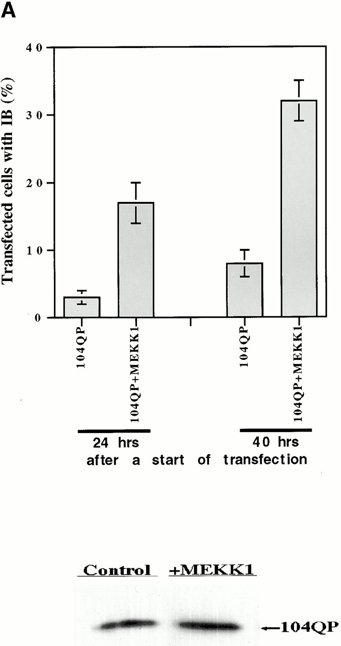
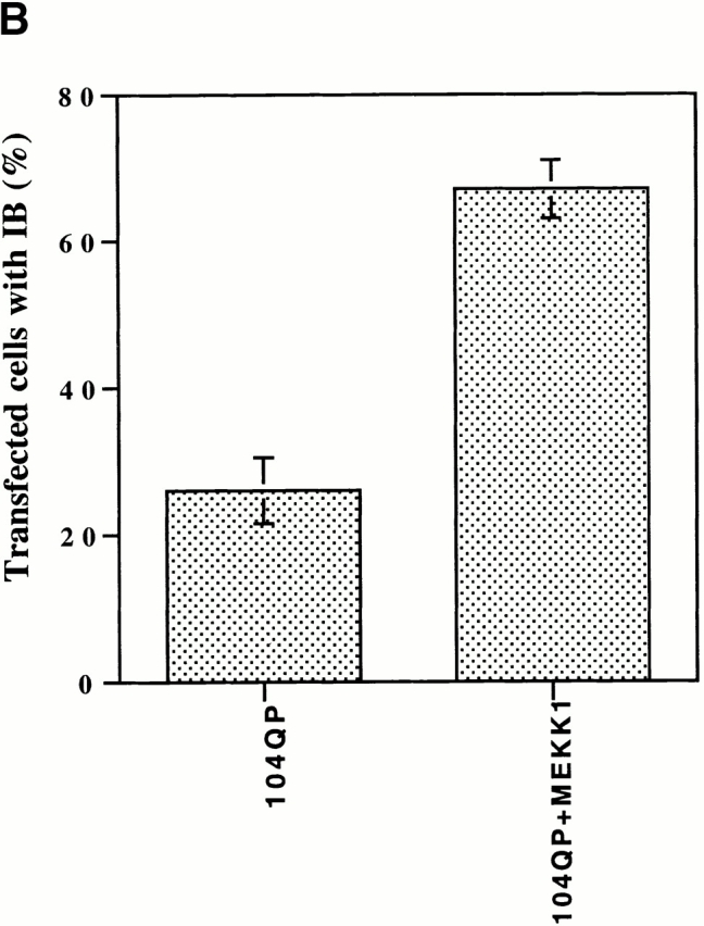
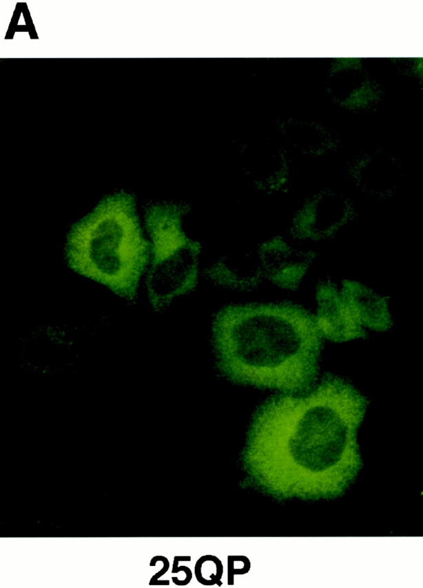
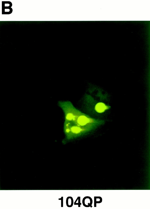
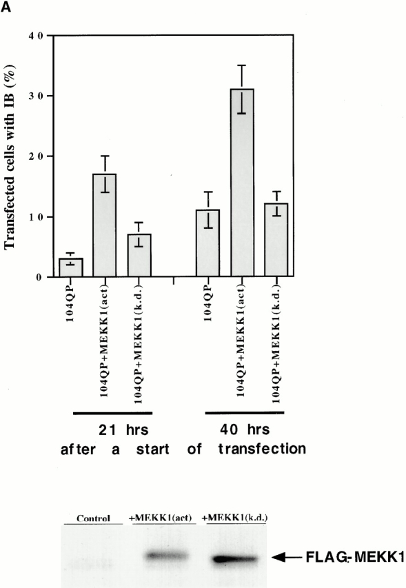
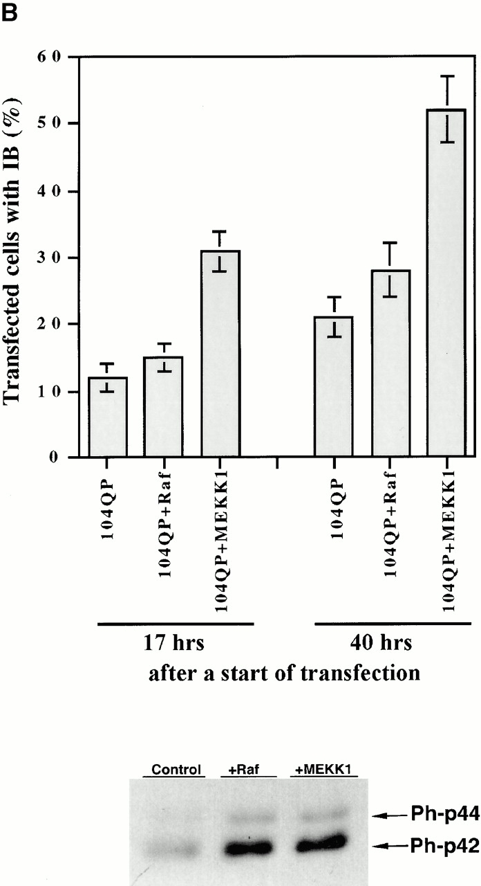
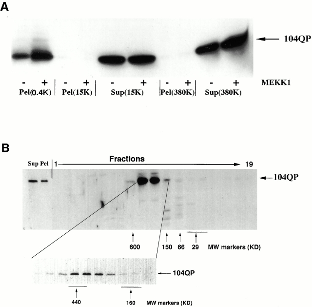
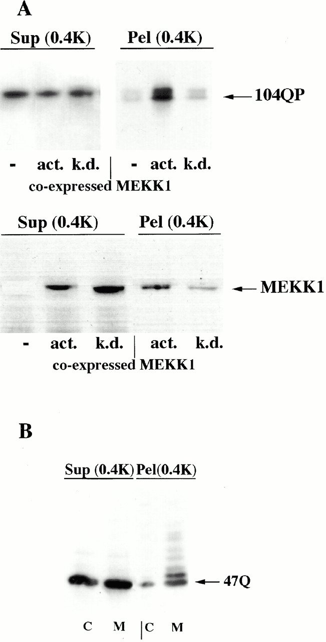
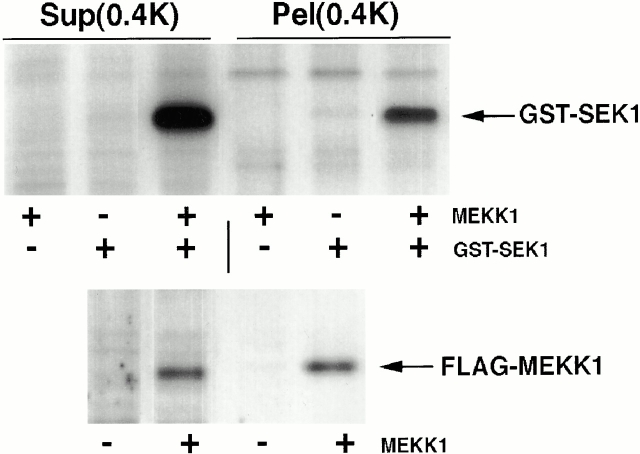
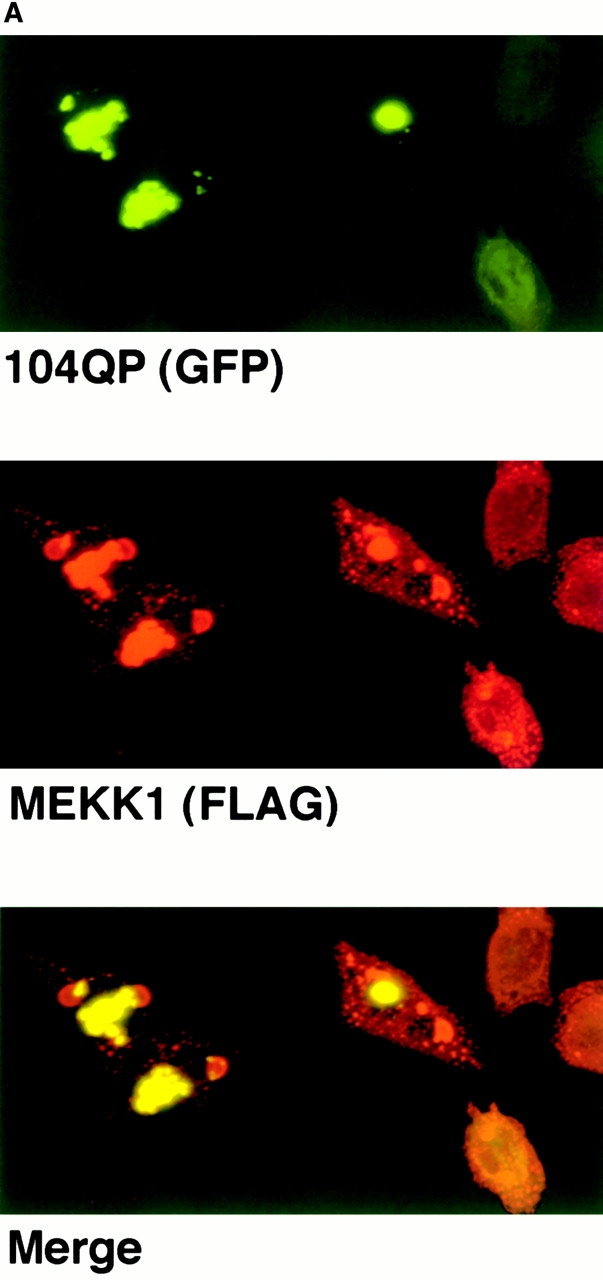
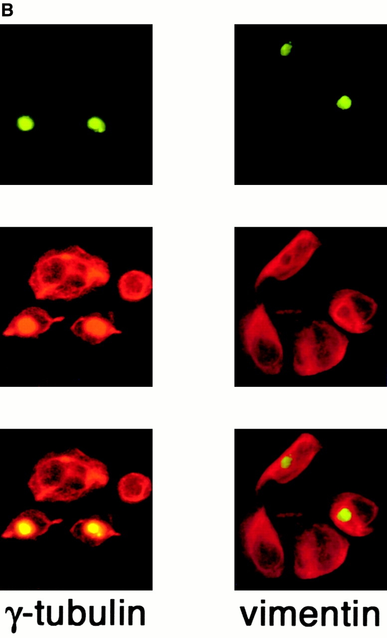
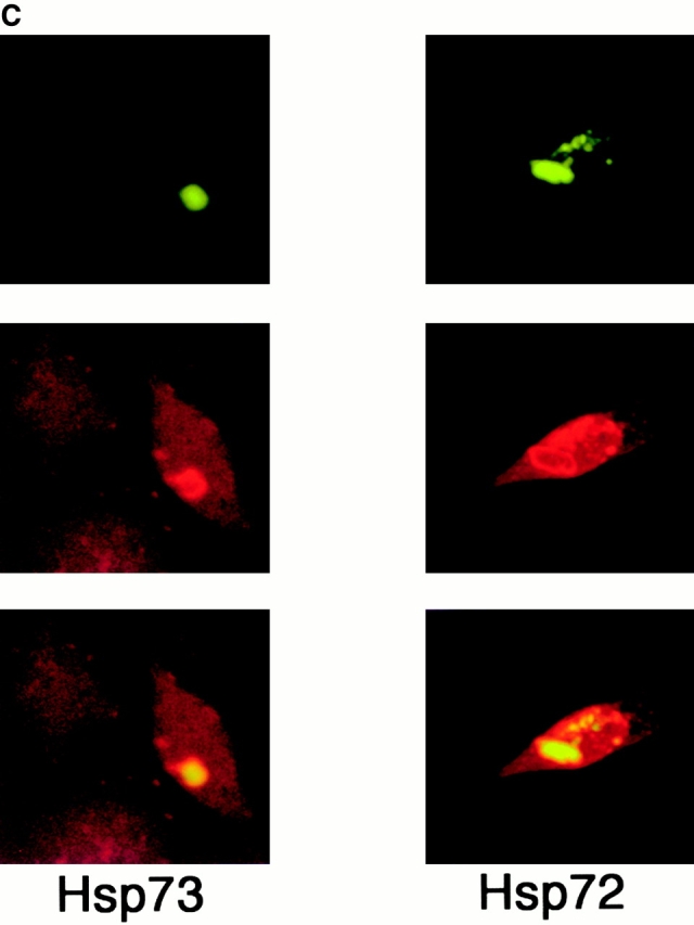
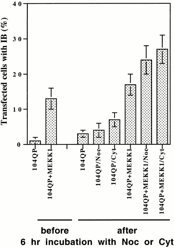
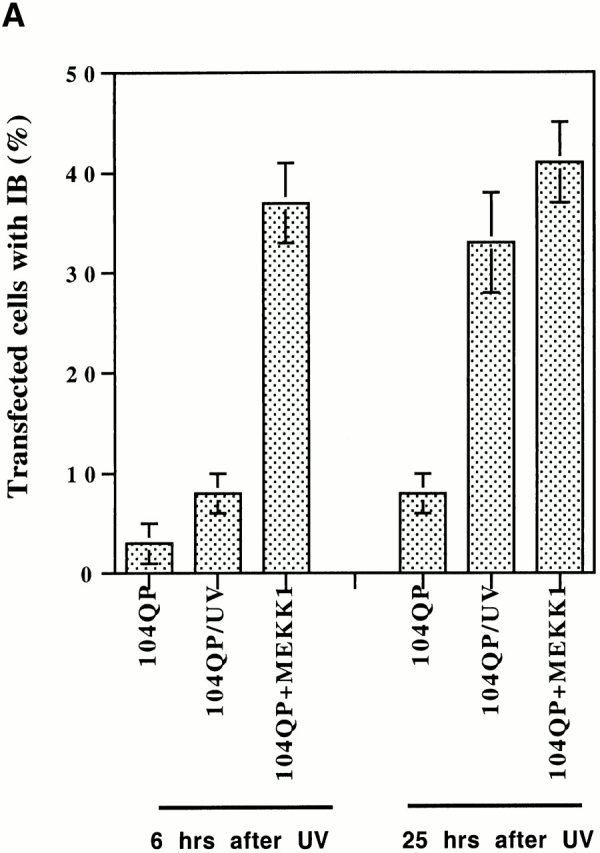
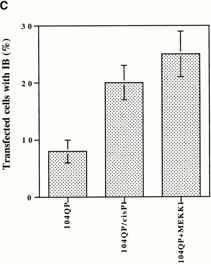
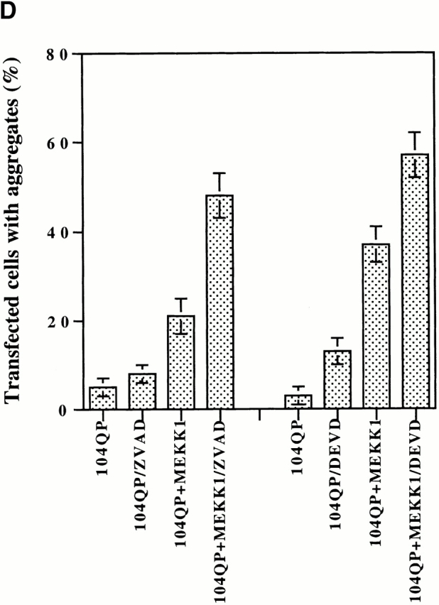
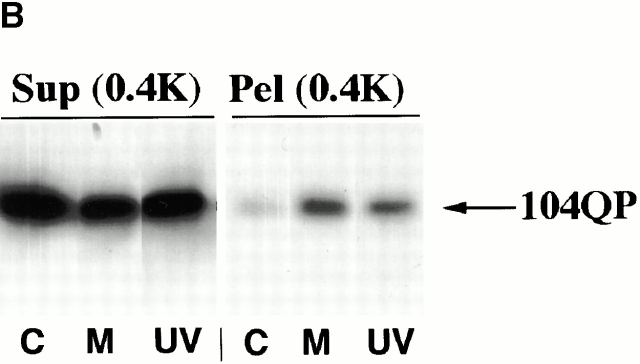
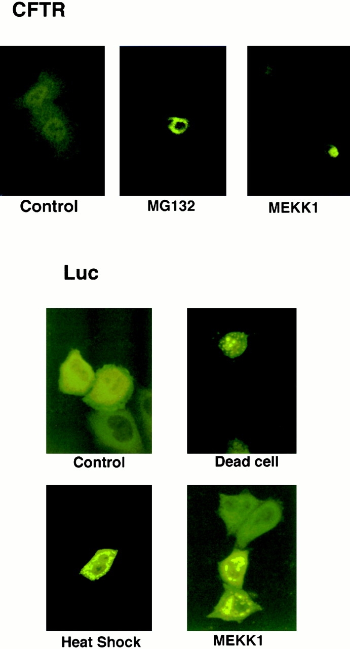
Abnormal proteins, which escape chaperone-mediated refolding or proteasome-dependent degradation, aggregate and form inclusion bodies (IBs). In several neurodegenerative diseases, such IBs can be formed by proteins with expanded polyglutamine (polyQ) domains (e.g., huntingtin). This work studies the regulation of intracellular IB formation using an NH(2)-terminal fragment of huntingtin with expanded polyQ domain. We demonstrate that the active form of MEKK1, a protein kinase that regulates several stress-activated signaling cascades, stimulates formation of the IBs. This function of MEKK1 requires kinase activity, as the kinase-dead mutant of MEKK1 cannot stimulate this process. Exposure of cells to UV irradiation or cisplatin, both of which activate MEKK1, also augmented the formation of IBs. The polyQ-containing huntingtin fragment exists in cells in two distinct forms: (a) in a discrete soluble complex, and (b) in association with insoluble fraction. MEKK1 strongly stimulated recruitment of polyQ polypeptides into the particulate fraction. Notably, a large portion of the active form of MEKK1 was associated with the insoluble fraction, concentrating in discrete sites, and polyQ-containing IBs always colocalized with them. We suggest that MEKK1 is involved in a process of IB nucleation. MEKK1 also stimulated formation of IBs with two abnormal polypeptides lacking the polyQ domain, indicating that this kinase has a general effect on protein aggregation.


Figures

Figure 2
Figure 2
Expression of active MEKK1 mutant stimulates formation of 104QP IBs. (A) HeLa cells were transfected with 104QP either alone (104QP or Control) or along with active MEKK1 (+MEKK1). Top, the fractions of transfected cells which form visible IB were counted at two time points. Bottom, immunoblot shows concentration of soluble 104QP in the same cells at the second time point. (B) HN33 cell line was transfected with 104QP either alone (104QP) or along with active MEKK1 mutant (+MEKK1). The fractions of the transfected cells which form visible IBs, were counted 26 h after start of transfection. (C) 293 cell line stably transfected with the 300Q–GFP fusion under the control of the muristerone-inducible promoter was incubated with muristerone for 24 h. Cells were then transfected with either MEKK1Δ gene (300Q+MEKK1) or carrier DNA alone (300Q) and further incubated in the presence of muristerone for an additional 24 h. The number of IBs in five randomly chosen microscope fields was counted.
Figure 2
Figure 2
Expression of active MEKK1 mutant stimulates formation of 104QP IBs. (A) HeLa cells were transfected with 104QP either alone (104QP or Control) or along with active MEKK1 (+MEKK1). Top, the fractions of transfected cells which form visible IB were counted at two time points. Bottom, immunoblot shows concentration of soluble 104QP in the same cells at the second time point. (B) HN33 cell line was transfected with 104QP either alone (104QP) or along with active MEKK1 mutant (+MEKK1). The fractions of the transfected cells which form visible IBs, were counted 26 h after start of transfection. (C) 293 cell line stably transfected with the 300Q–GFP fusion under the control of the muristerone-inducible promoter was incubated with muristerone for 24 h. Cells were then transfected with either MEKK1Δ gene (300Q+MEKK1) or carrier DNA alone (300Q) and further incubated in the presence of muristerone for an additional 24 h. The number of IBs in five randomly chosen microscope fields was counted.
Figure 1
Figure 1
Aggregation of 104QP in HeLa cells viewed by confocal microscopy. (A) Cells expressing 25QP construct; (B) cells expressing 104QP construct.
Figure 1
Figure 1
Aggregation of 104QP in HeLa cells viewed by confocal microscopy. (A) Cells expressing 25QP construct; (B) cells expressing 104QP construct.
Figure 3
Figure 3
Stimulation of 104QP IB formation depends on the MEKK1 activity. (A) HeLa cells were transfected with 104QP either alone (104QP or Control) or along with active MEKK1 mutant (+MEKK1 [act]), or along with kinase-dead MEKK1 mutant (+MEKK1 [k.d.]). Top, the fractions of transfected cells which form visible IBs were counted at two time points. Bottom, content of MEKK1 mutants in a soluble fraction of the same cells. Immunoblotting with anti-FLAG antibody. (B) HeLa cells were transfected with 104QP either alone (104QP or Control) or along with either active MEKK1 mutant (+MEKK1) or active Raf (+Raf) (to increase the amount of Raf DNA in transfection, it was done differently [see Materials and Methods]). Top, the fractions of transfected cells which form visible IBs were counted at two time points. Bottom, activation of ERK by transfected MEKK1 or Raf mutants in a soluble fraction of the same cells. Immunoblotting with anti–phospho-ERK (p42/44 MAPK) antibody.
Figure 3
Figure 3
Stimulation of 104QP IB formation depends on the MEKK1 activity. (A) HeLa cells were transfected with 104QP either alone (104QP or Control) or along with active MEKK1 mutant (+MEKK1 [act]), or along with kinase-dead MEKK1 mutant (+MEKK1 [k.d.]). Top, the fractions of transfected cells which form visible IBs were counted at two time points. Bottom, content of MEKK1 mutants in a soluble fraction of the same cells. Immunoblotting with anti-FLAG antibody. (B) HeLa cells were transfected with 104QP either alone (104QP or Control) or along with either active MEKK1 mutant (+MEKK1) or active Raf (+Raf) (to increase the amount of Raf DNA in transfection, it was done differently [see Materials and Methods]). Top, the fractions of transfected cells which form visible IBs were counted at two time points. Bottom, activation of ERK by transfected MEKK1 or Raf mutants in a soluble fraction of the same cells. Immunoblotting with anti–phospho-ERK (p42/44 MAPK) antibody.
Figure 4
Figure 4
104QP is present in transfected cells in two distinct forms: supernatant (Sup) and pellet (Pel). Immunoblots are shown. (A) Differential centrifugation. HeLa cells were transfected with 104QP either alone (−MEKK1) or along with active MEKK1 (+MEKK1). 40 h after the end of transfection cells were collected, cell lysates were diluted to equalize total protein concentration and subjected to consecutive centrifugations: 10 min at 400 g (0.4K), 10 min at 15,000 g (15K), and 40 min at 380,000 g (380K). (B) Soluble 104QP is present in a complex of a distinct size. HeLa cells transfected with the 104QP construct were lysed and subjected to low speed centrifugation at 400 g for 10 min (Sup and Pel). The supernatant was loaded onto a Superose 6 FPLC column and gel filtration was performed in lysis buffer (see Materials and Methods). Fractions were collected (numbering starts upon elution of a void volume), concentrated by TCA precipitation, and analyzed by immunoblotting. Inset shows the results of the gel filtration with the four-time decreased fraction size.
Figure 5
Figure 5
Expression of active MEKK1 mutant causes association of polyQ polypeptides with the insoluble fraction. Transfected HeLa cells were lysed and subjected to low-speed centrifugation at 400 g for 10 min. Immunoblots are shown. Pel(0.4K), pellet; Sup(0.4K), supernatant. (A) Cells were transfected with either 104QP alone (−) or along with active MEKK1 mutant (act.) or along with kinase-dead mutant (k.d.). Samples were collected 30 h after the end of transfection. Top, samples probed with anti-GFP antibody for 104QP. Bottom, samples probed with anti-FLAG antibody for MEKK1 mutants. (B) Cells were transfected with either 47Q alone (C) or along with active MEKK1 (M). Samples were collected 25 h after the end of transfection. In this immunoblot a pellet fraction was resuspended in half of the original volume.
Figure 6
Figure 6
MEKK1Δ in insoluble fraction is active. HeLa cells were transfected with 104QP either alone (− MEKK1) or along with active MEKK1 (+MEKK1). 20 h after the end of transfection cells were collected, and soluble and insoluble fractions were separated by centrifugations (10 min at 400 g): Sup(0.4K), supernatant; Pel(0.4K), pellet. Pellets were resuspended in lysis buffer and both fractions were tested for their ability to phosphorylate recombinant GST-SEK1. The autoradiogram shows phosphorylation of SEK1 in the samples (−GST-SEK1: no substrate was added to the reaction) (top). The immunoblot with anti-FLAG antibody shows distribution of FLAG-tagged active MEKK1 between soluble and insoluble fractions (bottom).
Figure 7
Figure 7
Colocalization of 104QP IB with active MEKK1 and cellular proteins. 24 h after the end of transfection HeLa cells were fixed in a way that mainly retained insoluble structures (Materials and Methods), including 104QP IB (green, top row). Samples were probed with antibodies to various proteins (red, middle row) to study their colocalization (yellow, bottom row) with 104QP. (A) Anti–γ-tubulin and antivimentin antibodies. (B) Anti-FLAG antibody to localize FLAG-tagged MEKK1 active mutant. (C) Anti-Hsp73 (constitutive member of the Hsp70 family) and anti-Hsp72 (inducible member of the Hsp70 family) antibodies.
Figure 7
Figure 7
Colocalization of 104QP IB with active MEKK1 and cellular proteins. 24 h after the end of transfection HeLa cells were fixed in a way that mainly retained insoluble structures (Materials and Methods), including 104QP IB (green, top row). Samples were probed with antibodies to various proteins (red, middle row) to study their colocalization (yellow, bottom row) with 104QP. (A) Anti–γ-tubulin and antivimentin antibodies. (B) Anti-FLAG antibody to localize FLAG-tagged MEKK1 active mutant. (C) Anti-Hsp73 (constitutive member of the Hsp70 family) and anti-Hsp72 (inducible member of the Hsp70 family) antibodies.
Figure 8
Figure 8
Disruption of microtubules or actin filaments does not abrogate stimulation of 104QP IB formation by MEKK1. HeLa cells were transfected with 104QP construct either alone (104QP) or along with active MEKK1 (104QP+MEKK1). After the end of transfection some cells were treated with 10 μg/ml of nocodazole (Noc) to disrupt microtubules, or with 40 μM cytochalasin B (Cyt) to disrupt actin filaments. The fractions of transfected cells which form visible IBs were counted either before or 6 h after the start of treatments.
Figure 9
Figure 9
Stresses and caspase inhibitors stimulate aggregation of 104QP. HeLa cells were transfected with 104QP either alone (104QP or C) or along with active MEKK1 (104QP + MEKK1 or M). (A) Cells after transfection were either UV-irradiated (150 J/m2) 104QP/UV or left untreated and the fractions of transfected cells which form visible IB, were counted at two time points. (B) Immunoblot of soluble supernatant (Sup[0.4K]) and insoluble pellet (Pel[0.4K]) fractions of the same (see Fig. 9 A) transfected cells collected at the second time point. (C) Cells after transfection were either exposed for 12 h to 40 μM cisplatin (104QP/cisPl) or left untreated and the fractions of transfected cells which form visible IBs were counted. (D) Cells marked 104QP/ZVAD from the start of transfection were exposed to 100 μM ZVAD and the fractions of transfected cells which form visible IBs were counted 17 h after the start. Cells marked 104QP/DEVD from the start of transfection were exposed to 100 μM DEVD and the fractions of transfected cells which form visible IBs were counted 21 h after the start.
Figure 9
Figure 9
Stresses and caspase inhibitors stimulate aggregation of 104QP. HeLa cells were transfected with 104QP either alone (104QP or C) or along with active MEKK1 (104QP + MEKK1 or M). (A) Cells after transfection were either UV-irradiated (150 J/m2) 104QP/UV or left untreated and the fractions of transfected cells which form visible IB, were counted at two time points. (B) Immunoblot of soluble supernatant (Sup[0.4K]) and insoluble pellet (Pel[0.4K]) fractions of the same (see Fig. 9 A) transfected cells collected at the second time point. (C) Cells after transfection were either exposed for 12 h to 40 μM cisplatin (104QP/cisPl) or left untreated and the fractions of transfected cells which form visible IBs were counted. (D) Cells marked 104QP/ZVAD from the start of transfection were exposed to 100 μM ZVAD and the fractions of transfected cells which form visible IBs were counted 17 h after the start. Cells marked 104QP/DEVD from the start of transfection were exposed to 100 μM DEVD and the fractions of transfected cells which form visible IBs were counted 21 h after the start.
Figure 10
Figure 10
MEKK1 stimulates aggregation of proteins lacking polyQ domain. Confocal microscopy. Top, HeLa cells were transfected with GFP-tagged CFTR either alone (Control or MG132) or along with active MEKK1 mutant (MEKK1). After transfection one sample transfected with CFTR alone was incubated with 5 μM of proteasome inhibitor MG132 for 15 h (MG132). Bottom, HeLa cells were transfected with GFP-tagged luciferase either along with active MEKK1 mutant (MEKK1) or alone (all other pictures). Cells transfected with luciferase alone after transfection were either left untreated (Control [living cells], and Dead cell) or heated at 44°C for 30 min followed by 15-h recovery (Heat Shock).

Similar articles

Cited by

References

    1. Agashe V.R., Hartl F.U. Roles of molecular chaperones in cytoplasmic protein folding. Semin. Cell Dev. Biol. 2000;11:15–25. - PubMed
    1. Anton L.C., Schubert U., Bacik I., Princiotta M.F., Wearsch P.A., Gibbs J., Day P.M., Realini C., Rechsteiner M.C., Bennink J.R., Yewdell J.W. Intracellular localization of proteasomal degradation of a viral antigen. J. Cell Biol. 1999;146:113–124. - PMC - PubMed
    1. Bao J., Sharp A.H., Wagster M.V., Becher M., Schilling G., Ross C.A., Dawson V.L., Dawson T.M. Expansion of polyglutamine repeat in huntingtin leads to abnormal protein interactions involving calmodulin. Proc. Natl. Acad. Sci. USA. 1996;93:5037–5042. - PMC - PubMed
    1. Bates G.P., Mangiarini L., Davies S.W. Transgenic mice in the study of polyglutamine repeat expansion diseases. Brain Pathol. 1998;8:699–714. - PMC - PubMed
    1. Chai Y., Koppenhafer S.L., Bonini N.M., Paulson H.L. Analysis of the role of heat shock protein (Hsp) molecular chaperones in polyglutamine disease. J. Neurosci. 1999;19:10338–10347. - PMC - PubMed

Publication types

MeSH terms