. 2001 May;13(5):1179-91.
doi: 10.1105/tpc.13.5.1179.

Small changes in the regulation of one Arabidopsis profilin isovariant, PRF1, alter seedling development

Affiliations

Small changes in the regulation of one Arabidopsis profilin isovariant, PRF1, alter seedling development

E C McKinney et al. Plant Cell. 2001 May.

Abstract

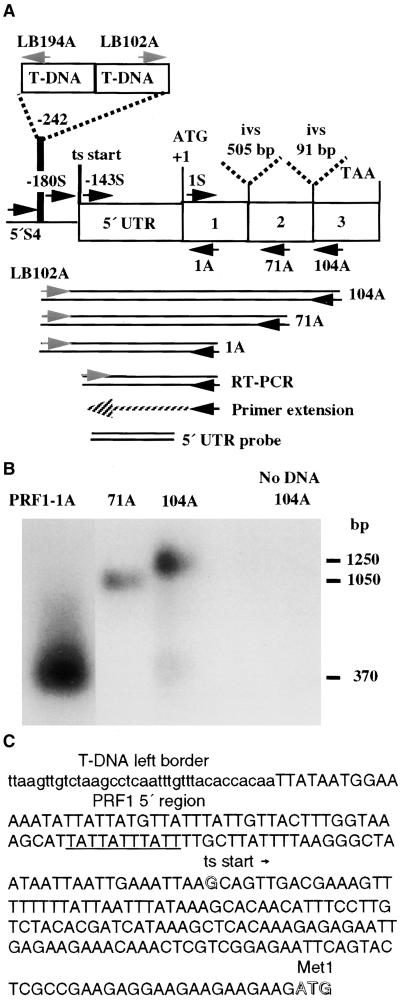
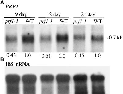
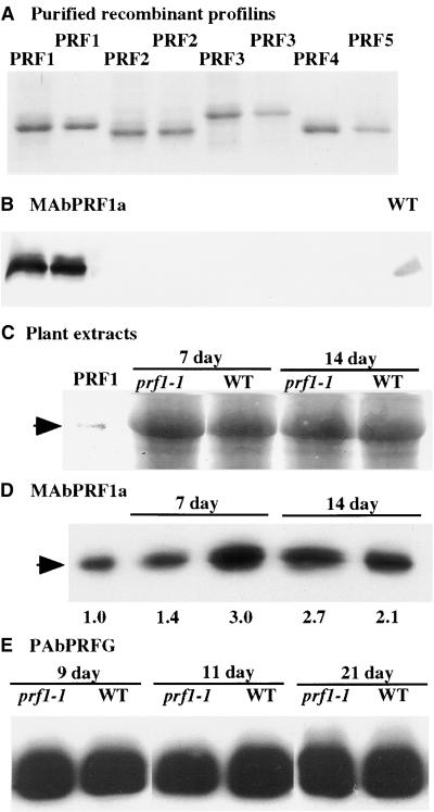
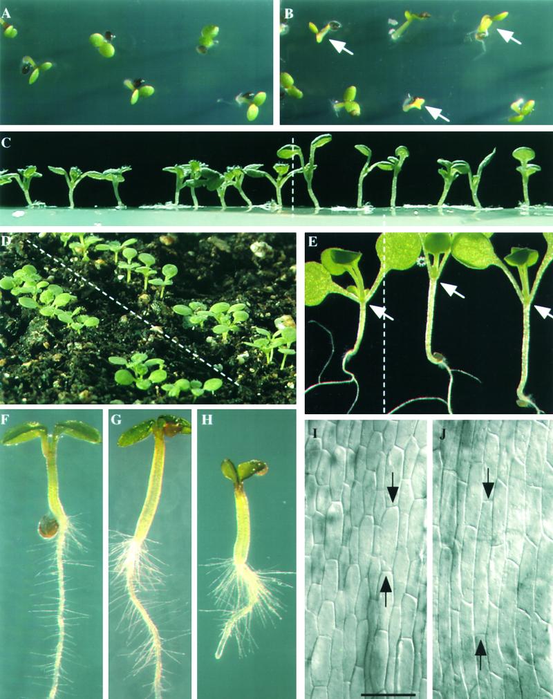
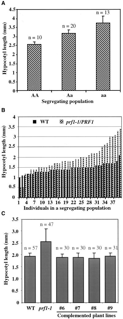
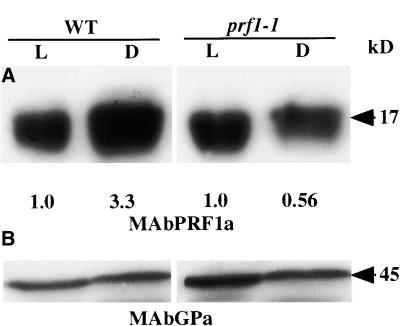
Profilin (PRF) is a low-molecular-weight actin binding protein encoded by a diverse gene family in plants. Arabidopsis PRF1 transcripts are moderately well expressed in all vegetative organs. A regulatory mutant in PRF1, prf1-1, was isolated from a library of T-DNA insertions. The insertion disrupted the promoter region of PRF1 100 bp upstream from the transcriptional start site. Although steady state levels of PRF1 transcripts appeared normal in mature prf1-1 plants, the levels in young seedlings were only one-half those observed in wild type. Reactions with a PRF1 isovariant-specific monoclonal antiserum and general anti-profilin antisera demonstrated that PRF1 protein levels also were one-half those found in wild-type seedlings, although total profilin levels were unaffected. Mutant seedlings no longer could downregulate PRF1 levels in the light, as did wild type. Consistent with their molecular phenotypes, young mutant seedlings displayed several morphological phenotypes but developed into apparently normal adult plants. Their initial germination rate and development were slow, and they produced excessive numbers of root hairs. Mutant seedlings had abnormally raised cotyledons, elongated hypocotyls, and elongated cells in the hypocotyl, typical of phenotypes associated with some defects in light and circadian responses. A wild-type PRF1 transgene fully complements the hypocotyl phenotypes in the prf1-1 mutant. The ability of profilin to regulate actin polymerization and participate directly in signal transduction pathways is discussed in light of the prf1-1 phenotypes.


Figures

Figure 1.
Figure 1.
Characterization of the prf1-1 Mutation and PRF1 Transcripts. (A) The location of the T-DNA insertion in the prf1-1 mutant is shown on a map of the Arabidopsis profilin gene PRF1. PRF1 encodes a 131–amino acid protein. The locations of primers used in this study are shown, with their 3′ ends indicated by arrows. The PCR products used to map the insertion, the primer extension product and RT-PCR products that were used to map the 5′ end of the PRF1 transcript (ts start), and the 5′ untranslated region (5′ UTR) used as a gene-specific probe are shown below the gene. Two introns (ivs) interrupt the coding region. (B) prf1-1 mutant plants were identified by a sequence-based screening strategy using a primer (see Table 1) located in the T-DNA left border (LB202) and a degenerate profilin primer, PRF104A, which produced a 1.25-kb product. A ladder of prf1-1/left border junction products is produced by a set of nested antisense profilin primers (PRF104A, 71A, and 1A). (C) Sequence of the 5′ region of the PRF1 gene (uppercase letters) in the prf1-1 mutant and location of the proximal left border of the T-DNA insertion (lowercase letters). The putative promoter TATA region (underlined), the G residue mapped as the start of transcription (open letter), and the ATG initiation codon (open letters) are indicated.
Figure 2.
Figure 2.
Transcript Levels in prf1-1 Mutant Seedlings. (A) RNA gel blot hybridized with a PRF1-specific DNA fragment from the 5′ untranslated region (see Figure 1). Total RNA samples from 9-, 12-, and 21-day-old wild-type (WT) and mutant (prf1-1) plants are shown. The level of PRF1 mRNA in the prf1-1 mutant is quantified relative to the level in the wild type after correcting for the loading of the 18S rRNA shown in (B). (B) The 18S rRNA is identified in the same blot shown in (A) to demonstrate equal loading and transfer of total RNA samples. Densitometric scanning was used to quantify the bands in (A) and (B).
Figure 3.
Figure 3.
Analysis of PRF1 Protein Levels in Seedlings. (A) Coomassie blue–stained 12.5% polyacrylamide gel resolves 2-μg samples of the recombinant profilins PRF1, PRF2, PRF3, PRF4, and PRF5. Membrane imprints of duplicate gels are examined by protein gel blotting in (B). (B) A duplicate of the same blot shown in (A) reacted with the PRF1-specific mouse monoclonal antibody MAbPRF1a. This antibody does not react with the other four recombinant Arabidopsis profilin proteins on a protein gel blot. A sample of 15 μg of total Arabidopsis leaf protein was run in the far right lane as a control (WT). (C) A Coomassie blue–stained SDS-PAGE gel in the region of profilin migration shows equal loading of plant protein extracts. Samples are as follows: lane 1, 0.1 μg of PRF1 standard protein; lanes 2 and 4, 30-μg total protein extracts from prf1-1 mutants; lanes 3 and 5, Ws wild type (WT). The image of the stained profilin standard, recombinant PRF1, is enhanced to show its position of migration. (D) Membrane imprints of a duplicate of the gel shown in (C) reacted with antibody MAbPRF1a to determine the levels of PRF1 protein in 7- and 14-day-old seedlings. Densitometric quantification of protein levels was performed relative to the signal obtained with the PRF1 loading control (left lane) that contained 0.1 μg of total PRF1 protein. (E) A rabbit polyclonal antibody (PAbPRFG) that reacts with all five Arabidopsis profilins was used to detect total profilin in extracts from wild type (WT) and prf1-1 mutant plants that were germinated and grown for 9, 11, and 21 days.
Figure 4.
Figure 4.
Morphology of prf1-1 Mutant Seedlings. (A) and (B) Many mutant prf1-1 seed are delayed in their germination, and the resulting seedlings are chlorotic after 2 to 3 days ([B], arrows) compared with wild type (A). (C) to (E) Seven- to 10-day-old mutant seedlings have raised cotyledons and elongated hypocotyls (right of dashed lines) compared with wild type (left of dashed lines). In (E), mutant cotyledons form an acute angle, whereas most wild-type cotyledons form an angle greater than 90° (arrows). These seedlings were harvested at 1 pm, 8 hr after subjective dawn, but mutants still show raised cotyledons. (F) to (H) Mutant prf1-1 seedlings ([G] and [H]) have increased root hair density and elongated root hairs compared with wild type (F). (I) and (J) Differential interference contrast microscopy of epidermal cells of 7-day-old hypocotyls from mutant (J) and wild-type (I) seedlings. Arrows indicate the ends of the longest cell in each frame. (I) and (J) represent same magnification. Bar in (I) = 85 μm.
Figure 5.
Figure 5.
Hypocotyl Lengths in prf1-1 Mutants. (A) Mean hypocotyl lengths in a population of 9-day-old seedlings segregating for the prf1-1 mutation from a heterozygous parent plant. Hypocotyl lengths were measured, and then the genotype of each plant was determined by PCR amplification of the wild-type PRF1 promoter region, the mutant junction sequences, or both sequences (heterozygotes) from each seedling. The mean length values from homozygous wild-type (AA), heterozygous mutant (Aa), and homozygous mutant (aa) seedlings are indicated. Standard errors from the mean and number of plants (n) are indicated for each genotype. The P value of 0.008 determined by analysis of variance supports the dependence of hypocotyl length on genotype. (B) Distribution of individual hypocotyl lengths from a 9-day-old wild-type population (WT) and a mutant population segregating from prf1-1/PRF1 parents. Each population contained 39 plants. (C) Mean hypocotyl lengths of 9-day-old homozygous prf1-1 mutant seedlings that were complemented with the wild-type PRF1 transgene. Populations derived from four different independent complemented transgenic lines (#6, #7, #8, and #9) were compared with wild-type (WT) and homozygous prf1-1 mutant populations. The number of individuals scored (n) and the standard errors from the mean are indicated for each population. Analysis of variance confirmed that the wild-type and complemented transgenic lines were all significantly different from the prf1-1 mutant (P < 0.0001) and indistinguishable from each other (formula image).
Figure 6.
Figure 6.
PRF1 Levels Are Light Regulated, and prf1-1 Mutants Are Defective in This Regulation. (A) Wild-type (WT) Arabidopsis seedlings show increased levels of PRF1 protein when grown in darkness (D) relative to being grown in the light (L), as measured by the PRF1-specific antibody, MAbPRF1a, on protein gel blots (left). Mutant prf1-1 seedlings have essentially the same low levels of protein when grown in light or darkness (right). Exposure intensities were adjusted to normalize the light-grown levels of PRF1 in wild-type and mutant plants. Densitometric quantification of each dark lane relative to the corresponding light lane is shown below the image. (B) The top half of the same membrane blot was reacted with an anti-actin monoclonal antibody (MAbGPa) that binds all eight Arabidopsis actins. This blot shows that similar levels of protein were loaded and transferred to the membrane from the light- and dark-grown samples that were compared.

Similar articles

Cited by

References

    1. Ahern-Djamali, S.M., Bachmann, C., Hua, P., Reddy, S.K., Kastenmeier, A.S., Walter, U., and Hoffmann, F.M. (1999). Identification of profilin and src homology 3 domains as binding partners for Drosophila enabled. Proc. Natl. Acad. Sci. USA 96 4977–4982. - PMC - PubMed
    1. An, Y.-Q., Huang, S., McDowell, J.M., McKinney, E.C., and Meagher, R.B. (1996). Conserved expression of the Arabidopsis ACT1 and ACT3 actin subclass in organ primordia and mature pollen. Plant Cell 8 15–30. - PMC - PubMed
    1. Bariola, P.A., MacIntosh, G.C., and Green, P.J. (1999). Regulation of S-like ribonuclease levels in Arabidopsis: Antisense inhibition of RNS1 or RNS2 elevates anthocyanin accumulation. Plant Physiol. 119 331–342. - PMC - PubMed
    1. Calman, B.G., and Chamberlain, S.C. (1992). Localization of actin filaments and microtubules in the cells of the Limulus lateral and ventral eyes. Vis. Neurosci. 9 365–375. - PubMed
    1. Carlier, M.F., Jean, C., Rieger, K.J., Lenfant, M., and Pantaloni, D. (1993). Modulation of the interaction between G-actin and thymosin beta 4 by the ATP/ADP ratio: Possible implication in the regulation of actin dynamics. Proc. Natl. Acad. Sci. USA 90 5034–5038. - PMC - PubMed

Publication types

MeSH terms

LinkOut - more resources