. 2001 Jun;21(11):3738-49.
doi: 10.1128/MCB.21.11.3738-3749.2001.

Pgc-1-related coactivator, a novel, serum-inducible coactivator of nuclear respiratory factor 1-dependent transcription in mammalian cells

Affiliations

Pgc-1-related coactivator, a novel, serum-inducible coactivator of nuclear respiratory factor 1-dependent transcription in mammalian cells

U Andersson et al. Mol Cell Biol. 2001 Jun.

Abstract

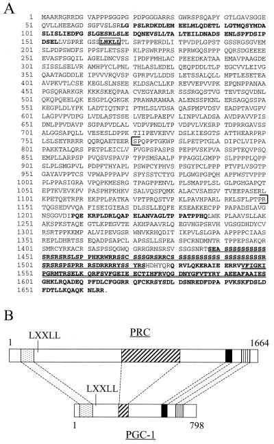
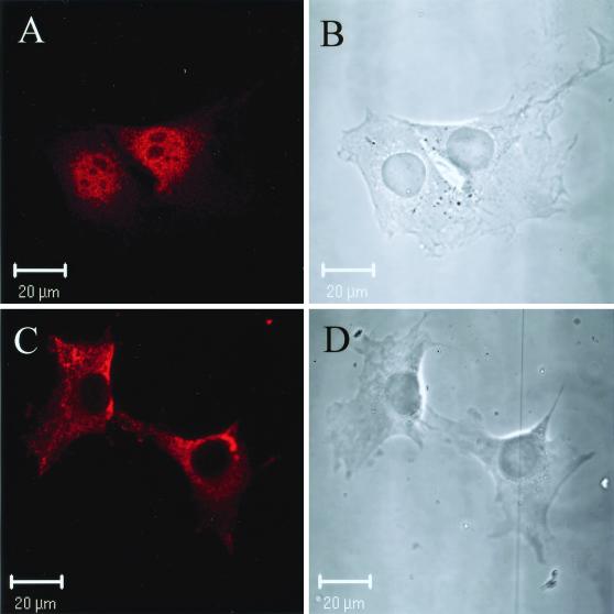
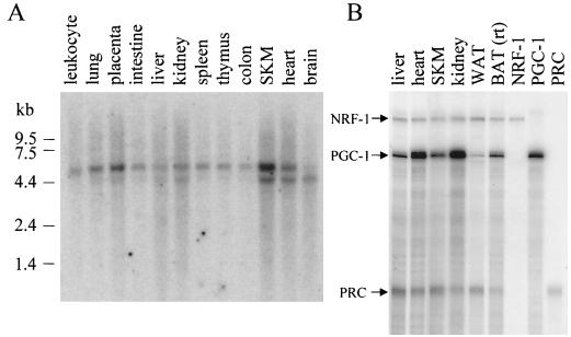
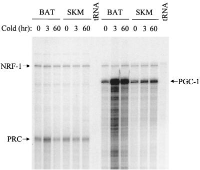
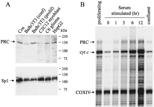
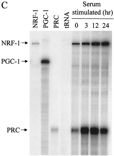
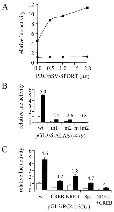
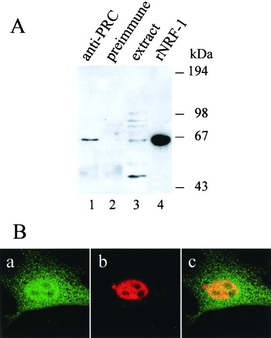
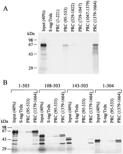
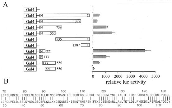
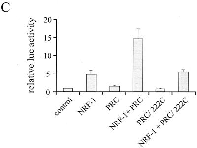
The thermogenic peroxisome proliferator-activated receptor gamma (PPAR-gamma) coactivator 1 (PGC-1) has previously been shown to activate mitochondrial biogenesis in part through a direct interaction with nuclear respiratory factor 1 (NRF-1). In order to identify related coactivators that act through NRF-1, we searched the databases for sequences with similarities to PGC-1. Here, we describe the first characterization of a 177-kDa transcriptional coactivator, designated PGC-1-related coactivator (PRC). PRC is ubiquitously expressed in murine and human tissues and cell lines; but unlike PGC-1, PRC was not dramatically up-regulated during thermogenesis in brown fat. However, its expression was down-regulated in quiescent BALB/3T3 cells and was rapidly induced by reintroduction of serum, conditions where PGC-1 was not detected. PRC activated NRF-1-dependent promoters in a manner similar to that observed for PGC-1. Moreover, NRF-1 was immunoprecipitated from cell extracts by antibodies directed against PRC, and both proteins were colocalized to the nucleoplasm by confocal laser scanning microscopy. PRC interacts in vitro with the NRF-1 DNA binding domain through two distinct recognition motifs that are separated by an unstructured proline-rich region. PRC also contains a potent transcriptional activation domain in its amino terminus adjacent to an LXXLL motif. The spatial arrangement of these functional domains coincides with those found in PGC-1, supporting the conclusion that PRC and PGC-1 are structurally and functionally related. We conclude that PRC is a functional relative of PGC-1 that operates through NRF-1 and possibly other activators in response to proliferative signals.


Figures

FIG. 1
FIG. 1
Primary structure of PRC. (A) Predicted amino acid sequence of PRC. Regions of significant sequence similarity with PGC-1 are in boldface letters. The coactivator signature LXXLL is boxed; the proline-rich region is within brackets. The underlined sequence represents the RS domain, and the double underlined sequence represents the RNA recognition motif. (B) Alignment of similar domains in PRC and PGC-1. Regions of similarity between the two proteins are as follows: activation domain (stippled box), proline-rich region (cross-hatched box), RS domain (solid box), and RNA recognition motif (vertically hatched box). Amino acid coordinates are indicated above and below.
FIG. 2
FIG. 2
Nuclear localization of PRC by confocal immunofluorescence microscopy. (A) BALB/3T3 cells were stained with anti-PRC serum as the primary antibody. (B) The phase-contrast image of the results shown in panel A. (C) BALB/3T3 cells were stained with anti-PRC serum that had been preadsorbed with recombinant PRC [PRC(95–533)/thioredoxin]. (D) The phase-contrast image of results shown in panel C. Rhodamine-conjugated goat anti-rabbit IgG was used as the secondary antibody in the results shown in panels A and C.
FIG. 3
FIG. 3
Expression profile of PRC mRNA. (A) A human multiple-tissue Northern blot (Clontech) probed with a 32P-labeled 1.6-kb PRC cDNA fragment comprising the 5′ end of the cDNA. Positions of RNA standards of known length in kilobases are indicated at the left. (B) Comparison of NRF-1, PGC-1, and PRC mRNA expression by RNase protection in various mouse tissues. Protected fragments (365, 297, and 192 bp for NRF-1, PGC-1, and PRC, respectively) from individual probes using liver RNA are shown in the last three lanes. WAT, white adipose tissue, rt, room temperature.
FIG. 4
FIG. 4
Response of NRF-1, PGC-1, and PRC transcripts to cold exposure. Transcripts were detected by RNase protection in BAT and SKM obtained from mice exposed to 4°C for the indicated times. Probes were the same as those used in the results shown in Fig. 3. tRNA serves as a negative control.
FIG. 5
FIG. 5
Cell cycle regulation of PRC expression. (A) Immunoblot of whole-cell extracts (30 μg per lane) from murine cell lines (BALB/3T3 fibroblasts, C2C12 myoblasts, and C6 glioma cells) and primate cell lines (Cos and HepG2) with rabbit anti-PRC antibodies or rabbit anti-Sp1 serum as indicated. Molecular mass standards in kilodaltons are indicated at the right. (B) BALB/3T3 cells were cultured, and total RNA was isolated from proliferating, serum-starved, serum-stimulated, or confluent cells as described in Materials and Methods. mRNA levels for PRC, cytochrome c (cyt c), and COXIV were analyzed by RNase protection assay. Protected fragments are 192, 175, and 130 bp for PRC, cyt c, and COXIV, respectively. (C) Comparison of NRF-1, PGC-1, and PRC transcripts upon serum stimulation of quiescent BALB/3T3 cells. Protected fragments (365, 297, and 192 bp for NRF-1, PGC-1, and PRC, respectively) from individual probes with mouse liver RNA are shown in the first three lanes with tRNA serving as a negative control. RNA samples were isolated at the indicated times following serum addition.
FIG. 5
FIG. 5
Cell cycle regulation of PRC expression. (A) Immunoblot of whole-cell extracts (30 μg per lane) from murine cell lines (BALB/3T3 fibroblasts, C2C12 myoblasts, and C6 glioma cells) and primate cell lines (Cos and HepG2) with rabbit anti-PRC antibodies or rabbit anti-Sp1 serum as indicated. Molecular mass standards in kilodaltons are indicated at the right. (B) BALB/3T3 cells were cultured, and total RNA was isolated from proliferating, serum-starved, serum-stimulated, or confluent cells as described in Materials and Methods. mRNA levels for PRC, cytochrome c (cyt c), and COXIV were analyzed by RNase protection assay. Protected fragments are 192, 175, and 130 bp for PRC, cyt c, and COXIV, respectively. (C) Comparison of NRF-1, PGC-1, and PRC transcripts upon serum stimulation of quiescent BALB/3T3 cells. Protected fragments (365, 297, and 192 bp for NRF-1, PGC-1, and PRC, respectively) from individual probes with mouse liver RNA are shown in the first three lanes with tRNA serving as a negative control. RNA samples were isolated at the indicated times following serum addition.
FIG. 6
FIG. 6
NRF-1-dependent coactivation by PRC. (A) Increasing amounts of the mammalian expression vector PRC/pSV-SPORT were cotransfected into BALB/3T3 cells with CMV/β-gal (Clontech) and reporter plasmid 4xNRF-1/luc. Graphs indicate the stimulation of luciferase expression in the presence (squares) or absence (circles) of NRF-1 expression vector pSG5/NRF1. Data are means ± standard deviations (SD) of two independent experiments and are expressed as the ratio of the luciferase and β-galactosidase activities. Control activity was normalized to a value of 1. (B) The δ-ALAS(-479)/pGL3 wild-type (wt) promoter plasmid or mutated derivatives having site-directed point mutations in either one (m1 or m2) or both (m1m2) NRF-1 recognition sites were cotransfected with either PRC/pSV-SPORT (black bars) or the pSV-SPORT control (gray bars). Data represent the means ± SD of the ratio of the luciferase and β-galactosidase activities for a representative experiment. Numbers above the bars refer to the fold induction achieved in the presence of PRC expression vector. (C) The pGL3/RC4 (-326) wild-type (wt) cytochrome c promoter plasmid or derivatives with mutations in CREB, NRF-1, Sp1, or CREB plus NRF-1 recognition sites were cotransfected with PRC/pSV-SPORT (black bars) or empty pSV-SPORT (gray bars). Data represent the means ± SD of the ratio of the luciferase and β-galactosidase activities for two independent experiments. Numbers above the bars refer to the fold induction achieved in the presence of PRC expression vector.
FIG. 7
FIG. 7
Interaction between PRC and NRF-1 in vivo. (A) Coimmunoprecipitation of PRC and NRF-1 from cell extracts. C2C12 myoblast whole-cell extracts (750 μg) were subjected to immunoprecipitation using either anti-PRC serum (lane 1) or preimmune serum (lane 2). Immune complexes were brought down with protein A-agarose, washed, and run on an SDS–7.5% PAGE gel. As positive controls, 50 μg of the extract and 3 ng of the recombinant NRF-1 were run in lanes 3 and 4, respectively. After transfer, the immunoblot was probed with affinity-purified goat anti-NRF1 antibody. Molecular mass standards in kilodaltons are indicated on the right. (B) Colocalization of PRC and NRF-1 by confocal laser scanning fluorescence microscopy. BALB/3T3 cells transfected with NRF-1–3xHA were stained with anti-PRC serum (green) (a) or anti-hemagglutinin antibody (red) (b). Green and red images were merged (panel c) to visualize nuclear colocalization. Panel dimensions are 66.5 by 66.5 μm. Confocal images were generated with an LSM 510 confocal microscope (Zeiss).
FIG. 8
FIG. 8
Molecular determinants required for interaction between PRC and NRF-1. (A) Mapping of the NRF-1 binding domains in PRC. Six different fragments of PRC were fused to S-tagged thioredoxin, purified, immobilized on protein S-agarose, and tested for binding with 35S-labeled NRF-1. After binding was complete, the complexes were washed five times, eluted in SDS-PAGE sample buffer, and separated on SDS–10% PAGE gels. Gels were dried, and bound proteins were visualized by autoradiography. As a negative control, S-tagged thioredoxin was used. Molecular mass standards are indicated on the left. (B) Mapping of the PRC binding domain of NRF-1. The two major NRF-1-interacting domains of PRC fused to S-tagged thioredoxin and thioredoxin alone were purified and immobilized on protein S-agarose and incubated with different 35S-labeled NRF-1 fragments as indicated. The bound proteins were analyzed as described in the legend to panel A.
FIG. 9
FIG. 9
Mapping of the PRC activation domain. (A) BALB/3T3 cells were transfected with a 5xGal4 luciferase reporter plasmid, CMV/β-gal, and either pSG424 control vector (Gal4) or the various PRC/pSG424 fusions, as indicated. Results are expressed as the ratio of the luciferase to β-galactosidase activities and are the means ± standard errors of the mean for at least three independent experiments, each performed in duplicate. (B) Comparison of the putative activation domains in PRC and PGC-1. A Lipman-Pearson protein alignment of aa 69 to 154 of PRC and aa 29 to 117 of PGC-1 is shown. Identical or homologous (by 1 distance unit) amino acids are indicated by a vertical line (|). (C) Comparison of wild-type PRC to the amino-terminal deletion PRC/222C in the coactivation of NRF-1-dependent transcription from a 4xNRF-1/luc reporter plasmid. Results are expressed as in shown in panel A.
FIG. 9
FIG. 9
Mapping of the PRC activation domain. (A) BALB/3T3 cells were transfected with a 5xGal4 luciferase reporter plasmid, CMV/β-gal, and either pSG424 control vector (Gal4) or the various PRC/pSG424 fusions, as indicated. Results are expressed as the ratio of the luciferase to β-galactosidase activities and are the means ± standard errors of the mean for at least three independent experiments, each performed in duplicate. (B) Comparison of the putative activation domains in PRC and PGC-1. A Lipman-Pearson protein alignment of aa 69 to 154 of PRC and aa 29 to 117 of PGC-1 is shown. Identical or homologous (by 1 distance unit) amino acids are indicated by a vertical line (|). (C) Comparison of wild-type PRC to the amino-terminal deletion PRC/222C in the coactivation of NRF-1-dependent transcription from a 4xNRF-1/luc reporter plasmid. Results are expressed as in shown in panel A.
FIG. 10
FIG. 10
Organization of the human PRC gene. A linear representation of the human PRC locus shows the relative positions and approximate sizes of 14 exons (open boxes) spanning 25 kb of chromosome 10. The PRC cDNA coordinates for the exons are as follows: 1, 1–192; 2, 193–381; 3, 382–528; 4, 529–630; 5, 631-3535; 6, 3536–3589; 7, 3590–3647; 8, 3648–3718; 9, 3719–4439; 10, 4440–4589; 11, 4590–4656; 12, 4657–4778; 13, 4779–4930; and 14, 4931–5332. The 5′ end of the cDNA (accession number AF325193) determined by rapid amplification of cDNA ends and PCR was designated 1.

Similar articles

Cited by

References

    1. Andersson U, Houstek J, Cannon B. ATP synthase subunit c expression: physiological regulation of the P1 and P2 genes. Biochem J. 1997;323:379–385. - PMC - PubMed
    1. Ausubel F M, Brent R, Kingston R E, Moore D D, Seidman J G, Smith J A, Struhl K. Current protocols in molecular biology. New York, N.Y: John Wiley and Sons; 1990.
    1. Becker T S, Burgess S M, Amsterdam A H, Allende M L, Hopkins N. not really finished is crucial for development of the zebrafish outer retina and encodes a transcription factor highly homologous to human nuclear respiratory factor 1 and avian initiation binding repressor. Development. 1998;124:4369–4378. - PubMed
    1. Braidotti G, Borthwick I A, May B K. Identification of regulatory sequences in the gene for 5-aminolevulinate synthase from rat. J Biol Chem. 1993;268:1109–1117. - PubMed
    1. Chau C A, Evans M J, Scarpulla R C. Nuclear respiratory factor 1 activation sites in genes encoding the gamma-subunit of ATP synthase, eukaryotic initiation factor 2α, and tyrosine aminotransferase. Specific interaction of purified NRF-1 with multiple target genes. J Biol Chem. 1992;267:6999–7006. - PubMed

Publication types

MeSH terms

Associated data

LinkOut - more resources